ISSN 2234-3806 • eISSN 2234-3814
http://dx.doi.org/10.3343/alm.2015.35.3.373 www.annlabmed.org 373
Ann Lab Med 2015;35:373-375
http://dx.doi.org/10.3343/alm.2015.35.3.373
Letter to the Editor
Diagnostic Hematology
A Case of Pentasomy 21 With Two Isochromosome 21s in Acute Megakaryoblastic Leukemia Associated With Down Syndrome
Yeongchun Park, M.D., Jinsook Lim, M.D., Yong Hyun Ko, M.D., Jimyung Kim, M.D., Gye Cheol Kwon, M.D., and Sun Hoe Koo, M.D.
Department of Laboratory Medicine, Chungnam National University Hospital, Daejeon, Korea
Dear Editor
Down syndrome (DS) patients exhibit a 50-fold increase in acute leukemia incidence during the first five years of life, compared with those without DS. Trisomy 21, with or without other cytoge- netic abnormalities, is reported in ~3% of AML and MDS cases;
however, tetrasomy 21 and pentasomy 21 are unusual [1, 2].
Isodicentric chromosome 21 is a rare but recurrent cytogenetic abnormality in acute leukemia patients [3]; however, pentasomy 21q with two isochromosome 21s is very rare and has not yet been reported in pediatric DS patients with acute megakaryo- blastic leukemia (AMKL).
Here, we report a first case of 19-month-old girl diagnosed as having DS, who was later diagnosed as having AMKL with penta- somy 21. The patient was admitted at 14 months of age, because bicytopenia was detected during a routine visit. Complete blood count (CBC) revealed the followings: Hb 8.6 g/dL; white blood cell (WBC) count, 4.6×10⁹/L; and platelet count, 27×109/L. Pe- ripheral blood smear showed anisopoikilocytosis with some ellip- tocytes and dacrocytes. DS was diagnosed at birth, and the pa- tient’s kayotype was 46,XX,+21c,der(21;21)(q10;q10)c. Although an attempted bone marrow aspiration resulted in a “dry tap,” a needle biopsy specimen showed moderate fibrosis and mega-
karyocytic hyperplasia (4.9/high power field) with dysmegakaryo- cytic features. She was diagnosed as having transient myeloprolif- erative disorder, received one unit of packed red blood cells, and was subsequently discharged.
Five months later, a follow-up CBC revealed leukoerythroblas- tic reaction, and a physical examination revealed hepatospleno- megaly. Bone marrow study revealed persistent megakaryocytic hyperplasia and grade 3 fibrosis (Fig. 1). Cytogenetic analysis re- vealed a 49,XX,+11,+19,+21c,der(21;21)(q10;q10)c,+der (21;21)[19]/46,XX,+21c,der(21;21)c[9] karyotype, per the Inter- national System for Human Cytogenetic Nomenclature (ISCN) 2013. FISH confirmed pentasomy 21 (Fig. 2). JAK2 mutation and BCR/ABL fusion were not detected. She received conserva- tive therapy, and one month later, the disease progressed such that the peripheral blood presented 15% blasts. The patient was transferred to another hospital where she was diagnosed as hav- ing DS-associated AML. The blast count was 30% on the bone marrow aspirate, and the immunophenotype of the neoplastic cells was characteristic of megakaryoblasts: CD13+, CD33+, CD34+, CD64w+, CD117+, CD41w+, CD7+, HLA-DR+, MPO-, and nTdT-. GATA1 mutation was not detected; however, an addi- tional trisomy 11-associated MLL gain was detected by FISH.
Received: October 1, 2014
Revision received: November 7, 2014 Accepted: February 7, 2015 Corresponding author: Sun Hoe Koo
Department of Laboratory Medicine, Chungnam National University Hospital, 33 Munhwa-ro, Jung-gu, Daejeon 301-721, Korea Tel: +82-42-280-7798, Fax: +82-42-257-5365
E-mail: [email protected]
© The Korean Society for Laboratory Medicine.
This is an Open Access article distributed under the terms of the Creative Commons Attribution Non-Commercial License (http://creativecommons.org/licenses/by-nc/3.0) which permits unrestricted non-commercial use, distribution, and reproduction in any medium, provided the original work is properly cited.
Park Y, et al.
Pentasomy 21 in acute megakaryoblastic leukemia
374 www.annlabmed.org http://dx.doi.org/10.3343/alm.2015.35.3.373 Calreticulin gene mutation test was not performed. The patient
underwent cytarabine plus daunorubicin induction chemother- apy but failed to respond to the treatment and expired six months later.
We identified pentasomy 21 with two isochromosome 21s in DS-associated transient abnormal myeloproliferative disorder, which was later transformed into AMKL. Transient myeloprolifer- ative disorder occurs in ~10% of newborns with DS, and non- transient AML develops one to three years later in ~20-30% of such cases [4]. Fifty percent of cases of DS-associated acute leukemia are usually AMKL. In -70% of AMKL cases preceded by a pre-leukemic phase, which is characterized by thrombocy-
topenia and dysplastic changes, and often accompanied by marrow fibrosis, the disease may last several months or even years [5]. The present patient showed a similar disease course, transforming eventually into AMKL a few months later. Trisomy 19 that was observed in this patient might be due to some un- known role played by the leukemic transformation, as the most common abnormality in AMKL [6], and in the AKT2, cyclin E, and MLL2 genes in the 19q12 region, which have been impli- cated in solid tumors and are under investigation in hematologic malignancies [7].
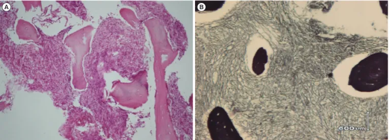
Genes on chromosome 21 play an important role in leukemo- genesis [8]. One gene ––RUNX1(AML1) at 21q22–– plays a Fig. 1. (A) Bone marrow needle biopsy specimen, showing grade 3 fibrosis (H&E stain, ×100). (B) Reticulin staining showing grade 3 fi- brosis (Reticulin stain, ×200).
A B
Fig. 2. Patient with Down syndrome exhibiting primary myelofibrosis with pentasomy 21. (A) Karyogram (Karyotype: 49,XX,+11,+19, +21c,der(21;21)(q10;q10)c, +der(21;21)[19]/46,XX,+21c,der(21;21)(q10;q10)c[9]. Arrows indicate abnormal chromosomes of 11, 19, and 21. (B) FISH analysis with a locus-specific probe for 21q22.13-q22.2, revealing pentasomy 21.
A B
Park Y, et al.
Pentasomy 21 in acute megakaryoblastic leukemia
http://dx.doi.org/10.3343/alm.2015.35.3.373 www.annlabmed.org 375
crucial role in hematopoiesis during embryonic development, and is frequently involved in chromosomal rearrangements ob- served in various myeloid malignancies [9]. Isochromosome 21q, formed as idic(21)(p10;p10), could result in an increased gene copy number on the entire long arm of chromosome 21 [10]. Furthermore, the duplication of idic(21)(p10;p10) gave rise to pentasomy 21q and to amplification of oncogenes at 21q, such as RUNX, ETS, and ERG, which may be involved in the de- velopment of leukemia. Moreover, the MLL gene gain due to tri- somy 11, might be associated with the poor prognosis in this particular patient. To date, 16 cases of pentasomy 21 with two isochromosomes have been reported in hematologic malignan- cies, but the clinical features common to these cases are un- known [3].
This case shows us that hematologic surveillance of peripheral blood is needed in DS newborns, and close follow-up as well as optimal molecular and cytogenetic studies should be performed in cases with abnormal peripheral blood findings. Clarification of the mechanism underlying myelofibrosis and leukemogenesis in pentasomy 21q with two isochromosomes, in myeloid leukemia associated with DS, requires further studies.
Authors’ Disclosures of Potential Conflicts of Interest
No potential conflicts of interest relevant to this article were re- ported.
REFERENCES
1. Cortes JE, Kantarjian H, O’Brien S, Keating M, Pierce S, Freireich EJ, et al. Clinical and prognostic significance of trisomy 21 in adult patients with acute myelogenous leukemia and myelodysplastic syndromes.
Leukemia 1995;9:115-7.
2. Ohsaka A, Hisa T, Watanabe N, Kojima H, Nagasawa T. Tetrasomy 21 as a sole chromo- some abnormality in acute myeloid leukemia. fluo- rescence in situ hybridization and spectral karyotyping analyses. Cancer Genet Cytogenet 2002;134:60-4.
3. Shimoyama M, Yamamoto K, Nishikawa S, Minagawa K, Katayama Y, Matsui T. Duplication of isodicentric chromosome 21, idic(21)(p11.2), leading to pentasomy 21q in acute myeloid leukemia with multilineage dysplasia. Cancer Genet Cytogenet 2009;194:38-43.
4. Baumann I, Niemeyer CM, Brunning RD, Arber DA, Porwit A. Myeloid proliferations related to Down syndrome. In: Swerdlow SH, Campo E, Harris NL, et al. eds. WHO classification of tumours of haematopoietic and lymphoid tissue. 4th ed. Lyon: IARC, 2008:142–4.
5. Roy A, Roberts I, Norton A, Vyas P. Acute megakaryoblastic leukaemia (AMKL) and transient myeloproliferative disorder (TMD) in Down syn- drome: a multi-step model of myeloid leukaemogenesis. Br J Haematol 2009;147:3-12.
6. Alvarez S, MacGrogan D, Calasanz MJ, Nimer SD, Jhanwar SC. Fre- quent gain of chromosome 19 in megakaryoblastic leukemias detected by comparative genomic hybridization. Genes Chromosomes Cancer 2001;32:285-93.
7. Nimer SD, MacGrogan D, Jhanwar S, Alvarez S. Chromosome 19 ab- normalities are commonly seen in AML, M7. Blood 2002;100:3838-9.
8. Fonatsch C. The role of chromosome 21 in hematology and oncology.
Genes Chromosomes Cancer 2010;49:497-508.
9. Hilgenfeld E, Padilla-Nash H, McNeil N, Knutsen T, Montagna C, Tchin- da J, et al. Spectral karyotyping and fluorescence in situ hybridization detect novel chromosomal aberrations, a recurring involvement of chro- mosome 21 and amplification of the MYC oncogene in acute myeloid leukaemia M2. Br J Haematol 2001;113:305-17.
10. Robinson HM, Harrison CJ, Moorman AV, Chudoba I, Strefford JC. In- trachromosomal amplification of chromosome 21 (iAMP21) may arise from a breakage-fusion-bridge cycle. Genes Chromosomes Cancer 2007;46:318-26.