서론
임플란트가 처음 개발된 이후 치과 치료는 혁신적인 발전을 이 루었다. 그러나 상악구치부에 임플란트 식립은 구치부의 골 흡 수, 상악동의 함기화(pneumatization), 불량한 골질 등으로 임플 란트 식립에 어려움을 안고 있다.1 특히 치조골의 수직 골량의 부 족은 임플란트의 식립에 어려움이 있으며 짧은 임플란트 식립은 낮은 성공률로 이어졌다.2,3
이러한 수직 골량의 부족을 극복하기 위한 방법으로 1977 년 Tatum이 Caldwell-Luc 수술법을 변형한 일명 측방 접근법 (lateral window method)를 제안했다.4 그러나 이러한 측방접근 법은 상악동 막의 천공, 동맥 천공에 따른 과다출혈, 감염, 상악 동염 등의 많은 합병증을 가지고 있었다.5-7
측방 접근법의 합병증들을 극복하고자 치조정 접근법(crestal approach method)인 osteotome 방법이 소개되었다.1,8 이러한 osteotome 방식은 상악동막을 거상하는 량에 한계가 있으며,
S-reamer와 겔 형태의 이식재를 이용한 치조정 접근법을 통한
상악동 거상술 임플란트의 성공률과 생존율: 5년 이상 추적 관찰을 통한 후향적 연구
김종진 조성암*
경북대학교 치과대학 치과보철학교실
Success and survival rate of the implant with crestal sinus lift using S-reamer and gel-type graft material: A retrospective study by more 5-years follow check up
Jong Jin Kim, Sung Am Cho*
Department of Prosthodontics, School of Dentistry, Kyungpook National University, Daegu, Republic of Korea
Purpose: The purpose of this retrospective study was to evaluate the method using the S-reamer and gel-type graft material by the success rate and survival rate. Materials and methods: Implantation period was from 2008 to 2014, Follow check up year is 2019. There were 59 patients and 117 implants. All implants were placed in the posterior maxilla with the sinus lift. The patients population consisted of 34 men and 25 women, ranging from 19 to 75 years. The residual bone heights were from 1 mm to 6 mm.
Sinus was perforated with S-reamer without membrane tearing and gel type bone graft material was used for membrane lifting and filling the space. all implants were placed simultaneously. Panoramic X-ray was taken. After 5 - 6 months healing period, final prostheses were restored. After more 5-years implant surgery, Panoramic X-ray was obtained and X-ray analysis and clinical examination were performed. Success criteria was referred to a Buser’s success critera. All implants were classified to success implant, survival implant, failed implant. A success implant was satisfying success criteria, a survival implant was a implant that was acute infection with suppuration and bone loss, a failed implant was a implant that was mobile, removed. Results: Five implants were removed, and 4 implants had infected with bone loss. Survival rate was 95.7% and success rate was 92.3%. Conclusion: This retrospective study presented that this method with S-reamer and gel-type graft material was a successful treatment without membrane tear in the condition of 1-6 mm residual bone height. (J Korean Acad Prosthodont 2020;58:23-9)
Keywords: Crestal approach; Gel-type graft material; Hydraulic sinus lift; S-reamer
*Corresponding Author: Sung-Am Cho
Department of Prosthodontics, School of Dentistry, Kyungpook National University,
#2175 Dalgubeoldae-ro, Jung-gu, Daegu 41940, Republic of Korea.
+82 (0)53 600 7672: e-mail, [email protected]
Article history: Received October 21, 2019 / Last Revision November 26, 2019 / Accepted December 16, 2019
2020 The Korean Academy of Prosthodontics
This is an Open Access article distributed under the terms of the Creative Commons Attribution Non-Commercial License (http://creativecommons.org/
licenses/by-nc/4.0) which permits unrestricted non-commercial use, distribution, and reproduction in any medium, provided the original work is properly cited.
c cc
Engelke과 Deckwer의 연구에서 상악동 막의 천공 없이 상악동 막을 최대 5 mm까지 거상할 수 있다고 보고 하였다.9 측방 접근 법은 상악동 막을 거상하면서 거상량과 형태를 볼 수 있고 가름 할 수 있는 반면 치조정 접근법은 상악동 막의 거상량과 모양과 형태를 볼 수도 가름할 수도 없다.
상악동 막의 찢김을 방지하고자 특수한 드릴의 개발, 생리 식 염수를 사용하여 수압으로 거상하는 수압 거상법, 생리 식염수 대신 생체적합 이식재를 사용하는 방법 등 다양한 방법들이 소 개되었다.10-12 이러한 방법중 이미 잘 알려지고 사용되어지는 겔 형태 이식재(gel type bone graft)를 사용하면 상악동 막을 거상 하는 방법이 상악동 막의 천공이 없고 환자의 불편감 감소등의 장점이 있다.12
치조정 접근법의 하나인 S-reamer와 이식재로 겔형태 이식재 를 사용하는 술식이 많은 장점을 가지고 있고 현재 널리 사용되 어지는 술식임에도 불구하고 5년 이상의 장기간의 결과를 관찰 한 연구는 부족하다. 본 연구는 연구자가 대구에서 개원하고 있 는 개인 치과의원에서 다양한 수직 골량을 가지는 환자에서 치 조정 접근법을 통한 술식으로 S-reamer와 이식재로 겔 형태 이식 재를 사용하여 상악동 막을 거상하면서 동시에 임플란트를 식립 한 후 5년 이상 추적 관찰(follow up check)를 통하여 성공률과 생존율을 바탕으로 치조정 접근법의 하나인 이 술식에 대한 결 과를 보고 하고자 한다.
대상 및 방법
1. 대상
2008년에서 2014년까지 상악동 거상술을 받은 환자를 추적 관찰하였다. 추적 관찰은 2019년 실시하였다. 전체 59명의 환자 에서 117개의 임플란트가 연구에 포함되었으며 대상과 시술에 대한 정보는 Table 1과 같다.
2. 수술방법
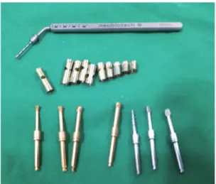
통법으로 마취를 시행 후 치은을 박리하여 상악 구치부 치조 골을 노출 시킨 후 SCA kit (Neo Biotech, Seoul, Korea)의 S- reamer (Fig. 1)를 이용하여 상악동 막의 천공없이 상악동을 거 상하였다. 그후 주사기에 담겨진 겔 형태의 이식재를 천공된 구 멍에 (Fig. 2) 압착한 후 이식재를 상악동으로 주입함으로 (Fig.
3) 상악동 막이 거상이 이루어지는 동시에 이식재를 거상된 상악
Fig. 1. S-reamer.
Table 1. Information of the patients and implants
Patients 59
Implant number 117
Implant type Internal submerged Implant manufacture B co, N co, D co, O co
Sex M: 34, F: 25
Age under 30’: 1, 30’: 1, 40’: 27, 50’: 19, 60’: 8, 70’: 3 Patient number in
operation year 08: 5, 09: 5, 10: 5, 11: 6, 12: 11, 13: 12, 14: 15 Implant number in
bone height under 2 mm: 13, 2 - 4 mm: 53, 4 - 6 mm: 51 Implant number in
implant length 8 mm: 3, 10 mm: 110, 12 mm: 4 Implant width 4.5 - 5.2 mm
Fig. 2. After drilling with S-reamer.
Fig. 3. Insertion of gel-type bone graft.
동에 채웠다. 이러한 술식의 원리는 과거 수압으로 상악동 막을 거상하던 방식과 유사하다. 수압법에서 생리 식염수가 균등한 압력으로 막을 거상하던 것을 겔 형태의 이식재가 생리 식염수 의 역할을 하게 된 것이다. 이식재는 Demineralized freeze-dried bone allograft 종류의 SureFuse (Hans Biomed, Seoul, Korea)를 사용하였다 (Fig. 4).
상악동 거상술과 동시에 임플란트를 식립하였다 (Fig. 5, Fig.
6). 식립 후 봉합하고 술 후 경구투약을 실시하였다. 이후 치유 기간 5 - 6개월 후에 최종 보철물을 수복하였다. 몇몇 경우에는 임시 보철물을 통한 점진적인 부하를 가한 후 최종 보철물을 수 복하였다.
3. 방사선적 분석과 임상검사
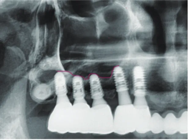
추적 관찰할 때 찍은 파노라마 방사선 사진 (Fig. 7)과 임플란 트 식립후 찍은 파노라마 방사선 사진을 비교하여 골 소실 여부 를 관찰하고 소실 골량을 측정하였다. 임상적으로 치은을 관찰 하고 탐침을 통하여 염증 여부, 화농 여부, 출혈 여부 등을 검사 하였다.
결과
성공률과 생존율에 대한 기준은 Buser의 성공 기준을 참고하 였다.13
Table 2를 참고로 하여 임플란트를 분류하였다. 성공 기준을 모두 만족하는 임플란트를 성공 임플란트로, 화농, 진행하는 골 소실이 있는 급성 염증을 가지는 임플란트 혹은 같은 증상의 과 거력이 있는 임플란트를 생존하는 임플란트로, 제거된 것, 움직 이는 임플란트를 실패한 임플란트로 분류하였다.
Table 2. Buser’s Success Criteria
No pain, no foreign body sensation, no dysesthesia No peri implant infection with suppuration No mobility
No radiolucency around implant Fig. 4. DFDBA bone graft.
Fig. 5. Pre-operation X-ray.
Fig. 6. Post-operation X-ray.
Fig. 7. Follow-up check X-ray.
Table 3. Result of follow up evaluation
Removed implant 5
Implant with infection and suppuration 4
Success rate 92.3%
Survival rate 95.7%
Patient and implant with bone loss 16, 24
Amount of bone loss average: 1.69 mm, max: 3.4 mm min: 1 mm
Patient with rehandled dome shape 12
고찰
상악 구치부에서 구치부 치조골의 소실, 상악동저 함기화 등 에 의해 수직골이 부족한 경우 Tatum에 의해 제안된 측방 접근 법이 성공적인 임플란트 치료였다.4,14-16 그러나 측방접근법은 상 악동 막 천공, 술 후 상악동염, 술 후 통증, 술 후 심한 부종 등 많 은 합병증을 유발한다.5,7,17 이러한 합병증 중에 상악동 막의 천 공이 3.6 - 56%에 이른다는 보고가 있다.5,18,19 또한 이러한 상악 동 막의 천공은 상악동염, 이식재 소실, 감염, 임플란트의 실패로 이어진다는 보고가 있다.5,7 이러한 보고를 보면 상악동 거상술을 시행할 때 막의 천공이 없이 상악동 막을 거상하는 것이 쉬운 일 이 아니며 임플란트 성공에 중요한 요인이라는 것을 알 수 있다.
측방접근법의 합병증을 극복하고자 Tatum에 의해 처음 개 념이 만들어지고 Summer에 의해 널리 알려진 치조정 접근법 인 Summer’s technique이 개발되어졌다. 이것은 osteotome이라 는 기구를 사용하여 치조정으로 상악동 막을 거상하는 방법이 다.8,20
이러한 osteotome법도 거상량에 한계가 있어서 막의 천공없이 5 mm 이상의 거상은 힘들었다.9 수직 골량이 아주 적은 경우에 는 적용하기가 힘들다. 그러나 최근의 연구에 의하면 치조정 접 근법을 통하여도 상악동의 수직 골이 4 mm 혹은 5 mm 이하인 경우에도 높은 성공률을 보인다. 이러한 것은 기구와 드릴의 발 전, 새로운 기술에 의한 것이다.21-23
가장 중요한 것은 막의 천공없이 상악동 막을 거상하는 것이 다. 여러 가지 연구 중 2005년에 Sotirakis 등이 소개한 생리식 염수와 주사기를 이용하여 수압으로 상악동 막을 거상하는 방 법이 소개되었다.11 그 후 이 술식의 어려움과 한계를 시정한 hydraulic sinus lift가 소개되었는데 이것은 특수한 기구를 사용 하여 이식재를 바로 주입함으로 상악동 막을 거상하며 동시에 이식재를 거상한 공간에 채우는 방식이다.12 본 연구에서도 이러 한 원리를 이용하였다.
이러한 원리에 기반하여 겔 형태의 이식재를 이용하면 균일한 압력을 상악동막에 주게 되어 막의 천공이 잘 일어나지 않는다.
또한 거상량에 제한을 받지 않는다. 본 연구에서 수직골이 2 mm 이하인 환자에서도 막의 천공 없이 거상을 시킬 수 있었다. 본 연 구에서 살펴본 모든 사례에서 상악동 막을 거상할 때 막의 천공 은 단 한 건도 발생하지 않았다.
이번 술식에서 막의 천공없이 상악동을 천공하는 술식 또한 중요하다. Osteotome법에 의한 방법은 상악동저 1 mm 하방에 서 기구와 망치를 이용하여 골을 파절 거상하는 방식을 사용하 는데 이러한 방식은 막의 천공의 쉽고 환자에게 불안감을 주게 된다. 본 연구에서 상악동을 천공하는데 사용한 기구는 SCA kit (Nobiotech)의 S-reame로써 막의 천공없이 상악동을 천공 시킨 다.23 이러한 새로운 술식과 기구의 발전을 통하여 환자는 치과에 대한 불안감없이 편안하게 진료를 받게 되고 술식의 용이함과 수술 시간의 단축을 알 수 있었다.
본 연구에서 성공률과 생존율은 Buser의 성공 기준을 참고하
여 성공기준으로 삼고, 각 임플란트를 성공 임플란트, 생존 임 플란트와 실패 임플란트로 분류하여 산출하였다.13 이러한 기준 에 의하여 성공률은 92.3%이며 생존율은 95.7%이다. 이것은 다 른 연구들의 생존율 85.7 - 97.9% 범위 안에 들어가는 것으로 보
인다.21,24-26 생존율은 95%이상이며 성공률도 90%이상을 보이고
있다. 따라서 본 연구의 술식이 생존율이 다른 술식과 비슷한 결 과를 보이면서 동시에 많은 장점을 가지고 있다고 말할 수 있다.
실패한 환자들을 살펴보면 대부분이 심한 흡연가였다. 하루 에 한 갑 이상을 피는 심한 흡연가였다. 이전 연구를 살펴보면 흡 연이 임플란트의 실패에 영향을 미친다는 문헌이 있다.27-29 하지 만 최근의 연구에서는 흡연이 임플란트의 실패와 무관하다는
연구도 있다.30-32 이러한 여려가지 견해 가운데 메타분석(meta-
analysis)을 통한 연구에 의하면 상악에서는 흡연이 임프란트의 실패에 영향을 미치고 하악에서 미치지 않는 것으로 나타났다.33 이러한 과거의 문헌을 참고로 볼 때 상악동 거상술을 통한 임플 란트의 실패는 흡연 여부에 영향을 받는 것으로 보여진다.
점진적인 부하(progressive loading)는 1980년대 Misch가 처 음 개념을 제안했다. 그 후 많은 연구를 통하여 점진적인 부하가 골의 밀도와 골질을 향상시키며 치조정의 골소실과 임플란트의 초기 실패를 막는다고 한다.34-36 환자 사례에는 점진적인 부하 없 이 최종 보철물을 한 환자도 있고 임시치아를 사용하여 점진적 인 부하를 가한 환자도 있었다. 점진적인 부하를 가한 환자 22명 중에 골 소실이 있는 환자는 10명이며 점진적인 부하를 가하지 않은 환자 37명중 골 소실이 있는 환자는 6명이었다. 위의 결과 로 평가할 수는 없는 것 같다. 왜냐하면 골질이 나쁘거나 수직 골 량이 적은 경우에 점진적인 부하를 가한 경우가 많아서 같은 조 건하에 비교가 어려웠다. 그리고 임플란트의 생존에 가장 영향 을 많이 미치는 인자가 수직 골량에 있기 때문에 여기서는 평가 가 어려울 것으로 보인다.24
상악동 거상술을 실시하고 즉시 임플란트를 식립한 후 치유 기간이 끝나고 보철물을 장착하고 기능을 한 후 추적 관찰을 하 는 동안에 상악동 거상술을 처음 실시했을 때와 다르게 거상된 상악동 막이 아래로 쳐진 형태를 보게 되었다. 이것은 치유과정 에서 겔 형태의 이식재가 흡수되고 골로 바뀌는 과정에서 흡수 가 많이 되었거나 치조정 접근법이 측방 접근법에 비해 볼 수 없 어서 거상형태와 거상량을 결정할 수 없다는 단점을 나타낸 것 으로 보인다. 2003년 문헌에서 흡수되어 재조정되는 양이 0.94 - 1.5 mm 정도 되므로 상악동 거상시 돔 형태가 임플란트 상방 2 mm 이상 되도록 하기를 권유하고 있다.37 이러한 돔 형태의 소실 은 이상적인 상악동 거상술식이라 할 수는 없다. 본 연구에서는 이러한 돔 형태의 소실이 전체 환자 59명중 12명에서 나타났다.
그 중에 골소실을 보인 환자는 5명으로 41.6%이며 돔 형태의 소 실이 없는 환자 47명중 골소실 환자는 11명으로 23.4%이다. 더 많은 연구가 필요한 듯하며 위의 내용처럼 상악동 거상시에 더 많은 이식재를 사용하여 거상량을 높일 필요는 있는 것 같다.
결론
S-reamer와 겔타입의 이식재를 이용한 상악동 거상술과 함께 식립된 임플란트의 성공률과 생존율은 각각 92.3%, 95.7%이며 이러한 상악동 거상술이 1 - 6 mm 잔존골이 있는 상황에서 막 천공이 없이 상악동을 거상할 수 있는 성공적인 술식임을 알 수 있었다.
ORCID
Sung-Am Cho https://orcid.org/0000-0002-8315-7833
References
1. Tatum H Jr. Maxillary and sinus implant reconstructions.
Dent Clin North Am 1986;30:207-29.
2. Misch CE, Steignga J, Barboza E, Misch-Dietsh F, Cianciola LJ, Kazor C. Short dental implants in posterior partial eden- tulism: a multicenter retrospective 6-year case series study. J Periodontol 2006;77:1340-7.
3. Petrie CS, Williams JL. Comparative evaluation of implant designs: influence of diameter, length, and taper on strains in the alveolar crest. A three-dimensional finite-element analysis.
Clin Oral Implants Res 2005;16:486-94.
4. Tatum OH. Maxillary sinus grafting for endosseous implant.
Presented at the annual meeting of the alabama implant study group. Birmingham, AL, April 1977.
5. Schwartz-Arad D, Herzberg R, Dolev E. The prevalence of surgical complications of the sinus graft procedure and their impact on implant survival. J Periodontol 2004;75:511-6.
6. Schwarz L, Schiebel V, Hof M, Ulm C, Watzek G, Pommer B.
Risk factors of membrane perforation and postoperative com- plications in sinus floor elevation surgery: Review of 407 aug- mentation procedures. J Oral Maxillofac Surg 2015;73:1275- 7. Katranji A, Fotek P, Wang HL. Sinus augmentation complica-82.
tions: etiology and treatment. Implant Dent 2008;17:339-49.
8. Summers RB1. A new concept in maxillary implant surgery:
the osteotome technique. Compendium 1994;15:152, 154-6, 158 passim; quiz 162.
9. Engelke W, Deckwer I. Endoscopically controlled sinus floor augmentation. A preliminary report. Clin Oral Implants Res 1997;8:527-31.
10. Cosci F, Luccioli M. A new sinus lift technique in conjunction with placement of 265 implants: a 6-year retrospective study.
Implant Dent 2000;9:363-8.
11. Sotirakis EG, Gonshor A. Elevation of the maxillary sinus floor with hydraulic pressure. J Oral Implantol 2005;31:197- 12. Andreasi Bassi M, Lopez MA. Hydraulic sinus lift: a new 204.
method proposal. J Osteology Biomaterials 2010;1:93-101.
13. Buser D, Weber HP, Lang NP. Tissue integration of non-sub- merged implants. 1-year results of a prospective study with 100 ITI hollow-cylinder and hollow-screw implants. Clin Oral Implants Res 1990;1:33-40.
14. Lee HW, Lin WS, Morton D. A retrospective study of com- plications associated with 100 consecutive maxillary sinus augmentations via the lateral window approach. Int J Oral Maxillofac Implants 2013;28:860-8.
15. Del Fabbro M, Rosano G, Taschieri S. Implant survival rates after maxillary sinus augmentation. Eur J Oral Sci 2008;116:497-506.
16. Pjetursson BE, Tan WC, Zwahlen M, Lang NP. A systematic review of the success of sinus floor elevation and survival of implants inserted in combination with sinus floor elevation. J Clin Periodontol 2008;35:216-40.
17. Pikos M. Complications of maxillary sinus augmentation. In Jensen OT (eds): The sinus graft. 2nd ed. Chicago, IL: Quin- tessence, 2006. p. 103-15.
18. Al-Dajani M. Incidence, risk factors, and complications of schneiderian membrane perforation in sinus lift surgery: A meta-analysis. Implant Dent 2016;25:409-15.
19. Wallace SS, Mazor Z, Froum SJ, Cho SC, Tarnow DP.
Schneiderian membrane perforation rate during sinus eleva- tion using piezosurgery: clinical results of 100 consecutive cases. Int J Periodontics Restorative Dent 2007;27:413-9.
20. Lalo J, Broukris G, Djemil M, Beleh M. Safe technique for sinus floor elevation through alveolar crest with stop sinus osteotomes. Implantodontie 2005;14:62-70.
21. Bernardello F, Righi D, Cosci F, Bozzoli P, Soardi CM, Spi- nato S. Crestal sinus lift with sequential drills and simultane- ous implant placement in sites with <5 mm of native bone: a multicenter retrospective study. Implant Dent 2011;20:439- 22. Lopez MA, Andreasi Bassi M, Confalone L, Carinci F. Max-44.
illary sinus floor elevation via crestal approach: the evolu- tion of the hydraulic pressure technique. J Craniofac Surg 2014;25:e127-32.
23. Kim YK, Lee JY, Park JW, Kim SG, Oh JS. Sinus membrane elevation by the crestal approach using a novel drilling sys- tem. Implant Dent 2017;26:351-6.
24. Rosen PS, Summers R, Mellado JR, Salkin LM, Shanaman RH, Marks MH, Fugazzotto PA. The bone-added osteotome sinus floor elevation technique: multicenter retrospective re- port of consecutively treated patients. Int J Oral Maxillofac Implants 1999;14:853-8.
25. Peleg M, Garg AK, Mazor Z. Predictability of simultaneous implant placement in the severely atrophic posterior maxilla:
A 9-year longitudinal experience study of 2132 implants placed into 731 human sinus grafts. Int J Oral Maxillofac Im- plants 2006;21:94-102.
26. Thor A, Sennerby L, Hirsch JM, Rasmusson L. Bone forma- tion at the maxillary sinus floor following simultaneous eleva- tion of the mucosal lining and implant installation without
graft material: an evaluation of 20 patients treated with 44 Astra Tech implants. J Oral Maxillofac Surg 2007;65:64-72.
27. Johnson GK, Hill M. Cigarette smoking and the periodontal patient. J Periodontol 2004;75:196-209.
28. Jones JK, Triplett RG. The relationship of cigarette smoking to impaired intraoral wound healing: a review of evidence and implications for patient care. J Oral Maxillofac Surg 1992;50:237-9; discussion 239-40.
29. Wallace RH. The relationship between cigarette smoking and dental implant failure. Eur J Prosthodont Restor Dent 2000;8:103-6.
30. Carlsson GE, Lindquist LW, Jemt T. Long-term marginal pe- riimplant bone loss in edentulous patients. Int J Prosthodont 2000;13:295-302.
31. Kumar A, Jaffin RA, Berman C. The effect of smoking on achieving osseointegration of surface-modified implants: a clinical report. Int J Oral Maxillofac Implants 2002;17:816-9.
32. Lambert PM, Morris HF, Ochi S. The influence of smoking
on 3-year clinical success of osseointegrated dental implants.
Ann Periodontol 2000;5:79-89.
33. Hinode D, Tanabe S, Yokoyama M, Fujisawa K, Yamauchi E, Miyamoto Y. Influence of smoking on osseointegrated im- plant failure: a meta-analysis. Clin Oral Implants Res 2006;
17:473-8.
34. Misch CE. Progressive bone loading. In: Misch CE, ed. Den- tal implant prosthetics. St. Louis: Mosby; 2005. p. 511-30.
35. Appleton RS, Nummikoski PV, Pigno MA, Cronin RJ, Chung KH. A radiographic assessment of progressive loading on bone around single osseointegrated implants in the posterior maxilla. Clin Oral Implants Res 2005;16:161-7.
36. Siadat H, Panjnoosh M, Alikhasi M, Alihoseini M, Bassir SH, Rokn AR. Does implant staging choice affect crestal bone loss? J Oral Maxillofac Surg 2012;70:307-13.
37. Ozyuvaci H, Bilgiç B, Firatli E. Radiologic and histomorpho- metric evaluation of maxillary sinus grafting with alloplastic graft materials. J Periodontol 2003;74:909-15.
S-reamer와 겔 형태의 이식재를 이용한 치조정 접근법을 통한
상악동 거상술 임플란트의 성공률과 생존율: 5년 이상 추적 관찰을 통한 후향적 연구
김종진 조성암*
경북대학교 치과대학 치과보철학교실
목적: 이번 후향적 연구의 목적은 수압을 이용하는 방식을 변형한 방식인 S-reamer 와 겔 형태의 이식재를 이용한 상악동 거상술을 시행한 임플란트의 5년 이상 추적 관찰을 함으로써 성공률과 생존률을 조사하여 이 술식에 대한 평가를 하는데 있다.
재료 및 방법: 2008년에서 2014년까지 환자들을 추적 관찰하였다. 관찰된 환자는 59명이었고 식립 임플란트는 117개였다. 남성 34명, 여성 25명이고 연령대는 다양하였다. 잔존 수직 골의 골 량은 1 - 6 mm로 다양하였다. 상악동 거상술은 막의 천공없이 상악동을 천공하는데 S-reamer를 사용하였고 막을 거상하는데 겔 형태의 이식재를 사용하여 상악동 거상술을 시행하였다. 모든 임플란트는 거상과 동시에 식립하였고 5 - 6개월 치유 기간 후 보철 물을 장착하였다. 방사선 검사는 추적 검사 할 때와 식립 후 바로 찍은 방사선 검사를 비교하였다 그리고 탐침을 통하여 화농여부, 염증여부, 출혈여부 등등 임상 검사를 통하여 검진 평가 하였다. Buser의 성공 기준을 참고하였고 모든 임플란트를 성공 임플란트, 생존 임플란트, 실패 임플란트로 분류하 였다.
결과: 실패하여 제거한 임플란트는 5개였고 골 소실을 동반한 급성 염증 치료를 했거나 염증을 보인 임플란트가 4개였다. 생존율은 95.7%이고 성공률 은 92.3%였다.
결론: S-reamer와 겔 형태의 이식재를 이용한 상악동 거상술이 1 - 6 mm 잔존 골이 있는 상황에서 막 천공이 없이 상악동을 거상할 수 있는 성공적인 술식임을 알 수 있었다. (대한치과보철학회지 2020;58:23-9)
주요단어: 치조정 접근법; 겔 형태 이식재; 수압 거상법; S-reamer
*교신저자: 조성암
41940 대구 중구 달구벌대로 2175 경북대학교 치과대학 치과보철학교실 053 600 7672: e-mail, [email protected]
원고접수일: 2019년 10월 21일 / 원고최종수정일: 2019년 11월 26일 / 원고채택일: 2019년 12월 16일
2020 대한치과보철학회
이 글은 크리에이티브 커먼즈 코리아 저작자표시-비영리 4.0 대한민국 라이선스에 따라 이용하실 수 있습니다.
c cc