Radiologic and Clinical Outcomes of Surgery in High Grade Spondylolisthesis Treated with
Temporary Distraction Rod
Farzad Omidi-Kashani, MD, Alireza Hootkani, MD, Lida Jarahi, MD*, Manizheh Rezvan, MD, Amir Moayedpour, MD
Orthopedic Research Center, Orthopedic Department, Imam Reza Hospital, Mashhad University of Medical Sciences, Mashhad,
*Addiction Research Center, Faculty of Medicine, Mashhad University of Medical Science, Mashhad, Iran
Received August 17, 2014; Accepted September 18, 2014 Correspondence to: Farzad Omidi-Kashani, MD
Orthopedic Research Center, Orthopedic Department, Imam Reza Hospital, Imam Reza Square, Mashhad University of Medical Sciences, Mashhad, Iran Tel: +98-915-514-9248, Fax: +98-51-38595023
E-mail: [email protected]
The patients with high grade lumbar spondylolisthesis (slip percentage > 50%) are usually treated by conservative treatment, but surgical decompression and stabilization are occasionally required.1,2) Surgical indications include progressive slip (especially in pediatric patients), signifi-
Background: Surgical techniques used in the treatment of patients with high grade lumbar spondylolisthesis (> 50% slippage) are usually associated with a great deal of controversies. We aim to evaluate the surgical outcomes of high grade spondylolisthesis treated with an intraoperative temporary distraction rod.
Methods: We retrospectively studied 21 patients (14 females and 7 males), aged 50.4 ± 9.2 years, who had high grade lumbar spondylolisthesis that was treated with intraoperative temporary distraction rods, neural decompression, pedicular screw fixation, and posterolateral fusion involving one more intact upper vertebra. The mean follow-up period was 39.2 months. Radiologic and clinical outcomes were measured by slip angle, slip percentage, correction rate, Oswestry Disability Index (ODI), visual analogue scale (VAS), patient’s satisfaction rate in the pre- and postoperative period. Data were analyzed by SPSS ver. 11.5.
Results: Analysis of the preoperative visits and final follow-up visits indicated that surgery could improve ODI, lumbar VAS, and leg VAS from 60.5% to 8.2%, from 6.7 to 2.2, and from 6.9 to 1.3, respectively. Slip angle and slip percentage were also changed from –8° to –15° and from 59.2% to 21.4%, respectively. Mean correction rate at the final follow-up visit was 64.1%. Loss of cor- rection was insignificant and a neurologic complication occurred in one patient due to misplacement of one screw. Excellent and good levels of satisfaction were observed in 90.5% of the patients.
Conclusions: In the surgical treatment of refractory high grade spondylolisthesis, the use of a temporary distraction rod to reduce the slipped vertebra in combination with neural decompression, posterolateral fusion, and longer instrumentation is associated with satisfactory clinical and radiologic outcomes.
Keywords: Spondylolisthesis, Surgery, Treatment outcome, Radiology
cant lumbosacral kyphotic deformity, neurologic deficit, intractable back pain, and refractory radicular pain.2) Many surgical approaches and techniques have been used for treating these special cases. In situ posterolateral fu- sion, in situ transsacral fusion, vertebral reduction and fusion via two separate anterior and posterior approaches or a single stage posterior approach are the commonly performed techniques.3-11) In operative management of high grade spondylolisthesis, the risk of neurologic com- plications and pseudoarthrosis is usually high.2,12) It would be ideal to find a simple method that can restore normal spinal alignment without causing any iatrogenic complica-
Copyright © 2015 by The Korean Orthopaedic Association
This is an Open Access article distributed under the terms of the Creative Commons Attribution Non-Commercial License (http://creativecommons.org/licenses/by-nc/3.0) which permits unrestricted non-commercial use, distribution, and reproduction in any medium, provided the original work is properly cited.
Clinics in Orthopedic Surgery • pISSN 2005-291X eISSN 2005-4408
tions and can provide satisfactory clinical outcomes.2,13) In this study, we aim to evaluate the surgical outcomes of high grade spondylolisthesis treated with an intraoperative temporary distraction rod, vertebral reduction, fusion and instrumentation, through the isolated posterior approach.
METHODS
After obtaining local Institutional Review Board approval
(registration number 899,534), we retrospectively stud- ied the patients with high grade (> 50% slippage) lumbar spondylolisthesis that was treated with an intraoperative temporary distraction rod at our Orthopedic Department in Imam Reza Hospital, Mashhad, Iran, from August 2009 to May 2012. We only included the patients with high grade spondylolisthesis who were clinically symptomatic and refractory to aggressive conservative treatment for more than three months, although in skeletally immature patients there was a high probability of slip progression;
asymptomatic patients with high grade slippage were also the surgical candidates. The patients who suffered from trauma, infection, or tumor-induced spondylolisthesis, the patients with significant underlying generalized diseases (like Paget disease, osteogenesis imperfecta, severe osteo- porosis, etc.), or the patients with less than two years of follow-up were excluded from this study. We also excluded very immature or anomalous patients in whom safe inser- tion of pedicle screws could not be guaranteed.
All of the mature patients signed the informed con- sent form. For the patients who had not reached the legal age of consent, the informed consent forms were signed by their legal guardians. Preoperatively, magnetic resonance imaging scans, standing anteroposterior and lateral ra- diographs were taken for all of the patients. Slip angle was measured as the angle formed by the L5 inferior endplate and the line perpendicular to the posterior aspect of S1.3) Slip percentage was calculated as a percentage of forward displacement of the upper vertebra on the top of the lower vertebra.1) Correction rate was measured by dividing the difference in slip percentage (pre- and postoperative slip- Fig. 1. Schematic representation of reduction stages. In stage one,
gentle distraction is applied to the upper and lower vertebrae. In stage two, gentle tightening of the intermediate (slipped) pedicle screws is performed.
Stage 1 Stage 2
Stage 1
L3
S1
Fig. 2. (A) A 48-year-old female with high grade spondylolisthesis (slip percentage 59%). She had a previous history of femoral shaft fracture. (B) This figure shows L5 laminectomy and the temporary distraction rod along with supra- and infralaminar hooks in S1 and L3, respectively. Pieces of cotton are placed on the dura mater for more protection. (C) Postoperative lateral radiograph shows a nearly perfect reduction (9% slip percentage with 85%
correction rate).
A B C
page) by preoperative slip percentage multiplied by 100.
Surgical Technique
All of the surgical procedures were performed by the se- nior author (FOK) using the isolated posterior approach and a similar technique. After exposing the posterior surfaces and laminectomy of the involved vertebrae, a midline temporary rod with its accompanying supralami- nar and infralaminar hooks was inserted into the lower and upper vertebrae for applying temporary intervertebral distraction to facilitate slippage reduction. Then bilateral pedicle screws and two lordotic molded rods were inserted bilaterally. Concurrent with screw tightening, the slipped vertebra gradually moved posteriorly and the slippage im- proved (Fig. 1). Neural elements were under direct visual control and neural impingement or excessive tension was avoided (Fig. 2). After vertebral reduction, the midline distraction rod along with its hooks was removed. In some cases, discectomy was also performed to promote vertebral reduction. Since reduction screws reduce the slipped ver- tebra, they are prone to loosening; therefore, we routinely included one more upper vertebra in the instrumentation construct. We did not use a lumbar interbody cage in any of the patients.
We evaluated patients’ disability and pain by using the Oswestry Disability Index (ODI) questionnaire ver.
2.1 and the visual analogue scale on a scale from zero to
ten.14,15) The ODI questionnaire was previously translated
and validated for Persian speaking patients by Mousavi et al.16) in 2006. Patient satisfaction with surgery was also as- sessed subjectively and graded as excellent (if the patient was willing to undergo the same procedure for the same problem), good (if the surgery improved the problem but
did not meet all expectations of the patient), fair (if the pa- tient’s status did not change with the operation), and poor (when the operation caused worsening of the patient’s symptoms). We also recorded the significant intra- or postoperative complications.
Statistical Analysis
Data were analyzed by SPSS ver. 11.5 (SPSS Inc., Chicago, IL, USA). For descriptive analysis, we used the one-sample Kolmogorov-Smirnov test for normality, frequency, per- centage, and interquartile range. For analytical analysis, we used the paired t-test, repeated measures analysis of vari- ance, and correlation (Pearson, Spearman). A p-value less than 0.05 was considered to be statistically significant.
RESULTS
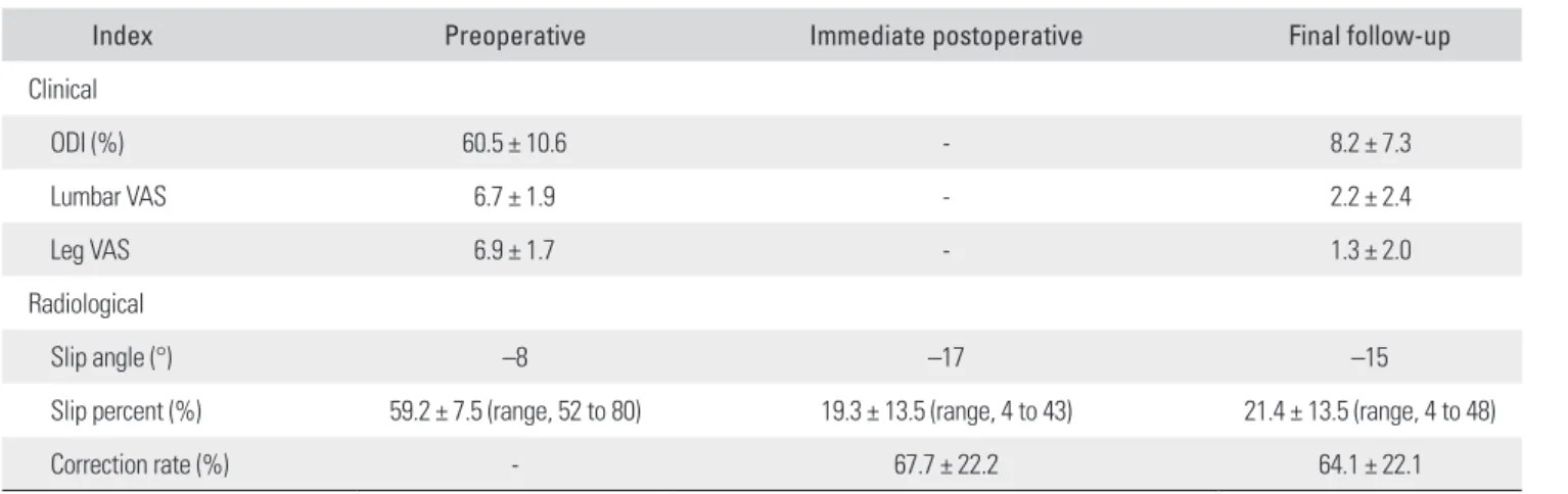
We finally evaluated 21 patients (14 females, 66.7% and 7 males, 33.3%) with a mean age of 50.4 ± 9.2 years (age range, 34 to 64 years). The location of slip was L5–S1 in 19 patients (90.5%) and L4–L5 in two patients (9.5%). The mean follow-up period was 39.2 ± 11.8 months (range, 25 to 60 months). Based on the one-sample Kolmogorov- Smirnov test, our data had a normal distribution. Clinical and radiologic data of our treated patients in the pre- and postoperative period are summarized in Table 1.
We did not exclude patients with spondyloptosis;
but there was no case of severe spondylolisthesis among our patients. Immediate and final correction rates showed no statistically significant difference (p = 0.470). Through- out the follow-up period, a 3.6% ± 3.7% loss of correc- tion was observed, and this amount of recurrence had no significant impact on the total correction rate (p = 0.091).
Table 1. Clinical and Radiologic Characteristics of the Study Patients
Index Preoperative Immediate postoperative Final follow-up
Clinical
ODI (%) 60.5 ± 10.6 - 8.2 ± 7.3
Lumbar VAS 6.7 ± 1.9 - 2.2 ± 2.4
Leg VAS 6.9 ± 1.7 - 1.3 ± 2.0
Radiological
Slip angle (°) –8 –17 –15
Slip percent (%) 59.2 ± 7.5 (range, 52 to 80) 19.3 ± 13.5 (range, 4 to 43) 21.4 ± 13.5 (range, 4 to 48)
Correction rate (%) - 67.7 ± 22.2 64.1 ± 22.1
Values are presented as mean ± standard deviation.
ODI: Oswestry Disability Index, VAS: visual analogue scale.
The amount of loss of correction also did not show any correlation with the duration of follow-up (r = 0.097, p = 0.677).
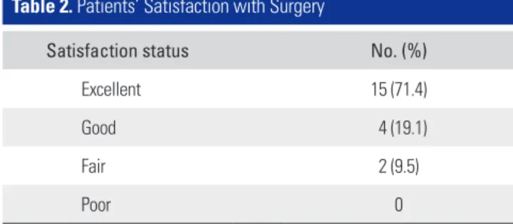
Intraoperative dural tear occurred in two patients, and it was repaired properly and it healed without any further complications. Due to intractable postoperative ra- dicular pain in one patient, re-insertion of a pedicle screw became necessary and then the pain improved immedi- ately. Postoperative worsening of the neurologic status did not occur in any of the patients. Two patients developed superficial wound infections that were cured with local wound care and oral antibiotics. Implant failure and non- union (at least symptomatically) did not occur in any of the patients, although we did not perform computed to- mography (CT) scanning routinely in all of the cases. Sub- jective satisfaction rate is shown in Table 2. There was no case with a poor surgical outcome and overall satisfaction (good and excellent results) was observed in 19 patients (90.5%). Although the amount of loss of correction ob- served throughout the follow-up was not significant, the satisfaction rate had a reverse correlation with the amount of loss of correction (r = –0.517, p = 0.16).
DISCUSSION
An assessment of the history of spine surgeries including those for spondylolisthesis indicates that most of the spine surgeons tend to perform spinal procedures via a posterior approach. The reasons that are usually cited include more familiarity with this approach, decreased risk of injury to great vessels or vital organs, greater ease of revision op- erations, ability to operate on multi-levels, and no need for assistance from a general or vascular surgeon.17-19) In surgical treatment of high grade spondylolisthesis, vari- ous approaches (posterior only, staged or simultaneous anterior and posterior approaches) have been reported in the literature, but it would be ideal to identify a familiar posterior approach that can achieve all surgical goals and provide appropriate long-term results.2,7) In this study, we think we could achieve this purpose with the use of tem-
porary intraoperative distraction technique.
In surgical treatment of high grade spondylolis- thesis, much controversy exists regarding the selection of vertebral reduction or in situ fusion among spine sur- geons. It seems that due to the availability of more power- ful and smaller implants that are more likely to achieve a successful fusion and provide better clinical satisfaction, reduction seems to be more popular.20) Hence, a variety of reduction techniques and tactics have been invented by numerous authors. Many of them have raised the issue of gradual reduction of the slipped vertebra.21,22) Karampalis et al.21) used Magerl’s external fixator for this purpose and then performed circumferential fusion in 9 patients with high grade spondylolisthesis and measured both clinical and radiological outcomes after about 11 years. The im- provement in slip magnitude (Meyerding classification), slip angle, lumbosacral angle, sacral rotation, and sacral inclination were 2.9 grades, 66%, 47%, 51%, and 47%, re- spectively. Although the patients’ expectations were met, solid fusion was achieved in 88.9% of the patients. Mean postoperative ODI and low back outcome scores were 8%
and 56.6%, respectively. The author finally recommended this technique as a powerful and safe technique without any neurologic complications that may be associated with other procedures. In comparison, the number of patients in our study was more than twice the number of patients in the above mentioned study and we had one patient with radicular pain who needed revision surgery for re- insertion of a pedicle screw. The mean improvement in slip magnitude and postoperative ODI in our study were 1.5 grades and 8.2%, respectively.
On the other hand, in 1996, Hu et al.5) reported the surgical results in 16 cases with high grade spondylolis- thesis that was treated with posterior decompression and reduction using the Edwards Modular Spine System. This technique could decrease slip percentage from 89% to 29%
and slip angle from 50° to 24°, respectively. Three cases developed a neurologic deficit and 4 patients had implant failure. Excellent, good, and fair results were obtained in 10, 5, and 1 patient, respectively. In conclusion, the authors themselves acknowledged that it was a complex technique and it may be associated with significant neurologic or hardware complications. In our study, the technique could improve slip percentage from 59% to 21% and slip angle from –8° to –15°, respectively. The comparison shows that the severity of vertebral slippage in our study was some- what lesser. In our study, there was only one case (4.8%) of neurologic complication and implant failure did not occur in any of the cases, but the overall surgical satisfaction was similar.
Table 2. Patients’ Satisfaction with Surgery
Satisfaction status No. (%)
Excellent 15 (71.4)
Good 4 (19.1)
Fair 2 (9.5)
Poor 0
In a study of 34 patients with high grade L5–S1 spondylolisthesis, Lengert et al.4) evaluated spondylolis- thesis reduction and maintenance over time with L4–S1 (25 patients) versus L5–S1 (9 patients) fusion. They used a lever-arm system and posterior fusion in combination with lumbosacral graft. In their patients, the mean Taillard spondylolisthesis index decreased from 64% to 37% and L5–S1 segmental lordosis increased from 11° to 18°. They observed a loss of reduction from 19° to 14° in the L5–
S1 group, while the reduction was maintained in the L4–
S1 group. These authors finally concluded that this system provided satisfactory reduction of spondylolisthesis, but in high grade L5–S1 spondylolisthesis, loss of reduction was better controlled with posterior L4–S1 fusion versus shorter L5–S1 fusion. In our study, we routinely included one upper intact vertebra in instrumentation to reduce the probability of implant failure and significant loss of correc- tion; therefore, we could not compare the results of short fusion versus long fusion but our results showed that long fusion was associated with non-significant loss of correc- tion.
Our study had some limitations. It had a retrospec-
tive design and it inevitably had the inherent limitations of retrospective studies. The number of patients was not significantly large, although it was not too small. CT scan- ning was not performed routinely in all of the cases for as- sessing the fusion rate, and this certainly reduced the accu- racy of fusion assessment. We suggest that in the future, a larger prospective study should be conducted by different surgeons using a similar technique and the results should be assessed by a neutral third person. In conclusion, in surgical treatment of refractory high grade spondylolis- thesis, the use of a temporary distraction rod to reduce the slipped vertebra in combination with neural decompres- sion, posterolateral fusion, and longer instrumentation is associated with satisfactory clinical and radiologic out- comes and this method can be the method of choice for surgical management of these difficult cases.
CONFLICT OF INTEREST
No potential conflict of interest relevant to this article was reported.
REFERENCES
1. Meyerding HW. Spondylolisthesis: surgical fusion of lumbo- sacral portion of spinal column and interarticular facets: use of autogenous bone grafts for relief of disabling backache. J Int Coll Surg. 1956;26(5 Part 1):566-91.
2. Kasliwal MK, Smith JS, Kanter A, et al. Management of high-grade spondylolisthesis. Neurosurg Clin N Am. 2013;
24(2):275-91.
3. Klockner C, Weber U. Correction of lumbosacral kyphosis in high grade spondylolisthesis and spondyloptosis. Ortho- pade. 2001;30(12):983-7.
4. Lengert R, Charles YP, Walter A, Schuller S, Godet J, Steib JP. Posterior surgery in high-grade spondylolisthesis. Or- thop Traumatol Surg Res. 2014;100(5):481-4.
5. Hu SS, Bradford DS, Transfeldt EE, Cohen M. Reduction of high-grade spondylolisthesis using Edwards instrumenta- tion. Spine (Phila Pa 1976). 1996;21(3):367-71.
6. Sailhan F, Gollogly S, Roussouly P. The radiographic results and neurologic complications of instrumented reduction and fusion of high-grade spondylolisthesis without decom- pression of the neural elements: a retrospective review of 44 patients. Spine (Phila Pa 1976). 2006;31(2):161-9.
7. Bridwell KH. Surgical treatment of high-grade spondylolis- thesis. Neurosurg Clin N Am. 2006;17(3):331-8.
8. Landi A, Marotta N, Mancarella C, Tarantino R, Delfini R.
Trans-sacral screw fixation in the treatment of high dyplas- tic developmental spondylolisthesis. World J Clin Cases.
2013;1(3):116-20.
9. Hart RA, Domes CM, Goodwin B, et al. High-grade spon- dylolisthesis treated using a modified Bohlman technique:
results among multiple surgeons. J Neurosurg Spine. 2014;
20(5):523-30.
10. Roca J, Ubierna MT, Caceres E, Iborra M. One-stage de- compression and posterolateral and interbody fusion for se- vere spondylolisthesis: an analysis of 14 patients. Spine (Phila Pa 1976). 1999;24(7):709-14.
11. Bohlman HH, Cook SS. One-stage decompression and pos- terolateral and interbody fusion for lumbosacral spondy- loptosis through a posterior approach: report of two cases. J Bone Joint Surg Am. 1982;64(3):415-8.
12. Kasliwal MK, Smith JS, Shaffrey CI, et al. Short-term com- plications associated with surgery for high-grade spondylo- listhesis in adults and pediatric patients: a report from the scoliosis research society morbidity and mortality database.
Neurosurgery. 2012;71(1):109-16.
13. Acosta FL Jr, Ames CP, Chou D. Operative management of adult high-grade lumbosacral spondylolisthesis. Neurosurg Clin N Am. 2007;18(2):249-54.
14. Wewers ME, Lowe NK. A critical review of visual analogue scales in the measurement of clinical phenomena. Res Nurs Health. 1990;13(4):227-36.
15. Fairbank JC, Pynsent PB. The Oswestry Disability Index.
Spine (Phila Pa 1976). 2000;25(22):2940-52.
16. Mousavi SJ, Parnianpour M, Mehdian H, Montazeri A, Mo- bini B. The Oswestry Disability Index, the Roland-Morris Disability Questionnaire, and the Quebec Back Pain Dis- ability Scale: translation and validation studies of the Iranian versions. Spine (Phila Pa 1976). 2006;31(14):E454-9.
17. Goya T, Morita Y. Chronological changes in the operative indications and approaches for the treatment of spondylosis deformans of the spine. Brain Nerve. 2009;61(6):627-35.
18. Bridwell KH. Indications and techniques for anterior-only and combined anterior and posterior approaches for thorac-
ic and lumbar spine deformities. Instr Course Lect. 2005;54:
559-65.
19. Xu GJ, Li ZJ, Ma JX, Zhang T, Fu X, Ma XL. Anterior versus posterior approach for treatment of thoracolumbar burst fractures: a meta-analysis. Eur Spine J. 2013;22(10):2176-83.
20. Hu SS, Tribus CB, Diab M, Ghanayem AJ. Spondylolisthesis and spondylolysis. J Bone Joint Surg Am. 2008;90(3):656- 71.
21. Karampalis C, Grevitt M, Shafafy M, Webb J. High-grade spondylolisthesis: gradual reduction using Magerl's external fixator followed by circumferential fusion technique and long-term results. Eur Spine J. 2012;21 Suppl 2:S200-6.
22. Wild A, Jager M, Webb JK. Staged reposition and fusion with external fixator in spondyloptosis. Z Orthop Ihre Grenzgeb. 2001;139(2):152-6.