Multiple myeloma is the most common primary bone malignancy with an unknown etiology. It occurs frequent- ly in older age groups and occurs twice as often in men as in women. The most common clinical signs and symp- toms of multiple myeloma include bone pain, fatigue, ane- mia, and infectious diseases, and it is characterized by multiple punched-out radiolucent lesions.1The clinical manifestations of the disease occur due to an expanding mass of plasma cells in the bone marrow and factors pro- duced by these cells, such as monoclonal immunoglobu- lin, Bence-Jones proteins, and osteoclast-activating fac- tors.2
Oral and maxillofacial manifestations of multiple myelo- ma very rarely present as an initial sign.1Jaw lesions, with an incidence of 8%-15%, may be a primary manifestation in the advanced stages of multiple myeloma.3A review of several case series and case studies indicated that the
maxillary involvement of multiple myeloma has been infre- quently reported.4-8 To the best of our knowledge, very few case reports of multiple myeloma with maxillary involvement have so far been published in the literature.
We report a case of multiple myeloma involving the maxilla in a 50-year-old man who experienced swelling and bone pain with mobile teeth in the left maxillary alveolar region. Computed tomography (CT) imaging of the skull and other radiographs showed multiple punched- out radiolucent lesions. A panoramic radiograph revealed an ill-defined osteolytic lesion in the left maxilla, eroding the walls and the floor of the maxillary sinus, which was eventually determined to be a rare presentation of multi- ple myeloma.
Case Report
A 50-year-old man presented with a diffuse swelling in the left maxillary alveolar region with mobility of the adjacent teeth. The upper left first and second molars had been extracted 15 days previously due to pain; following
Multiple myeloma presenting with a maxillary lesion as the first sign
Kiran Kumar Kotagudda Ramaiah1,*, Vajendra Joshi1, Shilpa Ravishankar Thayi1, Pathalapate Sathyanarayana1, Prashant Patil1, Zaheer Ahmed2
1Department of Oral Medicine and Radiology, Navodaya Dental College and Hospital, Raichur, India
2Department of Public Health Dentistry, Navodaya Dental College and Hospital, Raichur, India
ABSTRACT
Multiple myeloma is a clonal neoplastic proliferation of terminally differentiated B-lymphocytes involving the skele- tal system in a multifocal fashion. Its oral manifestations are less common in the maxilla than in the mandible due to the lower amount of hemopoietic bone marrow in the maxilla. We report the case of a 50-year-old man who pre- sented with a mass in the left maxillary alveolar region with tooth mobility. The mass had become enlarged after the teeth were extracted 15 days previously. Radiographs demonstrated multiple punched-out radiolucent lesions in the skull and pelvic region. Computed tomography images showed a soft tissue density mass in the left maxilla, eroding the floor and walls of the maxillary sinus. Although several analytical techniques were used to characterize the lesion, it was finally confirmed as multiple myeloma through immunohistochemistry. (Imaging Sci Dent 2015; 45:
55-60)
KEY WORDS: Multiple Myeloma; Maxilla; Plasma Cells; Plasmacytoma
Received July 29, 2014; Revised October 21, 2014; Accepted October 27, 2014
*Correspondence to : Dr. Kiran Kumar Kotagudda Ramaiah
Department of Oral Medicine and Radiology, Navodaya Dental College and Hospital, Raichur, Karnataka PIN-584103, India
Tel) 91-948-0407682, Fax) 91-85-32-223070, E-mail) krksmile@gmail.com
Copyright ⓒ 2015 by Korean Academy of Oral and Maxillofacial Radiology
This is an Open Access article distributed under the terms of the Creative Commons Attribution Non-Commercial License (http://creativecommons.org/licenses/by-nc/3.0) which permits unrestricted non-commercial use, distribution, and reproduction in any medium, provided the original work is properly cited.
Imaging Science in Dentistry∙pISSN 2233-7822 eISSN 2233-7830
the extraction, the lesion became enlarged (Fig. 1). Dif- fuse swelling in the left maxillary region had obliterated the left nasolabial fold extraorally. A sessile, soft, non-pul- satile, non-tender, non-hemorrhagic intraoral mass extend- ed from the left maxillary second premolar to the tubero- sity region, and extended medially up to the midpalatine raphe (Fig. 2). The lymph nodes were not palpable. The clinically considered differential diagnoses were carci- noma of the maxillary sinus and metastatic carcinoma.
A panoramic radiograph revealed an ill-defined osteo- lytic lesion in the left posterior maxilla with resorption of
the hard palate and the floor of the maxillary sinus (Fig.
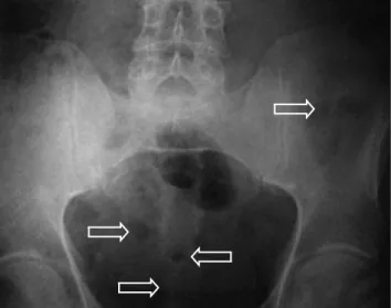
3). As the panoramic radiograph did not show any exten- sions of the lesion, a posterio-anterior view of skull was taken, which showed punched-out radiolucent lesions indicative of multiple myeloma as a radiological diagno- sis. In order to further confirm the diagnosis of multiple myeloma, a radiographic survey was carried out. An an- tero-posterior radiograph of the pelvis (Fig. 4) and a coro- nal section CT of the skull showed multiple punched-out radiolucent lesions (Fig. 5A). An axial section CT show- ed a soft tissue density mass measuring 4.2×4.1 cm in the left maxilla, eroding the medial and lateral walls and extending into the nasal cavity and the buccal space (Fig.
5B). The mass eroded the floor of the maxillary sinus and left maxillary alveolus in the coronal section CT (Fig.
5C). A sagittal section CT revealed erosion of the anterior wall of the maxillary sinus with involvement of the pre- maxillary tissues (Fig. 5D). The radiological differential
Fig. 2. An intraoral photograph shows a sessile mass in the left maxillary alveolus.
Fig. 1.An extraoral photograph shows diffuse swelling in the left middle third of the face.
Fig. 3. A panoramic radiograph shows an osteolytic lesion in the left posterior maxilla, with resorp- tion of the hard palate and the floor of the maxillary sinus.
diagnoses considered were multiple myeloma, carcinoma of the maxillary sinus, and metastatic carcinoma.
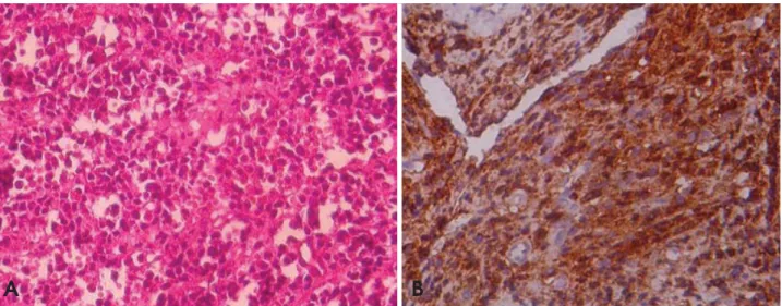
After the radiographic evaluation, an incisional biopsy was taken from the intraoral swelling. A histopathologi- cal examination of the specimen revealed atypical plasma cells with large hyperchromatic nuclei and large cytopla- sms, of which only few were binucleated (Fig. 6A). The report was suggestive of multiple myeloma. An oral patho- logist advised further confirmatory investigations for multi- ple myeloma. Immunohistochemically, the cells were positive for the CD138 marker (Fig. 6B). A bone marrow biopsy specimen showed 60% plasmacytosis. Serum elec- trophoresis revealed decreased albumin (2.78 g/dL), increas- ed globulin (5.22 g/dL), a reversed albumin : globulin ratio with increased β1-microglobulin (0.84 g/dL) and β2-micro- globulin (2.35 g/dL) levels, and an M-spike in the γ region
Fig. 4. An antero-posterior pelvic radiograph shows multiple punchedout radiolucent lesions.
Fig. 5.A. A coronal computed to- mography image of the skull shows punched-out radiolucent lesions. B.
An axial computed tomography image shows a soft tissue density mass in the left maxilla, eroding the medial and lateral walls. C. A coro- nal computed tomography image shows erosion of the left maxillary sinus floor and the maxillary alveo- lus. D. A sagittal computed tomo- graphy image shows erosion of the anterior wall of the maxillary sinus with involvement of the premaxil- lary tissues.
A B
C D
(1.86 g/dL). Bence-Jones proteins were absent in the urine.
Based on all the reports, a final diagnosis of multiple myeloma was made, the patient underwent dexametha- sone and thalidomide chemotherapy in a hospital speciali- zing in oncology, and is doing well.
Discussion
Multiple myeloma is the most aggressive primary bone malignancy.9It accounts for ~1% of all malignancies and
¤10% of hematologic malignancies.10The most common initial manifestations of multiple myeloma are bone pain (58%), fatigue (32%), and weight loss (24%).11Secondary invasion of the skeletal tissue is one of the most important characteristics of the disease.12The bone marrow reveals a large amount of abnormal plasma cells, which produce M-protein, light chain proteins (κ and λ), and cytokines.
Excessive production of M-protein causes hyperviscosity of the blood, which in turn leads to renal dysfunction.13,14
Oral manifestations of multiple myeloma have an incid- ence of 2%-70%, and rarely present as the first sign of the disease.15,16Swelling, orofacial pain, mobility of teeth, par- esthesia, hemorrhage, fracture, and root resorption are more frequently found in the mandible than the maxilla.4,12 In our case study, bone pain was the first sign, along with mobility of the adjacent teeth in the maxillary left alveo- lus. Swelling was seen following the extraction of the adja- cent teeth, and the patient had no history of hemorrhage or paresthesia.
Jaw involvement has been reported in as many as 30%
of multiple myeloma cases, although bony lesions are
less common in the maxilla than in the mandible because of the lower amount of hemopoietic marrow in the mandi- ble.4-8Bruce and Royer3and Miller et al.17have reported that 20%-30% of all cases had jaw involvement. Lambertenghi- Deliliers et al.12 showed that none of the 193 cases they examined involved the maxilla; on the contrary, Pisano et al.18reported four cases of maxillary involvement in 13 pati- ents, and Lae et al.19in 2003 reported that seven of the 33 cases they examined involved the maxilla. Similarly to the case reports of Pisano et al. and Lae et al., our case demon- strates the maxillary involvement.
Bony lesions in the jaws are directly attributed to the osteoclastic-activating factor, a lymphokine that is respon- sible for the development of these lesions.16Radiographi- cally bony lesions reveal multiple well-defined punched- out radiolucencies without a definitive cortical margin that often contain abnormal plasma cell proliferations.4These radiolucent lesions occur more frequently in the mandible than the maxilla, particularly in the posterior region, ramus, and condylar process.14 Our case study had multiple punched-out radiolucent lesions in the skull and pelvis, whereas the left maxilla had an ill-defined radiolucent lesion, which was a rare presentation of multiple myeloma.
Early multiple myeloma may not reveal observable ch- anges on plain radiographs. However, advanced imaging techniques can show evidence of active myeloma in ap- proximately 20% of patients with negative radiographs.20 A CT scan can visualize focal alterations of the bone mar- row before they can be detected with conventional radio- graphs.15Positron emission tomography and positive emis- sion tomography-CT with fluorodeoxyglucose may help
Fig. 6.Photomicrographs show the proliferating sheets of atypical plasma cells (A, H&E stain, 200×) and the immunohistochemically positive cells for CD138 (B, 200×).
A B
to detect new non-suspected lesions when staging the dis- ease, with important implications for treatment and asses- sing the response to treatment.21
The differential diagnoses of multiple myeloma in child- ren include multiple metastatic lesions and Langerhans’
cell disease. In adults, multiple myeloma and metastatic carcinoma are highly probable when several bones are in- volved. The clinically considered differential diagnoses in our case were carcinoma of the maxillary sinus and me- tastatic carcinoma. A panoramic radiograph revealed an ill-defined osteolytic lesion in the left maxillary sinus, which further indicated that carcinoma of the maxillary sinus was a relevant differential diagnosis. A further radio- logical examination showed multiple punched-out radiolu- cent lesions in the skull and pelvis, which led to a radiolo- gical diagnosis of multiple myeloma. Since multiple myel- oma is more common than multiple metastatic diseases, it should appear higher in the differential diagnosis. The dif- ferential diagnosis of these lesions requires a multidis- ciplinary approach to diagnosis. Asymptomatic multiple small radiolucencies in the jaws of an apparently healthy adult can represent multiple distinct bone marrow defects.22
The diagnosis of multiple myeloma depends on the iden- tification of abnormal monoclonal plasma cells, a full blood count, a bone marrow biopsy, levels of M-protein in the serum or urine, and a clinical image consistent with multiple myeloma. Serum electrophoresis identifies M- protein in ~93% of patients. Urine electrophoresis may identify M-protein in ~60% of patients. Additionally, ~70
% of myelomas secrete IgG, with κ light chains being more common (63%).23In the present case, serum electro- phoresis showed an IgG monoclonal spike of 1.86 g/dL with a κ light chain. Nevertheless, Bence-Jones proteins were not detected in the urine. Radiographs showed mul- tiple punched-out radiolucent lesions and a CT scan re- vealed a malignancy in the maxillary sinus. The histo- pathological and immunohistochemical reports led to the final diagnosis of multiple myeloma, which was support- ed by bone marrow biopsy and laboratory investigations.
The involvement of the jaw bones is a primary mani- festation that often occurs in the advanced stages of mul- tiple myeloma. In conclusion, we have reported a case of multiple myeloma presenting with bone pain and an ill- defined osteolytic radiolucency in the maxilla as a first sign, reinforcing the fundamental role of dentomaxillofa- cial radiologists in the early recognition of oral lesions that reflect underlying systemic disease, thus preventing or reducing morbidity and mortality in such cases.
References
1. Sharma V, Sharma A. Punched-out lesions in skull. Multiple myeloma. N Z Med J 2010; 123: 81-2.
2. Lee SH, Huang JJ, Pan WL, Chan CP. Gingival mass as the primary manifestation of multiple myeloma: report of two cases.
Oral Surg Oral Med Oral Pathol Oral Radiol Endod 1996; 82:
75-9.
3. Bruce KW, Royer RQ. Multiple myeloma occurring in the jaws: a study of 17 cases. Oral Surg Oral Med Oral Pathol 1953;
6: 729-44.
4. Witt C, Borges AC, Klein K, Neumann HJ. Radiographic manifestations of multiple myeloma in the mandible: a retro- spective study of 77 patients. J Oral Maxillofac Surg 1997;
55: 450-5.
5. Raubenheimer EJ, Lello GE, Dauth J, Fayman MS, Dvornak N, Senekal JC. Multiple myeloma presenting as localized ex- pansile jaw tumour. Int J Oral Maxillofac Surg 1988; 17: 382-5.
6. Raley LL, Granite EL. Plasmacytoma of the maxilla: report of case. J Oral Surg 1977; 35: 497-500.
7. Currie WJ, Hill RR, Keshani DK. An unusual case of maxil- lary tuberosity enlargement. Br Dent J 1994; 177: 60-2.
8. Albright RL, Finkelman A, Doner JM, Beaubien R. Multiple myeloma with manifestation of a maxillary bony lesion and plas- macytoma. Oral Surg Oral Med Oral Pathol 1968; 26: 167-72.
9. Lesmes D, Laster Z. Plasmacytoma in the temporomandibular joint: a case report. Br J Oral Maxillofac Surg 2008; 46: 322- 4.
10. Bird JM, Owen RG, D’Sa S, Snowden JA, Pratt G, Ashcroft J, et al. Guidelines for the diagnosis and management of multi- ple myeloma 2011. Br J Haematol 2011; 154: 32-75.
11. Kyle RA, Gertz MA, Witzig TE, Lust JA, Lacy MQ, Dispen- zieri A, et al. Review of 1027 patients with newly diagnosed multiple myeloma. Mayo Clin Proc 2003; 78: 21-33.
12. Lambertenghi-Deliliers G, Bruno E, Cortelezzi A, Fumagalli L, Morosini A. Incidence of jaw lesions in 193 patients with multiple myeloma. Oral Surg Oral Med Oral Pathol 1988; 65:
533-7.
13. Nau KC, Lewis WD. Multiple myeloma: diagnosis and treat- ment. Am Fam Physician 2008; 78: 853-9.
14. Ashcroft AJ, Davies FE, Morgan GJ. Aetiology of bone dise- ase and the role of bisphosphonates in multiple myeloma.
Lancet Oncol 2003; 4: 284-92.
15. Mozaffari E, Mupparapu M, Otis L. Undiagnosed multiple myeloma causing extensive dental bleeding: report of a case and review. Oral Surg Oral Med Oral Pathol Oral Radiol Endod 2002; 94: 448-53.
16. Witt C, Borges AC, Klein K, Neumann HJ. Radiographic mani- festations of multiple myeloma in the mandible: a retrospec- tive study of 77 patients. J Oral Maxillofac Surg 1997; 55:
450-3.
17. Miller CD, Goltry RR, Shenasky JH. Multiple myeloma involv- ing the mandible. Report of a case. Oral Surg Oral Med Oral Pathol 1969; 28: 603-9.
18. Pisano JJ, Coupland R, Chen S, Miller AS. Plasmacytoma of the oral cavity and jaws: a clinicopathologic study of 13 cases.
Oral Surg Oral Med Oral Pathol Oral Radiol Endod 1997; 83:
265-71.
19. Lae ME, Vencio EF, Inwards CY, Unni KK, Nascimento AG.
Myeloma of the jaw bones: a clinicopathologic study of 33 cases. Head Neck 2003; 25: 373-81.
20. Durie BG, Waxman AD, D’Agnolo A, Williams CM. Whole body (18)F-FDG PET identifies high-risk myeloma. J Nucl Med 2002; 43: 1457-63.
21. Schirrmeister H, Buck AK, Bergmann L, Reske SN, Bommer
M. Positron emission tomography (PET) for staging of solitary plasmacytoma. Cancer Biother Radiopharm 2003; 18: 841-5.
22. Wood NK, Goaz PW. Differential diagnosis of oral and maxil- lofacial lesions. 5th ed. St. Louis: Mosby; 1997. p. 387-9.
23. Zhao XJ, Sun J, Wang YD, Wang L. Maxillary pain is the first indication of the presence of multiple myeloma: a case report.
Mol Clin Oncol 2014; 2: 59-64.