Evaluation of zinc finger E-box binding homeobox 1 and transforming growth factor-beta2
expression in bladder cancer tissue in comparison with healthy adjacent tissue
Ali Mahdavinezhad1, Reza Yadegarazari2, Seyed Habibollah Mousavi-Bahar3, Jalal Poorolajal4, Mohammad Jafari5, Mohammad Ali Amirzargar3, Hosein Effatpanah6, Massoud Saidijam1
1Research Center for Molecular Medicine, Department of Genetics and Molecular Medicine, Hamadan University of Medical Sciences, Hamadan, 2Department of Molecular Medicine, Hamadan University of Medical Sciences, Hamadan, 3Department of Urology, Urology and Nephrology Research Center, Shaheed Beheshti Hospital, Hamadan University of Medical Sciences, Hamadan, 4Modeling of Non communicable Diseases Research Center, Department of Epidemiology & Biostatistics, School of Public Health, Hamadan University of Medical Sciences, Hamadan, 5Department of Pathology, Medical School, Hamadan University of Medical Sciences, Hamadan,
6Department of Public Health, Asadabad Faculty of Medical Sciences, Hamadan University of Medical Sciences, Hamadan, Iran
Purpose: The fifth most common cancer is allocated to bladder cancer (BC) worldwide. Understanding the molecular mechanisms of BC invasion and metastasis to identify target therapeutic strategies will improve disease survival. So the aim of this study was to measure expression rate of zinc finger E-box binding homeobox 1 (ZEB1) and transforming growth factor-beta2 (TGF-β2) mRNA in tissue samples of patients with BC and its healthy adjacent tissue samples and their association with muscle invasion, size and grade of the tumor.
Materials and Methods: Tissue samples were collected from 35 newly diagnosed untreated patients with BC from 2013 to 2014.
Total RNA was extracted from about 50-mg tissue samples using TRIzol reagent. TAKARA SYBR Premix EX Tag II was applied to de- termine the rate of mRNA expression by real-time polymerase chain reaction (PCR). To obtain final validation, PCR product of ZEB1 and TGF-β2 were sequenced. STATA 11 software was used to analyze the data.
Results: The expression level of ZEB1 in tumor samples was significantly more than of in healthy adjacent tissue samples. Up-regu- lation of TGF-β2 showed a strong association with muscle invasion (p=0.017). There was also demonstrated a relationship between over expression of ZEB1 with the tumor size (p=0.050).
Conclusions: It looks ZEB1 and TGF-β2 had a role in BC patients. In this study ZEB1 expression was higher in BC tissues than that of in healthy control tissues. There was demonstrated a markedly association between overexpression of TGF-β2 and muscle invasion.
Therefore, they are supposed to be candidate as potential biomarkers for early detection and progression of BC.
Keywords: Neoplasm grading; Transforming growth factor beta2; Urinary bladder neoplasms; ZEB1 protein
This is an Open Access article distributed under the terms of the Creative Commons Attribution Non-Commercial License (http://creativecommons.org/licenses/by-nc/4.0) which permits unrestricted non-commercial use, distribution, and reproduction in any medium, provided the original work is properly cited.
Received: 14 November, 2016 • Accepted: 22 December, 2016 Corresponding Author: Massoud Saidijam
Research Center for Molecular Medicine, Department of Genetics and Molecular Medicine, Hamadan University of Medical Sciences, Hamadan, Iran TEL: +98-912-132-4616, FAX: +98-81-38380-208, E-mail: [email protected]
ⓒ The Korean Urological Association, 2017
www.icurology.org
Investig Clin Urol 2017;58:140-145.
https://doi.org/10.4111/icu.2017.58.2.140 pISSN 2466-0493 • eISSN 2466-054X
INTRODUCTION
The fifth most common cancer is allocated to bladder cancer (BC) worldwide [1]. In Iran, 3,764 new cases of BC were reported in 2010 [2]. BC resulted from gathering of genetic and epigenetic alterations including uncontrolled cellular proliferation, invasion, promoted cell survival and metastatic distribution [3].
Majority portion of BCs (80%) are diagnosed as non
muscle invasive bladder cancers (NMIBCs) accompanied with rarely progression and good prognosis. The remaining cases (20%) are patients with muscle invasive bladder cancers (MIBCs) associated with poor prognosis due to high frequency of metastasis [4].
Cell motility increases through epithelialmesenchymal transition (EMT) process which is suggested to play crucial role in cancer invasion and metastasis [5]. Loss of epithelial and the acquisition of mesenchymal features happens during EMT process [6]. Morphology and motility of the cells will change to gain mesenchymal features. Loss of Ecadherin, which is controlled by several transcriptional repressors, has been demonstrated as a significant primary event in bladder tumorigenesis which bind to the promoter of Ecadherin [7]. Upregulation of transcriptional repressors such as zinc finger Ebox binding homeobox 1 (ZEB 1), Snail and twist interfere in EMT process [8].
ZEB1 mRNA is targeted by human miR141, miR200a, miR200b, and miR200c and these miRNAs are down
regulated by transforming growth factorbeta (TGFβ) thus TGFβ upregulates the expression level of ZEB1 [9] and activate EMT program [10].
On the other hand, different cell characteristics leading to metastasis could be regulated by TGFβ superfamily through over expression of matrix metalloproteinase2 [11].
Mammalian TGFβ superfamily consists of three members (TGFβ1, TGFβ2 and TGFβ3) with similar structure and function [12].
It is shown an association between higher level of TGFβ2 in glioma tumors, advanced disease stage and poor prognosis [13].
Some studies indicated an over expression of ZEB1 in MIBCs compared to NMIBCs [4,14,15] and some researchers reported expression level of TGFβ2 in colon cancer, glioma, gastric cancer and cervical lesions [12,1618] but on our knowledge TGFβ2 assessed in an old study [6] as well as in concomitant with ZEB1 in bladder tumor and healthy tissue samples. Consequently, understanding the molecular mechanisms of BC progression is important to identify target therapeutic strategies and to improve disease survival
[4] and prevent cancer progression [7].
The aim of this study was to evaluate TGFβ2 and ZEB1 mRNA expression in tissue samples of patients with BC and healthy adjacent tissue samples and their association with muscle invasion, grade and the size of the tumor.
MATERIALS AND METHODS
1. Setting
This casecontrol study was conducted in School of Medicine, Hamadan University of Medical Sciences from 2013 to 2014. Thirtyfive patients with newly diagnosed BC at the Beheshti and Buali hospitals (Hamadan, Iran) enrolled in the study if they met the eligibility criteria. Study protocol approved by the Ethics Committee of the Hamadan University of Medical Sciences and Health (Hamadan, Iran) (approval number: 9112154556). All procedures have been carried out in accordance with Declaration of Helsinki and informed consent was obtained from all participants.
2. Case and control groups
Patients who their disease clinically and pathologically confirmed irrespective of sex and age were evaluated.
Transurethral resection tissue samples were collected from 35 newly diagnosed untreated patients with BC from 2013 to 2014. Control samples consisted of adjacent normal urothelium resected about 10 cm far from the tumor lesion of patients.
3. Exclusion criteria
The following patients were excluded from the study:
(1) patients with other organ or genitourinary cancers, (2) genitourinary infection, and (3) history of radiotherapy or chemotherapy.
4. Tissue samples collection and RNA extraction After explaining the purpose of the study, written informed consent obtained from all participants. Then, cystoscopy and clear description of tumor done by an expert urologist under spinal anesthesia. The entire tumor was resected as deep as possible. After that, bladder and urethra were washed with normal saline three times, and then a normal urothelium area sample was taken at least 10 cm far from tumor bed. Immediately, the samples were washed with RNasefree cold saline solution, snapfrozen in liquid nitrogen and stored at 80oc until pathologic confirmation and further analysis. All samples, observed by 2 pathologists, diagnosed transitional cell carcinomas with the proportion of tumor cells greater than 80%. The International Union
against Cancer, World Health Organization/International Society of Urological Pathology criteria of 2004 was applied to assess tumor staging and histological grading, respectively.
Total RNA was extracted from 50 mg of tissue sample using TRIZOL Reagent (Invitrogen, Carlsbad, CA, USA), according to the manufacturer’s protocol. Depending on the quantity of the precipitation, the extracted RNA was dissolved in 20 to 50µL RNasefree water. Concentration and purity of total RNA was determined by optical density measurement using a NanoDrop spectrophotometer (BioTeK, Winooski, VT, USA). Integrity of extracted RNA was evaluated by agarose electrophoresis.
5. cDNA synthesis and quantification of mRNA expression
Five microgram of total RNA was applied to synthesize cDNA using Reverse Transcription kit (Fermentas, Walt
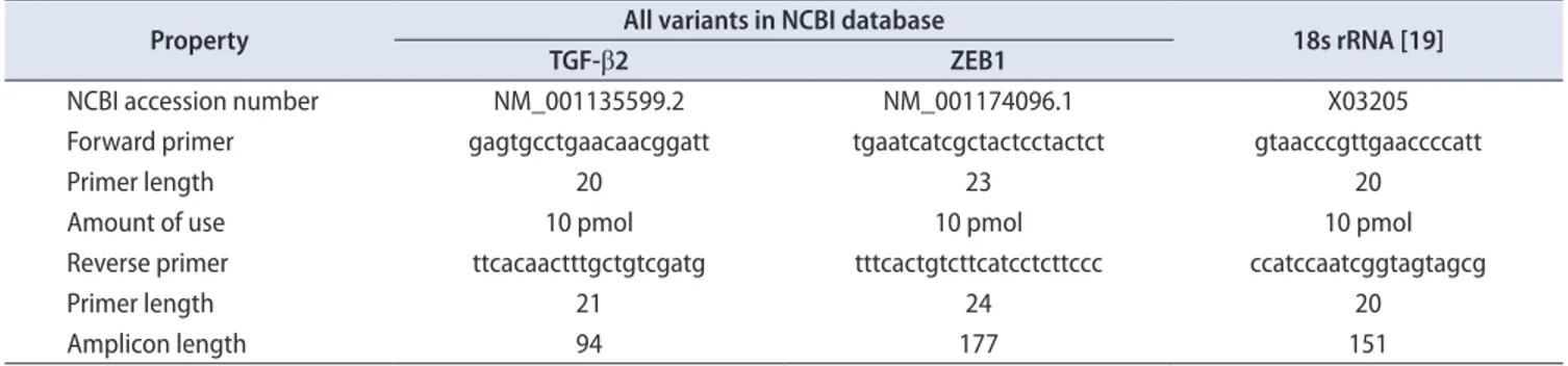
ham, MA, USA). Quantification of mRNA expression level was performed by applying TAKARA SYBR Premix EX Tag II in a CFX96 realtime polymerase chain reaction (PCR) detection system (BioRad, Hercules, CA, USA) in duplicate. Melting curve analysis was performed to evaluate specificity of primers. To design TGFβ2 mRNA and ZEB1 mRNA specific primers, we applied allele ID6 software and the designed primers was synthesized by Bioneer company (Daejeon, Korea) (Table 1) [19].
ZEB1 mRNA and TGFβ2 mRNA expression was normalized to 18s rRNA as internal control which there was no difference between case and control groups. No
template control was included in each PCR run to evaluate contaminations which all were negative. Relative expression of the studied genes were calculated by the 2-ΔΔct method [20].
Findings greater and less than 1 were determined to classify over expression and lower expression, respectively. In order to final validation, sequencing of PCR products for ZEB1 and TGFβ2 were applied.
6. Statistical analysis
Data were analyzed with analysis of variance, Paired ttest and ttest at 0.05 significant levels using STATA11 (StataCorp LP., College Station, TX, USA).
RESULTS
Seventy tissue samples from 35 patients with BC evaluated in present study (35 from malignant site and 35 from adjacent normal tissue). Two in the 35 patients were female and the remaining 33 were male. Mean age of patients was 71±11 (a range of 44 to 91 years old). In pathological examination, 63% of cases had muscle invasion categorized as MIBC, the rest was NMIBC. Sixteen of BC were low grade carcinoma as well as 17 were highgrade carcinoma. The 2 remaining tumor specimens were papillary urothelial neoplasm of low malignant potential.
There was indicated significant upregulated ZEB1 mRNA in tumor tissue compared with healthy adjacent tissue (p=0.042) (Table 2). Interestingly, it was observed that ZEB1 mRNA was overexpressed in 57% of malignant tissue
Table 1. Characteristics of TGF-β2 and ZEB1 mRNA primers designed with allele ID6 software
Property All variants in NCBI database
18s rRNA [19]
TGF-β2 ZEB1
NCBI accession number NM_001135599.2 NM_001174096.1 X03205
Forward primer gagtgcctgaacaacggatt tgaatcatcgctactcctactct gtaacccgttgaaccccatt
Primer length 20 23 20
Amount of use 10 pmol 10 pmol 10 pmol
Reverse primer ttcacaactttgctgtcgatg tttcactgtcttcatcctcttccc ccatccaatcggtagtagcg
Primer length 21 24 20
Amplicon length 94 177 151
TGF-β2, transforming growth factor-beta2; ZEB1, zinc finger E-box binding homeobox 1; NCBI, National Center for Biotechnology Information.
Table 2. ∆CT values of ZEB1 and TGF-β2 between case and control groups
Variable Case Control Difference
p-valuea
No. Mean±SD No. Mean±SD Mean±SD
ZEB1 35 9.36±2.49 35 10.49±2.39 1.13±3.16 0.042
TGF-β2 35 10.17±2.54 35 9.80±1.67 0.37±3.10 0.499
ΔCT=(CT of the mRNA target–CT of the reference gene).
CT, cycle threshold; ZEB1, zinc finger E-box binding homeobox 1; TGF-β2, transforming growth factor-beta2; SD, standard deviaiton.
a:Paried t-test.
samples. The folding change of ZEB1 mRNA expressions was 2.19 in cancer group compared with control group. No relationship showed between TGFβ2 mRNA expression and BC (p=0.499) (Table 2).
It was not observed significant association between ZEB1 mRNA and TGFβ2 mRNA expression and smoking state (p=0.749 and p=0.599, respectively) (Table 3).
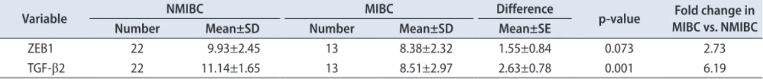
There was demonstrated higher expression level of TGFβ2 in MIBC group compared to NMIBC (p=0.001). The expression level of ZEB1 in MIBC group was higher than that of ZEB1 in NMIBC group. However, it was statistically nonsignificant (p=0.073). Mean standard deviation of ZEB1 and TGFβ2 mRNA is presented in Table 4. As indicated in Table 3, upregulation of TGFβ2 showed a strong association with muscle invasion.
In tumor specimens, we did not see any significant association between grading and ZEB1 mRNA and TGFβ2 mRNA deregulation (p=0.363 and p=0.127, respectively).
However, in according to CT of the mRNA target – CT of the reference gene in case and control group, separately (ΔCT) values, in a parallel direction with the poorly differentiation of tumor cells, we observed higher expression of TGFβ2 and ZEB1 (Table 5).
The tumor samples was subdivided based on the surface of the tumor, which was reported in 2 dimensional ultrasonography by radiologist, into greater and less than 10 cm2, the expression of ZEB1 mRNA was 3.5 fold higher in tumor samples with a size ≥10 cm2 compared to tumor samples with a size of <10 cm2 (p=0.039) (Table 6). In spite of 1.65 fold higher expression of TGFβ2 in tumor with size ≥10
Table 5. Association between grading and ZEB1 and TGF-β2 expression
Variable PUNLMP LG HG
p-valuea
Number Mean±SD Nmber Mean±SD Number Mean±SD
ZEB1 2 11.72±1.73 16 9.39±2.59 17 9.04±2.41 0.363
TGF-β2 2 12.75±0.56 16 10.64±1.62 17 9.41±3.09 0.127
ZEB1, zinc finger E-box binding homeobox 1; TGF-β2, transforming growth factor-beta2; SD, standard deviation; PUNLMP, papillary urothelial neoplasm of low malignant potential; LG, low grade carcinoma; HG, high grade carcinoma.
a:Analysis of variance.
Table 4. ∆CT values and relative quantification between nonmuscle invasive bladder cancer and muscle invasive bladder cancer
Variable NMIBC MIBC Difference
p-value Fold change in MIBC vs. NMIBC
Number Mean±SD Number Mean±SD Mean±SE
ZEB1 22 9.93±2.45 13 8.38±2.32 1.55±0.84 0.073 2.73
TGF-β2 22 11.14±1.65 13 8.51±2.97 2.63±0.78 0.001 6.19
ΔCT=(CT of the mRNA target–CT of the reference gene).
CT, cycle threshold; MIBC, muscle invasive bladder cancer; NMIBC, nonmuscle invasive bladder cancer; ZEB1, zinc finger E-box binding homeobox 1; TGF-β2, transforming growth factor-beta2; SD, standard deviation; SE, standard error.
Table 3. Expression state of ZEB1 and TGF-β2 in according to smoking
Variable Nonsmoker Smoker Difference
p-valuea
Number Mean±SD Nmber Mean±SD Mean±SE
ZEB1 12 9.17±2.83 23 9.46±2.35 0.29±0.90 0.749
TGF-β2 12 9.85±2.85 23 10.33±2.41 0.48±0.91 0.599
ZEB1, zinc finger E-box binding homeobox 1; TGF-β2, transforming growth factor-beta2; SD, standard deviation; SE, standard error.
a:t-test.
Table 6. ZEB1 and TGF-β2 mRNA expression and relative quantification between tumors with size<10 cm2 and size≥10 cm2
Variable Size<10 cm2 Size≥10 cm2 Difference
p-valuea Fold change size≥10 cm2 vs.
size<10 cm2
Number Mean±SD Number Mean±SD Mean±SE
ZEB1 23 9.98±2.62 12 8.17±1.73 1.81±0.84 0.039 3.50
TGF-β2 23 10.41±2.52 12 9.69±2.61 0.72±0.91 0.431 1.65
ZEB1, zinc finger E-box binding homeobox 1; TGF-β2, transforming growth factor-beta2; SD, standard deviation; SE, standard error.
a:t-test.
cm2, we did not obtain statistically significant relationship between tumor size and TGFβ2 upregulation (p=0.431) (Table 6).
DISCUSSION
BC has high mortality and 70% recurrence rate. More over it is the ninth prevalent and the second most common genitourinary tract malignant tumor [21,22].
Early detection, prevention and treatment of cancer are very important. In this study we evaluated TGFβ2 and ZEB1 mRNA expression in BC and normal adjacent tissue and their relationship with muscle invasion, grade and the size of the tumor.
The level of TGFβ2 expression was not statistical significant between BC and control group. Normal tissue samples obtained from healthy adjacent tissue therefore;
field effect may be a potential confounding variable in present study [23]. It was observed a 6.19 times up regulation of TGFβ2 in MIBC. The expression of TGFβ2 was not related to tumor size.
Our data supported by Gupta et al. [24] research which they confirmed the role of TGFβ in BC cell invasion. In a study, opposite to our results, expression of TGFβ2 in bladder tumor epithelial cells was higher than that of in normal epithelial cells [6].
Ma et al. [16] showed a higher expression of TGFβ2 in patients with early and advanced cancers compared to controls and it was detected a correlation between TGFβ2 mRNA expression and prognosis.
TGFβ2 mRNA and protein expression were significantly increased during colon carcinoma progression [12]. Gupta et al. [24] also showed over expression of TGFβ isoforms in invasive highgrade BC. In our study TGFβ2 expression was increasing as tumor grade increased. TGFβ2 also was diagnosed as a potential biomarker in cervical intraepithelial neoplasia [18]. In according to our findings TGFβ2 could be considered as a potential progression biomarker in BC.
The results showed significant ZEB1 mRNA up
regulation in case group. Although ZEB1 expression in MIBC group was 2.73 times higher than that of in NMIBC group but this difference was not significant. The expression also was greater in tumor samples with a size of ≥10 cm2.
In agree with this study, it was shown over expressed ZEB1 mRNA has been related to invasion in cancer patients [4].
Another study evaluated ZEB1 expression in bladder tumorigenesis and showed slightly low level ZEB1 expression in NMIBC and grades I/II compared to MIBC and tumor
grade III respectively. They also observed no significant correlation, between tumor stage and grade, nodal involvement, vascular invasion, metastasis and survival [14].
It was not detected any significant association between ZEB1 expression, muscle invasion and grade of BC in our study.
Another study evaluated ZEB1 expression in noninvasive and invasive bladder tumor tissue samples. They reported ZEB1 was more frequently detected in high grade than in lowgrade cancers [15]. In spite of higher expression in high
grade samples, significant over expressed ZEB1 did not obtain in present study. However; ZEB1 was up regulated in tumor tissues generally.
Remarkable higher expression of ZEB1 associated with tumor size has not been reported in other investigations.
There were some limitations in our research including:
small study population, which future studies with larger sample size should be suggested. The next, in this study tissue samples were obtained using an invasive procedure.
It is very helpful to design future studies based on non invasive biologic urine or serum samples to discover biomarkers. The last, grossly normal mucosa may has characteristics of tumor cells especially at molecular level and plays as confounding variable; therefore it would be better to design two control groups and introduce both normal tissue and healthy adjacent tissue samples as control group in such investigations.
Other studies should be done with large sample size in the future to evaluate the relation between ZEB1 and clinical followup and treatment out come.
CONCLUSIONS
ZEB1 and TGFβ2 play a role in BC. In this study expression of ZEB1 was higher in BC tissues than that of in healthy control tissues. There was demonstrated a markedly association between overexpression of TGFβ2 and muscle invasion. Therefore; they could be supposed to be potential biomarkers for early detection and progression of BC. Further studies should be performed about ZEB1 and TGFβ2 alterations in BC to understand role of these genes in tumor initiation, progression and metastasis.
CONFLICTS OF INTEREST
The authors have nothing to disclose.
ACKNOWLEDGMENTS
This study was funded by the Vicchancellor of Research
and Technology, Hamadan University of Medical Sciences.
REFERENCES
1. Siegel R, Naishadham D, Jemal A. Cancer statistics, 2012. CA Cancer J Clin 2012;62:10-29.
2. Mousavi SM et al. Guideline: National cancer registry (per- sian). 2nd ed. Tehran (IR): Ministry of health and medical education,office of Deputy minister for health, center for dis- ease control and prevention,cancer office; 2012.
3. Adam L, Zhong M, Choi W, Qi W, Nicoloso M, Arora A, et al. miR-200 expression regulates epithelial-to-mesenchymal transition in bladder cancer cells and reverses resistance to epidermal growth factor receptor therapy. Clin Cancer Res 2009;15:5060-72.
4. Wu K, Fan J, Zhang L, Ning Z, Zeng J, Zhou J, et al. PI3K/Akt to GSK3β/β-catenin signaling cascade coordinates cell coloni- zation for bladder cancer bone metastasis through regulating ZEB1 transcription. Cell Signal 2012;24:2273-82.
5. Wu K, Ning Z, Zeng J, Fan J, Zhou J, Zhang T, et al. Silibinin inhibits β-catenin/ZEB1 signaling and suppresses bladder cancer metastasis via dual-blocking epithelial-mesenchymal transition and stemness. Cell Signal 2013;25:2625-33.
6. Eder IE, Stenzl A, Hobisch A, Cronauer MV, Bartsch G, Klock- er H. Expression of transforming growth factors beta-1, beta 2 and beta 3 in human bladder carcinomas. Br J Cancer 1997;75:
1753-60.
7. Matsui Y, Assi K, Ogawa O, Raven PA, Dedhar S, Gleave ME, et al. The importance of integrin-linked kinase in the regula- tion of bladder cancer invasion. Int J Cancer 2012;130:521-31.
8. Shan Y, Zhang L, Bao Y, Li B, He C, Gao M, et al. Epithelial- mesenchymal transition, a novel target of sulforaphane via COX-2/MMP2, 9/Snail, ZEB1 and miR-200c/ZEB1 pathways in human bladder cancer cells. J Nutr Biochem 2013;24:1062- 9.
9. Katoh Y, Katoh M. Hedgehog signaling, epithelial-to-mesen- chymal transition and miRNA (review). Int J Mol Med 2008;
22:271-5.
10. Hurt EM, Saykally JN, Anose BM, Kalli KR, Sanders MM. Ex- pression of the ZEB1 (deltaEF1) transcription factor in human:
additional insights. Mol Cell Biochem 2008;318:89-99.
11. Dehnavi E, Soheili ZS, Samiei S, Ataei Z, Aryan H. The effect of TGF-beta2 on MMP-2 production and activity in highly metastatic human bladder carcinoma cell line 5637. Cancer Invest 2009;27:568-74.
12. Bellone G, Carbone A, Tibaudi D, Mauri F, Ferrero I, Smirne C,
et al. Differential expression of transforming growth factors- beta1, -beta2 and -beta3 in human colon carcinoma. Eur J Cancer 2001;37:224-33.
13. Hau P, Jachimczak P, Schlaier J, Bogdahn U. TGF-β2 signaling in high-grade gliomas. Curr Pharm Biotechnol 2011;12:2150- 7.
14. Kenney PA, Wszolek MF, Rieger-Christ KM, Neto BS, Gould JJ, Harty NJ, et al. Novel ZEB1 expression in bladder tumori- genesis. BJU Int 2011;107:656-63.
15. Lee H, Jun SY, Lee YS, Lee HJ, Lee WS, Park CS. Expression of miRNAs and ZEB1 and ZEB2 correlates with histopathologi- cal grade in papillary urothelial tumors of the urinary bladder.
Virchows Arch 2014;464:213-20.
16. Ma GF, Miao Q, Zeng XQ, Luo TC, Ma LL, Liu YM, et al.
Transforming growth factor-β1 and -β2 in gastric precancer and cancer and roles in tumor-cell interactions with peripheral blood mononuclear cells in vitro. PLoS One 2013;8:e54249.
17. Qiu B, Zhang D, Wang C, Tao J, Tie X, Qiao Y, et al. IL-10 and TGF-β2 are overexpressed in tumor spheres cultured from hu- man gliomas. Mol Biol Rep 2011;38:3585-91.
18. Xu XC, Mitchell MF, Silva E, Jetten A, Lotan R. Decreased ex- pression of retinoic acid receptors, transforming growth factor beta, involucrin, and cornifin in cervical intraepithelial neopla- sia. Clin Cancer Res 1999;5:1503-8.
19. Schmittgen TD, Zakrajsek BA. Effect of experimental treat- ment on housekeeping gene expression: validation by real- time, quantitative RT-PCR. J Biochem Biophys Methods 2000;46:69-81.
20. Schmittgen TD, Livak KJ. Analyzing real-time PCR data by the comparative C(T) method. Nat Protoc 2008;3:1101-8.
21. Yoshino H, Enokida H, Chiyomaru T, Tatarano S, Hidaka H, Yamasaki T, et al. Tumor suppressive microRNA-1 mediated novel apoptosis pathways through direct inhibition of splicing factor serine/arginine-rich 9 (SRSF9/SRp30c) in bladder can- cer. Biochem Biophys Res Commun 2012;417:588-93.
22. Shirodkar SP, Lokeshwar VB. Potential new urinary markers in the early detection of bladder cancer. Curr Opin Urol 2009;19:
488-93.
23. Jones TD, Wang M, Eble JN, MacLennan GT, Lopez-Beltran A, Zhang S, et al. Molecular evidence supporting field effect in urothelial carcinogenesis. Clin Cancer Res 2005;11:6512-9.
24. Gupta S, Hau AM, Al-Ahmadie HA, Harwalkar J, Shoskes AC, Elson P, et al. Transforming growth factor-β is an upstream regulator of mammalian target of rapamycin complex 2-depen- dent bladder cancer cell migration and invasion. Am J Pathol 2016;186:1351-60.