Received on July 7, 2012. Revised on July 25, 2012. Accepted on August 2, 2012.
CC This is an open access article distributed under the terms of the Creative Commons Attribution Non-Commercial License (http://creativecommons.org/licenses/by-nc/3.0) which permits unrestricted non-commercial use, distribu- tion, and reproduction in any medium, provided the original work is properly cited.
*Corresponding Author. Tel: 82-33-248-2564; Fax: 82-33-241-8250; E-mail: [email protected] Keywords: Endotoxin shock, Cytokines, Inflammation, Signal transduction
Abbreviations: CaMK, calcium/calmodulin-dependent protein kinase; HMGB1, high mobility group box 1; IRF3, interferon regulatory factor 3; TBK1, TANK-binding kinase 1; TRIF, Toll/IL-1 receptor domain-containing adaptor inducing interfer- on-β
Calcium/Calmodulin-Dependent Protein Kinase is Involved in the Release of High Mobility Group Box 1 Via the Interferon-β Signaling Pathway
Lijuan Ma, Seon-Ju Kim and Kwon Ik Oh*
Department of Pathology, Hallym University College of Medicine, Chuncheon 200-702, Korea
Previously, we have reported that high mobility group box 1 (HMGB1), a proinflammatory mediator in sepsis, is released via the IFN-β-mediated JAK/STAT pathway. However, de- tailed mechanisms are still unclear. In this study, we dis- sected upstream signaling pathways of HMGB1 release us- ing various molecular biology methods. Here, we found that calcium/calmodulin-dependent protein kinase (CaM kinase, CaMK) is involved in HMGB1 release by regulating IFN-β production. CaMK inhibitor, STO609, treatment inhibits LPS-induced IFN-β production, which is correlated with the phosphorylation of interferon regulatory factor 3 (IRF3).
Additionally, we show that CaMK-I plays a major role in IFN-β production although other CaMK members also seem to contribute to this event. Furthermore, the CaMK inhibitor treatment reduced IFN-β production in a murine endoto- xemia. Our results suggest CaMKs contribute to HMGB1 re- lease by enhancing IFN-β production in sepsis.
[Immune Network 2012;12(4):148-154]
INTRODUCTION
HMGB1 is one of the most abundant and highly conserved nonhistone chromosomal proteins in eukaryotes (1). HMGB1 interacts with several transcription factors, thereby allowing them to perform their cellular roles such as transcription, rep-
lication, and cellular differentiation (2). In addition to its roles within the nucleus, immune cells such as macrophages stimu- lated with LPS export nuclear HMGB1 to the cytoplasm and subsequently secrete it, and extracellular HMGB1 acts as an inflammatory mediator in various ways (2). It has been re- ported that HMGB1 facilitates diverse aspects of proin- flammatory responses, including chemotaxis (3,4), increased permeability of cell monolayer (5), and the release of various proinflammatory cytokines such as TNF, IL-1, IL-6 and macro- phage inflammatory protein-1 (6). Two recent papers demon- strated that HMGB1 can enhance proinflammatory activity by binding to LPS (7) and cytokines (8). These observations sug- gest HMGB1 can be involved in inflammation in direct and indirect manners.
In contrast to the clarity of the functions of HMGB1, the molecular mechanisms by which macrophages release HMGB1 are still unclear. Although many cytokines and sig- naling molecules are shown to be involved in this process (1) and modifications such as hyperacetylation and phosphor- ylation are needed for extracellular release (9), the relation and hierarchy between them are poorly understood. Recently, we have demonstrated that IFN-β plays a role in LPS- in- duced HMGB1 release as an intermediating molecule (10).
However, we did not show the relation between IFN-β and other players involved in HMGB1 release.
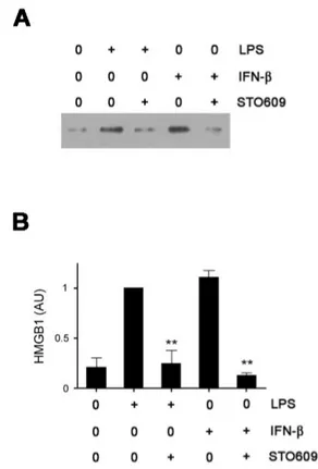
Figure 1. CaMKs are indispensable to HMGB1 secretion. (A) RAW cells were pre-incubated with STO609 for 30 minutes and then treated with LPS (1 mg/ml) or IFN-β (500 U/ml) overnight. The levels of HMGB1 in supernatants were measured by western blotting. (B) The relative densitometric values of HMGB1 bands are shown as arbitrary units (AU). Densitometric value from the LPS-treated cells was taken as 1 and the mean values±SEM fold changes relative to the control (the LPS-treated cells) are shown (n=3). **p<0.01 compared with LPS (or IFN-β)-treated control samples.
In this paper, we show that CaMK, one of the mediators in HMGB1 release (11), exploits IFN-β pathway to regulate LPS-induced HMGB1 release. We also demonstrate that these signaling pathways are also needed in vivo. Our study sug- gests that the regulatory roles of CaMK in IFN-β production are indispensible for optimal release of HMGB1 in inflamma- tory conditions.
MATERIALS AND METHODS
Cells and reagents
Both mouse macrophage cell line, RAW 264.7 and human embryonic kidney cell line, 293 (American Type Culture Collection, Rockville, MD), were maintained in DMEM sup- plemented with 10% FBS and 0.1% penicillin/streptomycin (Life Technologies Korea, Seoul, Korea). LPS (O55:B5) were purchased from Sigma (St. Louise, MO). CaMK inhibitor, STO609, was obtained from Calbiochem (San Diego, CA).
IFN-β was purchased from PBL Interferon Source (Piscata- way, NJ). The following antibodies were used in this study:
anti-HMGB1 (Abcam, Cambridge, MA), anti-phospho-IRF3 (Cell Signaling, Danvers, MA), anti-phospho-TANK-binding kinase 1 (TBK1) (BD Biosciences, San Jose, CA), anti-TBK1 (Cell signaling).
Mice
C57BL/6 (B6) mice were purchased from Daehan Biolink (Daejeon, Korea). All mice were housed under specific pathogen-free conditions at the animal facility of the Hallym University College of Medicine. Experiments were performed after the approval of the Animal Experimentation Committee at Hallym University (Hallym2009-57-1).
Measurement of cytokines
Concentrations of IFN-β were measured using ELISA kits (PBL Interferon Source) according to the manufacturer's instructions.
Real-time PCR
Total RNA from macrophages was extracted using Trizol re- agents (Life Technologies Korea), according to the manu- facturer's instruction and subjected to reverse transcription us- ing Superscript II (Life Technologies Korea). Primers were pur- chased from Ambion Korea (Seoul, Korea) except TRIF (forward, AACCTCCACATCCCCTGTTTT; reverse, GCCCTGGCATGGATAA- CCA). Quantitative PCR was performed using SYBR green master
mix (Qiagen Korea, Seoul, Korea). Genes were normalized to housekeeping gene, actin.
Western blot analysis
The level of HMGB1 was determined by western blotting.
Samples of culture supernatant were concentrated with cen- tricon (Milipore, Billerica, MA) and then separated on 12%
SDS-PAGE gels, transferred to nitrocellulose membranes. The membranes were probed with anti-HMGB1 antibodies, and subsequently incubated with a HRP-conjugated secondary antibody. Bands were detected using SuperSignal West Femto kit (Thermo scientific, Rockford, IL). The amounts of HMGB1 were quantified by densitometric scanning of the exposed X-ray film. The relative densitometric value of HMGB1 of each sample shown as AU (arbitrary units) in the figures was
Figure 2. Inhibition of CaMKs blocks IFN-β production. (A and B) CaMK inhibitor STO609 inhibits IFN-β production in LPS-treated RAW cells (A) and macrophages (B) in a dose-dependent manner (n=3). RAW cells or peritoneal macrophages were stimulated with LPS (1 μg/ml) in the presence of vehicle (DMSO) or various doses of STO609 for 2 hours. The mRNA were extracted from each cells and subjected to real time PCR.
The value of the LPS-treated cells was arbitrarily assigned a value of 1. (C and D) 293 cells were transfected with TRIF (C) or TBK1 (D) plasmids.
After 24 hours of culture, IFN-β production was measured by real time PCR and the normalized values of IFN-β were analyzed the same as in A (n=3). Data were expressed as mean±SEM. AU, arbitrary unit. *p<0.05; **p<0.01; ***p<0.001 compared with each control.
calculated by arbitrarily assigning that of control (a LPS-treat- ed sample) as 1 (10).
RNA interference
Small interfering RNA (siRNA) targeting CaMK-I and CaMK-IV and control siRNA were purchased from Ambion and Bioneer Korea (Seoul, Korea). siRNA duplexes were transfected into RAW cells using Nucleofactor (Lonza, Allendale, NJ) accord- ing to the manufacturer’s instructions.
Statistical analysis
All values in the figures and the text are expressed as mean±SEM, and p<0.05 was considered to be statistically significant. Survival data were analyzed by the log-rank test, and all other data were evaluated by unpaired t-test (non- parametric, two-tailed).
RESULTS
CaMK inhibitor, STO609 inhibits LPS-induced IFN-β production
To investigate the mechanisms on LPS-induced HMGB1 re- lease, we searched candidate molecules that seem to be in- volved in HMGB1 release. Among several candidates, we found that CaMK inhibitor, STO609, dramatically reduced LPS-induced HMGB1 release in RAW cells as previously re- ported (11). Since we previously demonstrated that IFN-β signal pathway is essential in HMGB1 release and IFN-β it- self like LPS induces HMGB1 (10), we treated RAW cells with STO609 plus IFN-β and measured extracellular HMGB1.
STO609 treatment reduced HMGB1 levels induced by IFN-β as well as LPS (Fig. 1), suggesting that STO609 could inhibit both up- and down-stream molecules of IFN-β. Since it was
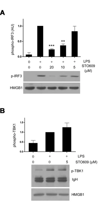
Figure 3. Inhibition of CaMK interferes with IRF3 phosphorylation induced by LPS. (A) STO609 treatment reduced IRF3 phospho- rylation. RAW cells were treated with LPS in the presence of various doses of STO609 for 1 hour, lysed and then subjected to phospho- IRF3 western blotting (middle). The same blot was subjected again to HMGB1 western blotting as a loading control. A graph shows the relative densitometric values of each sample in arbitrary unit (AU). Densitometirc value from the LPS-treated cells was taken as 1. (B) TBK1 phosphorylation was not affected by STO609. Raw cells were treated with LPS plus STO609 and the cell lysates were imm- unoprecipitated with anti-phospho-TBK1 antibody and then detected with anti-total TBK1 antibody (middle). Lower panel, equal amounts of each lysate were subjected to HMGB1 western blotting as a loading control. **p<0.01; ***p<0.001 compared with LPS alone.
published that CaMKs are critical in type I IFN-induced JAK-STAT1 signaling (12), however, we turned our attention into upstream signals of IFN-β and investigated how CaMK is involved in LPS-induced IFN-β production. To test this, we treated RAW cells and primary macrophages with LPS plus various doses of STO609 and compared the levels of IFN-β using real-time PCR. STO609 reduced IFN-β pro- duction in a dose dependent manner in both cases (Fig. 2A and B). To dissect the action mechanism of STO609, we transfected 293 cells with Toll/IL-1 receptor domain-contain- ing adaptor inducing interferon-β (TRIF) or TBK1 plasmids and treated them with STO609. TRIF or TBK1 transfected cells actively produced IFN-β mRNA without any stimulation.
However, the transcription of IFN-β was dramatically re- duced by STO609 in TRIF (Fig. 2C) or TBK1 (Fig. 2D) trans- fected cells.
Inhibition of CaMK blocks the phosphorylation of IRF3
TLR4 uses two different adaptors, MyD88 and TRIF, and TRIF signals are relayed through TRIF-TBK1-IRF3 axis to produce IFN-β. In addition, our transfection assay suggested that STO609 does not affect TRIF-TBK1 themselves but their com- mon effectors. These observations led us to test whether IRF3 is a target of STO609. RAW cells were treated with LPS plus various doses of STO609 and subjected to western blotting using anti-phospho-IRF3 antibody. Interestingly, STO609 re- duced the phosphorylation of IRF3 in a dose dependent man- ner (Fig. 3A). Since the phosphorylation levels reflect the ac- tivity of IRF3, these data suggest that STO609 inhibits LPS-in- duced IRF3 function. Consistent with the above results, the phosphorylation of TBK1 was not reduced (Fig. 3B).
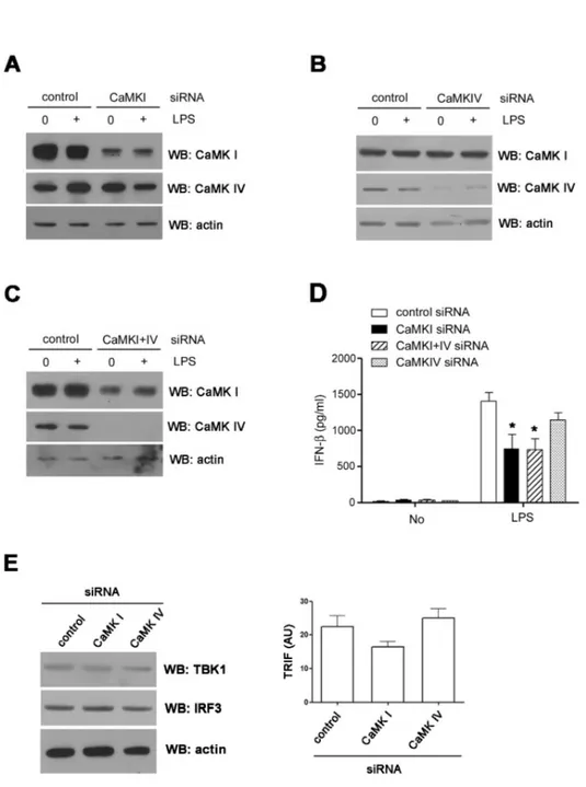
CaMKI is important in LPS-induced IFN-β production Since STO609, a selective inhibitor of CaMK kinase (13), can effectively block the activation of the downstream CaMKs (CaMKI and IV), we examined whether CaMKI and/or IV knock- down could also down-regulate IFN-β production. As shown in Fig. 4, endogenous CaMKI and IV expression in RAW cells was down-regulated by siRNA transfections (Fig. 4A∼C). Com- pared with control siRNA, CaMKI siRNA significantly de- creased LPS-induced production of IFN-β in RAW cells.
Although CaMKIV siRNA also decreased IFN-β production, these inhibitory effects were not statistically significant. To test the synergistic effects of CaMKI and IV, we transfected RAW cells with both CaMKI and IV siRNAs simultaneously
Figure 4. CaMK1 plays a role in IFN-β production. (A∼C) RAW cells were transfected with CaMKI (A), IV (B) or I and IV (C) siRNAs.
After 48 hours, the expressions of each CaMK isoform were detected by western blotting. (D) The levels of IFN-β in the supernatant were measured by ELISA. (E) The levels of TBK1, IRF3 proteins (left) and TRIF transcripts (right) in siRNA treated cells were analyzed using western blotting (left) or real time PCR. *p
<0.05 compared with LPS treated cells transfected with control siRNA.
but did not find any synergistic effects (Fig. 4D). Next, we checked the expression levels of TRIF, TBK1, and IRF3 using CaMKI or IV knockdown cells and found no significant differ- ence between control and knockdown cells (Fig. 4E).
CaMK signals are important to LPS-induced IFN-β release in vivo
To evaluate the role of CaMKs in IFN-β production in vivo, we utilized the murine endotoxemia model. To test whether STO609 treatment reduces IFN-β levels, we administered LPS
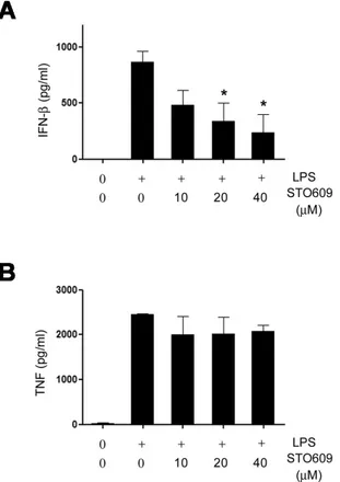
plus STO609 and measured blood IFN-β levels. Consistent with in vitro studies, the IFN-β levels of the STO609 treated mice were significantly lower than the control mice (Fig. 5A).
However, the difference in mortality (data not shown) and blood TNF levels (Fig. 5B) were not seen. These mortality re- sults seem to be a contrast to the previous reports that IFN-β deficiency improved the sepsis survivals (14,15). However, since a significant amount of IFN-β still remained in STO609 treated mice and the level of TNF is comparable to control mice (Fig. 5B), we assume that the protective effect of
Figure 5. The release of IFN-β depends on CaMK signaling pathway in vivo. (A and B) Serum levels of IFN-β (A) and TNF (B) in B6 mice treated with LPS (50 mg/kg) plus various doses of STO609 or vehicle (DMSO) were measured by ELISA (n=4). B6 mice were treated various doses of STO609 twice (18 and 0.5 hours) before LPS infusion. Mice were sacrificed 2 hours after LPS administration and sera were collected from each mouse for ELISA. Data are presented as mean±SEM. *p<0.05 compared with LPS plus vehicle-treated mice.
STO609 could be neglected in our experimental condition.
DISCUSSION
In this study, we have shown that CaMK regulates HMGB1 release via IFN-β and how CaMK is involved in IFN-β production. IFN-β synthesis was impaired in not only LPS treated macrophages but also TRIF or TBK1 transfected 293 cells by CaMK inhibitor, STO609 (Fig. 2). In addition, STO609 treatment reduced the levels of IRF3 but not TBK1 phosphor- ylation (Fig. 3) induced by LPS. These above findings suggest that the phosphorylation of IRF3 may be a target of CaMK.
In knockdown study, we found that CaMKI played a major role (Fig. 4). To elucidate detailed interactions between
CaMK isoforms and IRF3, we performed co-immunoprecip- itation assay. However, we failed to detect any physical inter- action between them (data not shown). This result can be interpreted in several ways. First, CaMK affects IRF3 activa- tion in an indirect way. Second, the affinity between CaMK and IRF3 is relatively weak. And finally, other CaMK isoform like CaMKII (16) might be essential for IRF3 activation.
Currently, we speculate that several CaMK isoforms and adap- tors might form big complexes and induce IRF3 activation with low binding affinities as they do in the regulation of IFN- α-induced JAK-STAT activation (12).
ACKNOWLEDGEMENTS
This research was supported by the Hallym University Research Fund (HRF-2008-037) and Basic Science Research Program through the National Research Foundation of Korea (NRF) funded by the Ministry of Education, Science and Technology (20110003455).
CONFLICT OF INTEREST
The authors have no conflicting financial interest.
REFERENCES
1. Sims, G. P., D. C. Rowe, S. T. Rietdijk, R. Herbst, and A.
J. Coyle. 2010. HMGB1 and RAGE in inflammation and cancer. Annu. Rev. Immunol. 28: 367-388.
2. Lotze, M. T. and K. J. Tracey. 2005. High-mobility group box 1 protein (HMGB1): nuclear weapon in the immune arsenal.
Nat. Rev. Immunol. 5: 331-342.
3. Degryse, B., T. Bonaldi, P. Scaffidi, S. Müller, M. Resnati, F. Sanvito, G. Arrigoni, and M. E. Bianchi. 2001. The high mobility group (HMG) boxes of the nuclear protein HMG1 induce chemotaxis and cytoskeleton reorganization in rat smooth muscle cells. J. Cell Biol. 152: 1197-1206.
4. Rouhiainen, A., J. Kuja-Panula, E. Wilkman, J. Pakkanen, J.
Stenfors, R. K. Tuominen, M. Lepäntalo, O. Carpén, J.
Parkkinen, and H. Rauvala. 2004. Regulation of monocyte migration by amphoterin (HMGB1). Blood 104: 1174-1182.
5. Sappington, P. L., R. Yang, H. Yang, K. J. Tracey, R. L.
Delude, and M. P. Fink. 2002. HMGB1 B box increases the permeability of Caco-2 enterocytic monolayers and impairs intestinal barrier function in mice. Gastroenterology 123:
790-802.
6. Andersson, U., H, Wang, K. Palmblad, A. C. Aveberger, O.
Bloom, H. Erlandsson-Harris, A. Janson, R. Kokkola, M.
Zhang, H. Yang, and K. J. Tracey. 2000. High mobility group 1 protein (HMG-1) stimulates proinflammatory cytokine syn- thesis in human monocytes. J. Exp. Med. 192: 565-570.
7. Youn, J. H., Y. J. Oh, E. S. Kim, J. E. Choi, and J. S. Shin.
2008. High mobility group box 1 protein binding to lip- opolysaccharide facilitates transfer of lipopolysaccharide to CD14 and enhances lipopolysaccharide-mediated TNF-alpha production in human monocytes. J. Immunol. 180: 5067- 5074.
8. Sha, Y., J. Zmijewski, Z. Xu, and E. Abraham. 2008. HMGB1 develops enhanced proinflammatory activity by binding to cytokines. J. Immunol. 180: 2531-2537.
9. Bonaldi, T., F. Talamo, P. Scaffidi, D. Ferrera, A. Porto, A.
Bachi, A. Rubartelli, A. Agresti, and M. E. Bianchi. 2003.
Monocytic cells hyperacetylate chromatin protein HMGB1 to redirect it towards secretion. EMBO J. 22: 5551-5560.
10. Kim, J. H., S. J. Kim, I. S. Lee, M. S. Lee, S. Uematsu, S.
Akira, and K. I. Oh. 2009. Bacterial endotoxin induces the release of high mobility group box 1 via the IFN-beta signal- ing pathway. J. Immunol. 182: 2458-2466.
11. Zhang, X., D. Wheeler, Y. Tang, L. Guo, R. A. Shapiro, T.
J. Ribar, A. R. Means, T. R. Billiar, D. C. Angus, and M. R.
Rosengart. 2008. Calcium/calmodulin-dependent protein kin- ase (CaMK) IV mediates nucleocytoplasmic shuttling and re- lease of HMGB1 during lipopolysaccharide stimulation of macrophages. J. Immunol. 181: 5015-5023.
12. Wang, L., I. Tassiulas, K. H. Park-Min, A. C. Reid, H.
Gil-Henn, J. Schlessinger, R. Baron, J. J. Zhang, and L. B.
Ivashkiv. 2008. 'Tuning' of type I interferon-induced Jak- STAT1 signaling by calcium-dependent kinases in macro- phages. Nat. Immunol. 9: 186-193.
13. Tokumitsu, H., H. Inuzuka, Y. Ishikawa, M. Ikeda, I. Saji, and R. Kobayashi. 2002. STO-609, a specific inhibitor of the Ca(2+)/calmodulin-dependent protein kinase kinase. J. Biol.
Chem. 277: 15813-15818.
14. Karaghiosoff, M., R. Steinborn, P. Kovarik, G. Kriegshäuser, M. Baccarini, B. Donabauer, U. Reichart, T. Kolbe, C.
Bogdan, T. Leanderson, D. Levy, T. Decker, and M. Müller.
2003. Central role for type I interferons and Tyk2 in lip- opolysaccharide-induced endotoxin shock. Nat. Immunol. 4:
471-477.
15. Weighardt, H., S. Kaiser-Moore, S. Schlautkötter, T.
Rossmann-Bloeck, U. Schleicher, C. Bogdan, and B. Hol- zmann. 2006. Type I IFN modulates host defense and late hyperinflammation in septic peritonitis. J. Immunol. 177:
5623-5630.
16. Liu, X., M. Yao, N. Li, C. Wang, Y. Zheng, and X. Cao. 2008.
CaMKII promotes TLR-triggered proinflammatory cytokine and type I interferon production by directly binding and acti- vating TAK1 and IRF3 in macrophages. Blood 112: 4961- 4970.