CASE REPORT
팽대부 주위 게실로 인해 담관 삽관이 어려운 환자에서 캡 장착 내시경적 역행성 담췌관 조영술의 유용성
김준환, 이준수, 김의주, 김연석, 조재희
가천대학교 길병원 소화기내과
The Usefulness of Cap-assisted Endoscopic Retrograde Cholangiopancreatography for Cannulation Complicated by a Periampullary Diverticulum
Joonhwan Kim, Jun Soo Lee, Eui Joo Kim, Yeon Suk Kim and Jae Hee Cho
Division of Gastroenterology, Department of Internal Medicine, Gachon University Gil Medical Center, Incheon, Korea
Endoscopic retrograde cholangiopancreatography (ERCP) is an advanced therapeutic procedure to manage choledocholithiasis and pancreatobiliary malignancy. On occasion, ERCP failure is encountered due to difficulties in cannulation. We assessed the safety and feasibility of cap-assisted ERCP via analyzing cases in which cannulation was complicated by periampullary diverticulum.
Between November 2013 and March 2014, ERCP procedures were performed in 346 patients in our tertiary medical center. Among the 73 patients who had a periampullary diverticulum, conventional ERCP failed in 5 patients due to hidden papilla (n=3) or use of tangential approach (n=2). As a rescue method, needle knife fistulotomy and selective biliary cannulation using cap-fitted for- ward-viewing endoscopy were successfully used in 4 patients without major complications. Based on our experience, cap-fitted for- ward-viewing endoscopy was relatively easy to measure the exact position of papilla and to perform biliary cannulation properly.
Therefore, we recommend using cap-assisted ERCP by forward-viewing endoscopy as a useful and safe alternative to manage patients in whom cannulation is complicated by periampullary diverticulum. (Korean J Gastroenterol 2018;71:168-172)
Key Words: Cap-assisted endoscopy; Endoscopic retrograde cholangiopancreatography; Diverticulum
Received August 31, 2017. Revised January 12, 2018. Accepted February 8, 2018.
CC This is an open access article distributed under the terms of the Creative Commons Attribution Non-Commercial License (http://creativecommons.org/licenses/
by-nc/4.0) which permits unrestricted non-commercial use, distribution, and reproduction in any medium, provided the original work is properly cited.
Copyright © 2018. Korean Society of Gastroenterology.
교신저자: 조재희, 21565, 인천시 남동구 남동대로 774번길 21, 가천대학교 길병원 소화기내과
Correspondence to: Jae Hee Cho, Division of Gastroenterology, Department of Internal Medicine, Gachon University Gil Medical Center, 21 Namdong-daero 774beon-gil Namdong-gu, Incheon 21565, Korea. Tel: +82-32-460-3778, Fax: +82-32-460-3408, E-mail: [email protected]
Financial support: None. Conflict of interest: None.
INTRODUCTION
Endoscopic retrograde cholangiopancreatography (ERCP) is a widely used procedure that is not only useful to evaluate the anatomy and the condition of pancreatobiliary tract, but also to plan proper mode of treatment, including the local sur- gical approach, for achieving internal drainage.1
Successful biliary cannulation is an essential step in ther- apeutic ERCP that plays an important role in avoiding and
minimizing complications, such as post-ERCP pancreatitis.2 However, we frequently encounter difficulties with ERCP as- sociated with abnormal anatomy, presence of a diverticulum, lack of space, or bulky papillae. In cases of difficult cannula- tion, other methods are needed to properly perform cannula- tion and ERCP procedure.1-4 Various methods depending on the preference of the endoscopist have been developed to overcome such difficulties. Among them, the cap-assisted technique has been widely adopted in various endoscopic
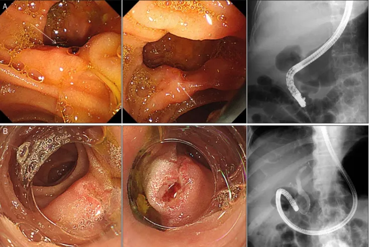
A
B
Fig. 1. Biliary cannulation with cap-fitted forward-viewing endoscope in patients with periampullary diverticulum. (A) Biliary cannulation by side-viewing endoscope failed due to tangential approach toward papilla. (B) Observation of papilla and biliary cannulation with needle-knife infundibulotomy overcoming tangential approach by cap-fitted forward-viewing endoscope.
procedures. For example, the use of cap-fitted forward-view- ing endoscopy for ERCP in patients with anatomical varia- tions after Billroth II gastrectomy has been proposed.5-7
Herein, we report a retrospective case series of patients in whom cannulation was complicated by periampullary diver- ticulum and assess the utility and safety of cap-assisted ERCP for biliary cannulation.
CASE REPORT
Between November 2013 and March 2014, ERCP proce- dures were performed in 346 patients at a tertiary medical center. Among these patients, 73 had periampullary divertic- ulum, of whom 5 (6.8%) underwent therapeutic ERCP using a cap-fitted forward-viewing endoscope as a rescue method due to difficult biliary cannulation. The inclusion criteria were as follows: (a) a documented periampullary diverticulum and (b) use of cap-assisted ERCP as a rescue method during the
first endoscopic encounter after failed attempts to perform ERCP using a standard side-viewing endoscope. The size of diverticulum was estimated by measuring the longest diame- ter on computed tomography image.
The ERCP procedure was performed initially using a stand- ard side-viewing endoscope (Olympus TJF Q260; Olympus Optical Co., Ltd., Tokyo, Japan). All procedures were performed by two endoscopists with more than five years of experience at a single tertiary center. Periampullary diverticulum was de- fined as extraluminal outpouchings of the mucosa occurring within a 2-3 cm from the ampulla of Vater or the hepatopancre- atic ampulla.8 Difficult biliary cannulation was defined as a cannulation time exceeding five minutes or as five or more passes or injections into the pancreatic duct. If the conven- tional endoscopic method failed due to hidden papilla or tan- gential approach, cap-fitted forward-viewing endoscopy (Olympus GIF Q260; Olympus Optical Co., Ltd.) with selective cannulation was performed (Fig. 1). The channel size of scope
cap-assisted method, a cap (distal attachment; D-201-10704;
Olympus Optical Co., Ltd.) was attached to the tip of the endoscope.
DISCUSSION
A total of 5 patients were included in our case review; three males and two females, with a median age of 69 years (range 53-90 years). The indications for ERCP were bile duct stones (n=4) and common bile duct stricture (n=1) (Table 1). The first attempt using the conventional ERCP failed in these five patients due to a periampullary diverticulum; after the first attempt, therapeutic ERCP was completed using a cap-fitted forward-viewing endoscope in four patients. The main caus- es of difficulty were hidden papillae (n=3) and tangential ap- proach (n=2). In case of hidden papilla, because papilla was located behind the wall of periampullary diverticulum, it was difficult to measure the exact position of the papilla, and con- sequently, accurate cannulation was not possible. In cases of tangential approach, it was also difficult to make frontal view of pailla or angular approach even if papilla was ob- served in the diverticulm. In all five patients, the ampulla of Vater was located at the 6 o’clock position. The mean diame- ter of the long axis of periampullary diverticula was 30 mm (range 15-50 mm). As a rescue method, needle knife fistulot- omy and selective biliary cannulation using a cap-fitted for- ward-viewing endoscopy were successfully achieved in 4 patients. As the policy of our clinic, fistulotomy was per- formed after more than five attempts for cannulation with a standard cannula. The mean number of ERCP sessions was 1.8 per patient, and the mean procedure time was 38 minutes.
Repeated procedures were performed in three cases. In two cases, fistulotomy under cap-fitted forward scope was per- formed in the first session, and biliary drainage was com- pleted through the second session. In one case, which failed in first session, successful cannulation with cap fitted for- ward-viewing endoscope was achieved under the rendezvous method with a percutaneous transhepatic cholangiography.
There were no major complications, including bleeding or perfo- ration, and planned procedures (fistulotomy and stone re- moval in all cases, ERBD insertion in two cases, balloon dila- tation in one case) were successfully completed. Only two pa- tients experienced complications of mild post-ERCP pan-
creatitis and hyperamylasemia. However, these complica- tions were self-limited and not deemed clinically significant.
In this present case review, we present 5 patients who ex- perienced difficulty in cannulation caused by periampullary diverticulum. The main causes of difficulty were hidden papil- lae (n=3) and tangential approach (n=2). In two patients with difficulty in biliary cannulation caused by tangential ap- proach, a cap-assisted forward-viewing endoscope allowed for a better approach to get to the papilla at the inner brim of the periampullary diverticulum. The cap fitted onto the endo- scope is useful for pushing aside the mucosal fold to enable a better view of the papilla and to stabilize the tip of the endoscope. By approaching the papilla at the right angle, en- doscopic fistulotomy was performed successfully. In three patients with hidden papillae, a cap-fitted forward-viewing endoscope provided a clear view of the papillary roof. Biliary cannulation was achieved by regular cannulation via endo- scopic fistulotomy.
Cap-fitted forward-viewing endoscopy is widely used for en- doscopic submucosal dissection or endoscopic hemostasis to achieve a clear view and proper approach.9,10 The efficacy of the cap-fitted forward-viewing endoscope was achieved by providing a better angle to approach the tangentially-posi- tioned or blinded lesions as well as providing improved stabi- lization of the endoscope tip by positioning it against the mu- cosa or lesion. It also minimized the effects of movement dur- ing respiration and enabled proper estimation of the dis- tance between the scope and lesion.11
Structural changes, including surgically altered anatomy and acquired lesions, such as a periampullary diverticulum, present a challenge for therapeutic endoscopists. Despite advances in the ERCP techniques over the past 25 years, bili- ary cannulation fails in about 5-20% of all cases.12 Difficulty in cannulation can increase the risk of post-ERCP complica- tions, such as pancreatitis.2,13 As previously reported, the risk of post-ERCP pancreatitis after difficult cannulation is in- creased compared with that after standard cannulation.14,15 Possible explanations for this include excessive manipu- lation that result in mechanical trauma, edema of the pancre- atic sphincter, and repeated contrast injections into the pan- creatic duct.2,13
Periampullary diverticulum is defined by extraluminal mu- cosal outpouchings of the duodenum arising near or encom- passing the ampulla of Vater (major papilla), including the in-
traluminal portion of common bile duct within 2-3 cm of the ampulla of Vater.16 The prevalence of duodenal diverticulum in the general population varies between 0.16 and 27% and increases with age, as indicated by previous reports.16-19 It is usually acquired during middle ages as a result of duodenal wall motility disorders, advancing age, weakening of in- testinal smooth muscle, and increased intraluminal pres- sure, all of which may lead to outpouching of the duodenum at a defect in the duodenal musculature.16,20 A periampullary diverticulum hinders biliary cannulation by limiting the visual field caused by mucosal folds or inadequate angle between the ampulla and the scope, particularly during ERCP procedures.
Previous reports by Myung et al. suggest that diverticula lead- ing to a hidden papilla and tangential approach hamper bili- ary cannulation using a conventional side-viewing endoscope.3 For these reasons, a cap-assisted forward-viewing endo- scope was used as an alternative, and therapeutic ERCP was performed successfully.
The main limitations of this study are its single-center pop- ulation, which was relatively small, and the lack of a control group. Differences in proficiency among physicians may also affect the efficacy of the procedure. Although long-term stud- ies involving a greater number of cases are needed, our expe- riences suggest that cap-assisted endoscopy can be helpful to allow for proper cannulation at least in patients compli- cated with periampullary diverticulum.
As a rescue method, cap-assisted ERCP using forward- viewing endoscopy could be a useful and safe alternative in patients experiencing cannulation difficulty due to peri- ampullary diverticulum.
REFERENCES
1. Chu YC, Su SJ, Yang CC, Yeh YH, Chen CH, Yueh SK. ERCP plus pap- illotomy by use of double-balloon enteroscopy after Billroth II gastrectomy. Gastrointest Endosc 2007;66:1234-1236.
2. Udd M, Kylänpää L, Halttunen J. Management of difficult bile duct cannulation in ERCP. World J Gastrointest Endosc 2010;
2:97-103.
3. Myung DS, Park CH, Koh HR, et al. Cap-assisted ERCP in patients with difficult cannulation due to periampullary diverticulum.
Endoscopy 2014;46:352-355.
4. Park TY, Kang JS, Song TJ, et al. Outcomes of ERCP in Billroth II gastrectomy patients. Gastrointest Endosc 2016;83:1193-1201.
5. Anastassiades CP, Salah W, Pauli EM, Marks JM, Chak A. Cap-as- sisted ERCP with a forward-viewing gastroscope as a rescue en- doscopic intervention in patients with Billroth II anatomy. Surg
6. Easler JJ, Sherman S. Cap-assisted pancreaticobiliary endos- copy in Billroth II anatomy: ERCP "through the looking glass".
Gastrointest Endosc 2016;83:1202-1204.
7. Park CH, Lee WS, Joo YE, Kim HS, Choi SK, Rew JS. Cap-assisted ERCP in patients with a Billroth II gastrectomy. Gastrointest Endosc 2007;66:612-615.
8. Li X, Zhu K, Zhang L, et al. Periampullary diverticulum may be an important factor for the occurrence and recurrence of bile duct stones. World J Surg 2012;36:2666-2669.
9. Kim JI, Kim SS, Park S, et al. Endoscopic hemoclipping using a transparent cap in technically difficult cases. Endoscopy 2003;
35:659-662.
10. Wang KK, Prasad G, Tian J. Endoscopic mucosal resection and endoscopic submucosal dissection in esophageal and gastric cancers. Curr Opin Gastroenterol 2010;26:453-458.
11. Yap CK, Ng HS. Cap-fitted gastroscopy improves visualization and targeting of lesions. Gastrointest Endosc 2001;53:93-95.
12. Swan MP, Bourke MJ, Williams SJ, et al. Failed biliary cannulation:
clinical and technical outcomes after tertiary referral endoscopic retrograde cholangiopancreatography. World J Gastroenterol 2011;
13. Cheon YK, Fogel EL. ERCP topics. Endoscopy 2006;38:1092- 1097.
14. Freeman ML, DiSario JA, Nelson DB, et al. Risk factors for post- ERCP pancreatitis: a prospective, multicenter study. Gastrointest Endosc 2001;54:425-434.
15. Vandervoort J, Soetikno RM, Tham TC, et al. Risk factors for com- plications after performance of ERCP. Gastrointest Endosc 2002;56:652-656.
16. Lobo DN, Balfour TW, Iftikhar SY, Rowlands BJ. Periampullary di- verticula and pancreaticobiliary disease. Br J Surg 1999;86:
588-597.
17. Kirk AP, Summerfield JA. Incidence and significance of juxtapapillary diverticula at endoscopic retrograde cholangiopancreatography.
Digestion 1980;20:31-35.
18. Løtveit T, Skar V, Osnes M. Juxtapapillary duodenal diverticula.
Endoscopy 1988;20 Suppl 1:175-178.
19. Vaira D, Dowsett JF, Hatfield AR, et al. Is duodenal diverticulum a risk factor for sphincterotomy? Gut 1989;30:939-942.
20. Ackermann W. Diverticula and variations of the duodenum. Ann Surg 1943;117:403-413.