www.krspine.org
Factor Analysis Affecting the Leakage of Bone Cement After Vertebroplasty
Jae-Hoon Kim M.D., Kyung-Jin Song M.D.*, Tai-Seung Kim M.D.†, Jae-Lim Cho M.D.† , Ye-Soo Park M.D.
J Korean Soc Spine Surg 2010 Mar;17(1):13-17.
Originally published online March 31, 2010;
doi: 10.4184/jkss.2010.17.1.13
Korean Society of Spine Surgery
Department of Orthopaedic Surgery, Ewha Womans University Collge of Medicine
#911-1 Mok-dong, Yangcheon-gu, Seoul, 158-710, Korea Tel: 82-2-2646-6808 Fax: 82-2-2646-6804
©Copyright 2010 Korean Society of Sping Surgery pISSN 2093-4378 eISSN 2093-4386
The online version of this article, along with updated information and services, is located on the World Wide Web at:
http://www.krspine.org/DOIx.php?id=10.4184/jkss.2010.17.1.13
This is an Open Access article distributed under the terms of the Creative Commons Attribution Non-Commercial License (http://
creativecommons.org/licenses/by-nc/3.0) which permits unrestricted non-commercial use, distribution, and reproduction in any medium, provided the original work is properly cited.
Journal of Korean Society of
Spine Surgery
Received: December 12, 2008; Accepted: February 16, 2010 Corresponding auther: Ye-Soo Park M.D.
Department of Orthopaedic Surgery, Hanyang University Kuri Hospital, Hanyang University College of Medicine
249-1 Kyomoon-dong, Guri-City, Kyunggi-do, 471-701, Korea TEL: 82-31-560-2316, FAX : 82-31-557-8781
E-mail: [email protected] available online March 31, 2010
“This is an Open Access article distributed under the terms of the Creative Commons Attribution Non-Commercial License (http://creativecommons.
org/licenses/by-nc/3.0/) which permits unrestricted non-commercial use, distribution, and reproduction in any medium, provided the original work is
Factor Analysis Affecting the Leakage of Bone Cement After Vertebroplasty
Jae-Hoon Kim M.D., Kyung-Jin Song M.D.*, Tai-Seung Kim M.D.
†, Jae-Lim Cho M.D.
†, Ye-Soo Park M.D.
Department of Orthopaedic Surgery, Guri Hospital, Hanyang University College of Medicine Department of Orthopaedic Surgery, Chonbuk University College of Medicine*
Department of Orthopaedic Surgery, Hanyang University College of Medicine†
Study Design: Retrospective study
Objective: This study examined the causative factors of cement leakage in an osteoporotic compression fracture that had received percutaneous vertebroplasty.
Summary of Literature Review: Percutaneous vertebroplasty is simple and safe for the treatment of osteoporotic compression fractures. However, serious complications, such as pulmonary emboli and paraplegia, can occur if the bone cement leaks into the pulmonary artery or spinal canal.
Materials and Methods: Between Oct. 2002 and Apr. 2008, 95 patients (148 vertebral bodies) underwent percutaneous vertebroplasty for the treatment of an osteoporotic compression fracture. The presence of cement leakage was evaluated by plain radiography and computed tomography. The correlations between cement leakage and gender, age, level of fractured vertebra, fracture type, bone density, procedure, injecting amount, preoperative vertebral body compression rate, timing of surgery, and the existence of an intravertebral cleft on magnetic resonance imaging (MRI) were analyzed.
Results: Leakage was found in 37 bodies on plain radiography and 56 on the CT-scan. A comparison of the leakage and non-leakage groups revealed the bone density (p=0.046) and amount injected (p=0.000) to be related to cement leakage. Multivariate logistic regression showed that injecting more than 4.0ml was related to cement leakage with an odds ratio of 2.23(95% CI, 1.476~3.377).
Conclusions: Cement leakage after percutaneous vertebroplasty is associated with the amount injected. Therefore, the cement volume should be restricted to the amount required for pain relief.
Key words: Vertebroplasty, Cement leakage
서론
경피적 척추 성형술은 1984년 Galibert1) 등에 의해 처음 소 개된 이후 혈관종, 전이성 암, 골수종, 임파종, 골다공증에 의 한 압박골절 등의 치료에 이용되고 있다. 특히 골다공증을 동 반한 척추체 압박골절에서 술 후 즉각적인 통증의 완화와 척 추의 안정성을 얻을 수 있어 조기 거동이 가능하여 점차 사 용 빈도가 증가하고 있는 추세이다. 그러나 고령의 환자에서 비교적 안전하고, 결과 또한 양호하다는 많은 보고에도 불구 하고 부작용 또한 많은 것이 사실이며,2,3) 부작용은 대부분 골 시멘트의 유출과 연관이 있다고 보고되고 있다. 이에 본 저 자들은 골다공증성 척추 압박 골절의 치료에 있어 흔히 시행 되고 있는 경피적 척추 성형술 후 골 시멘트 유출의 원인을 분석해 보고자 하였다.
연구 대상 및 방법
2002년 10월부터 2008년 4월까지 본원 정형외과에서 골다
Jae-Hoon Kim et al Volume 17 • Number 1 • March 2010
www.krspine.org
14
공증성 압박 골절로 경피적 척추 성형술을 시행 후 3개월 이 상 추시 관찰이 가능하였던 95례, 148추체를 대상으로 후향 적 방법으로 연구하였다. 술 전 진단은 단순 방사선 사진, 조 영증강 자기공명영상검사를 이용하였으며, 골다공증의 진 단은 이중 방사선 에너지 흡수법(Dual energy X-ray absorp- tiometry)을 이용하였다. 골절 양상에 따른 즉 피질골의 손상 부위에 따른 골 시멘트의 유출을 보기 위하여 골절의 형태를 Denis의 분류법4)을 이용하여 분류하였고, 술 전 압박률은 측 면 단순 방사선 사진을 기준으로 압박된 추체의 전방 추체높 이와 후방 추제높이를 기준으로 그 차이값의 백분위로 계산 하였다.
시술은 본원의 일인 술자에 의해 시행되었으며, C형 방 사선 투시기하에 일측 혹은 양측 척추경 도달법을 이용 하였다. 시술시에는 모든 예에서 투관 침을 추체의 전 방 1/3부까지 삽입하고. 바늘의 경사면이 골절면을 향 하지 않도록 조절하였으며, 조영제를 이용하여 척수 강 및 신경 공으로의 유출을 C형 방사선 투시기로 확인 후 polymethylmethacrylate(PMMA, DePuy CMW 3 bone cement, Blackpool, England)를 엷은 치약과 같은 굳기의 상 태로 즉 골 시멘트의 점도가 굳어지는 상태에서 주입을 시행 하였다. 주입 중 척추체외로 골 시멘트 유출이 발견되면 즉 시 주입을 멈추고 정맥을 통한 색전 유무, 척수 강 또는 신경 공으로의 유출 유무를 확인 후 나사방식의 주입기를 이용하 여 일정 속도로 주입하였다.
술후 단순 방사선 소견 및 전산화 단층 촬영으로 골 시멘
트 유출을 확인하였으며, 성별, 나이, 골절 추체, 골절 양상, 술 전 압박률, 골밀도, 주입량, 술식, MRI 상 골절 추체 내 cleft의 존재 여부, 수술 시기(증상 발현 2주 전과 2주 후)에 따른 골 시멘트 유출의 차이 및 유의성을 분석하였다. 통계 학적 분석은 Pearson chi-square test, Fisher’s exact test, logistic regression method를 이용하여 단순 로지스틱 회귀 분석(simple logistic regression) 및 다중 로지스틱 회귀분석 (multiple logistic regression)을 시행하였다.
결과
95례 중 여자 86례, 남자 9례 였으며, 평균 연령은 69.6 세(50~87) 이었다. 술전 시행한 골밀도 검사의 평균 T 점 수는 -3.4(-1.2~-5.8)이었고, 골 시멘트의 평균 주입량은 4.3ml(1.5~8.0)이었다.
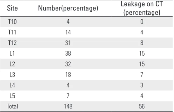
골 시멘트의 유출은 단순 방사선 소견상으로는 148추체중 37 추체로 25%에서 확인할 수 있었고, 전산화 단층 촬영에서는 56 추체로 38%에서 유출을 확인할 수 있었으나(Table 1)(Fig. 1), 유 출이 있었던 모든 환자들에서 신경증상, 혈관 색전 등의 합병증 은 발생하지 않았다. 골 시멘트 유출은 그 경로에 따라 전방 척 추관외 정맥총(anterior external vertebral venous plexus)을 통한 유출, 기처 추체 정맥(basivertebral vein)을 통한 유출, 기저 추체 정맥을 넘어서 전방 척추관내 정맥총(anterior internal vertebral venous plexus)까지의 유출, 피질골 결손을 통한 유출로 분류하 였으며 피질골 결손을 통한 유출은 다시 추체 전방이나 측방, 추
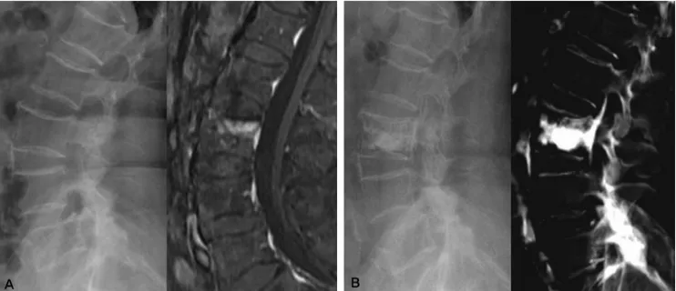
Fig. 1. (A) An 66-year-old female has recent osteoporotic compression fracture of L3. (B) Postoperative Lateral roentgenogram and Computed tomography (CT) show leakage of PMMA.
간판 사이, 척추관이나 신경공으로의 유출로 분류하였다. 유출 이 있었던 56추체 중 10추체에서는 두 곳에서의 유출이 확인되 어 총 누출 건수는 66례 이었다(Table 2).
단순 회귀 분석 결과를 보면 골 시멘트의 유출은 남자의 경 우 9례 중 4례, 여성의 경우 86례 중 41례에서 유출이 발생하였 으며, 유출이 있었던 환자군의 평균 연령은 69.5세(50~82), 유 출이 없었던 환자군의 평균 연령은 69.6세(56~87)로, 환자의 성별(p=0.569) 및 연령(p=0.889)은 골 시멘트 유출과의 연관 성은 없었다. 골절은 흉요추부에서 가장 많았으며, 각각의 추 체별로 시멘트의 유출의 차이는 없었으며(p=0.425)(Table 1), Denis의 분류를 이용하여 A, B, C, D로 골절 양상을 구분하였 으나, 시멘트 유출과의 연관성은 없었다(p=0.376)(Table 3). 골 시멘트 유출이 있었던 환자군에서의 평균 골밀도는 -3.33(- 1.2~-5.8)이었으며, 유출이 없었던 환자군에서의 평균 골밀 도는 -3.53(-1.2~-5.2)으로 통계적으로 유의한 차이를 보였 다(p=0.046). 골 시멘트 유출이 있었던 환자군에서의 평균 주 입량은 4.85ml(3~8)이었으며, 유출이 없었던 환자군에서의 평균 주입량은 4.06ml(1.5~6.5)로 통계적 유의성이 있었다
(P=0.000). 골 시멘트 유출이 있었던 환자군에서의 술 전 평균 압박률은 22.4%(0~51), 유출이 없었던 환자군에서의 압박률 은 20.5%(0~62)로 통계적 유의성은 없었다. 술전 MRI 소견상 추체내 cleft가 있었던 경우 17추체중 7추체에서 유출이 있었으 나 cleft유무와 유출간에 통계적인 유의성은 없었다(p=0.763).
수술 시기상으로 분석해 본 결과 2주 이내 수술의 경우 44추체 중 21추체에서, 2주 이후 수술한 경우에는 104추체중 35추체 에서 유출이 발생하여 2주 전,후를 기준으로 했을 때 수술시기 와 유출간의 통계적 유의성은 없었다(p=0.107). 술식에 따른 분 석은 양측 척추경 사용시 140체중 54 추체, 일측 8추체중 2추체 에서 유출이 발생하여 술식과 유출은 통계적 유의성은 없었다 (p=0.593).
다변량 분석 시행 결과 주입량만이 골 시멘트의 유출과 통계 적으로 의미 있는 관련 요소이었으며(p=0.000) 주입량이 4ml 이상이었을 경우 비교 위험도는 2.23(95% CI, 1.476~3.377)이 었다.
고찰
경피적 척추 성형술은 고령 및 장시간 수술에 따른 합병 증 없이 술 후 즉각적인 통증의 완화와 척추의 안정성을 얻을 수 있다는 장점으로 최근 수년 사이에 활발히 사용되기 시작 한 수술로 심한 동통을 유발하는 골다공증성 척추 압박골절 의 치료에 있어서 80~95%에서 우수한 치료 결과를 보인다 는 많은 연구 결과가 있다.5,6,7,8) 그러나 척추 성형술은 아직 까지 장기 추시 결과가 없으며, 드물지만 시멘트 유출로 인
한 마비9,10) 나 폐색전증11)과 같은 심각한 합병증을 유발할 수
있다는 단점을 가지고 있다. 대부분의 경우 합병증은 시멘트 의 유출과 연관이 있으며 추간판 내로 누출될 경우에는 인접 추체의 후속 골절을 유발할 수 있고 척추 주위 정맥으로의 누출은 폐색전증의 합병증이 발생할 수 있으며, 척추 강이나 Table 1. Incidence of cement leakage according to vertebral body
Site Number(percentage) Leakage on CT (percentage)
T10 4 0
T11 14 4
T12 31 8
L1 38 15
L2 32 15
L3 18 7
L4 4 3
L5 7 4
Total 148 56
Table 2. Classification of cement leakage
Types of leakage Number(Percentage)
Anterior external venous plexus 18(27%)
Anteriro internal venous plexus 23(35%)
Basivertebral vein 9(14%)
Cortical defect 16(24%)
Intervertebral disc space 12(18%)
Anterior or lateral to the vertebral body 4(6%)
Spinal canal 0(0%)
Neural foramen 0(0%) Total 66(100%)
Jae-Hoon Kim et al Volume 17 • Number 1 • March 2010
www.krspine.org
16
추간 공으로의 누출은 방사통, 척수 손상을 유발할 수 있는 것으로 보고되고 있다.11) 또한 경피적 추체 성형술 중 원인 모를 저혈압, 호흡 곤란, 급성 호흡 곤란 증후군 등도 골 시멘 트의 유출이 주요한 원인으로 생각 되고 있다.12) 경피적 척추 성형술 후 골 시멘트의 유출은 30% 이상으로 보고5,12,13)되 고있으며, 저자들의 경우에서도 유출이 최소화 될 수 있도 록 골 시멘트를 주입하고자 노력하였음에도 불구하고 골 시 멘트의 유출은 전산화 단층 촬영을 통하여 38%에서 확인할 수 있었다. 이는 대부분의 환자들에서 심한 골다공증을 가지 고 있고, 상당수가 흉요추 이행부의 골절로 C형 방사선 투과 기의 화질이 미세한 유출을 확인하기에는 충분치 않았고, 술 후 골 시멘트의 유출의 판정을 전산화 단층 촬영을 이용하였 기 때문인 것으로 생각된다.
그러나 시멘트의 유출에 의한 신경학적 이상, 부정맥, 호 흡 곤란, 저혈압 등의 중대한 합병증은 발생하지 않았다. 시 멘트 색전의 경우 액상 상태의 골 시멘트가 하대정맥을 통하 여 폐혈관 및 뇌혈관까지 이동시 발생할 수 있으며, 대부분 의 신경학적 이상은 척추 강 및 신공 공 주위로의 시멘트 유 출에 의하여 발생하는 것으로 알려져 있다. 저자들은 골 시 멘트의 주입시 엷은 치약과 같은 굳기의 상태로 주입하였기 때문에 혈관 색전 등의 합병증은 피할 수 있었으며, 골 시멘 트의 유출 중 척추체 후면의 골피질 손실을 통한 경막 외 공 간과 신경 공 주위로의 직접적인 골 시멘트의 유출은 없었으 며, 전방 척추관내 정맥총으로의 유출이 23례에서 있었으나 그 크기 및 위치가 신경학적 이상을 일으킬 만한 원인이 되 지 않았기 때문으로 생각된다.
Ryu 등14)은 347명의 골다공증성 압박 골절 환자에서 경피 적 척추 성형술을 시행한 결과 주입량이 많을수록 그리고 제 7 흉추 이상에서 시술하였을 때 비교 위험도가 높았음을 보 고하였다. 하지만 본 연구에서 각 추체에 따른 유출의 차이 는 없었는데, 이는 본 연구에서 골다공증성 압박 골절이 심 했던 제 10 흉추 4례를 제외하고는 제 10 흉추 이상 추체의 압박 골절은 보존적 치료를 원칙으로 하여 척추 성형술 대상 에서 제외하였기 때문이며, 제 10 흉추에서 제 5 요추사이에 서는 해부학적으로 척추체와 척추경의 크기가 큰 차이가 없
어 추체에 따른 유출이 연관이 없었던 것으로 보인다. 만약 Ryu 등의 연구에서와 같이 제 7 흉추 이상의 상부 흉추를 대 상으로 했다면 결과가 달랐을 것으로 생각된다. 골절 양상 즉 피질골의 손상부위에 따른 골 시멘트 유출을 보고자 비록 외상성 압박골절은 아니지만 Denis 분류를 사용하여 골절 형 태를 분류하였으나 골 시멘트 유출과의 연관성은 확인할 수 없었다.
본 연구에서 수술 시기를 2주 전,후로 보았을 때 유출과 통계 학적 연관성이 없었는데, 수술 시기의 경우 국내에서 보존적 치 료 3주 이후에나 척추 성형술 치료가 가능하도록 함으로 해서 수술적 치료의 남용을 방지하고자 하는 긍정적인 면도 있으나, 경우에 따라 불필요한 입원 비용의 증가, 초기 적극적 재활의 미 비로 인한 골의 약화 등이 초래되어 이러한 점들을 고려한다면 환자에게 도움이 되는 방향으로 시술을 하는 것이 좋을 것으로 생각된다.
주입량의 경우 적은 양을 주입하는 경우 유출이 적었다고 보 고를 하고 있지만14) 그 양을 통계적으로 제시한 문헌은 흔치 않 았다. 저자들의 경우 시멘트 주입시 추체의 강성(stiffness) 복원 을 위해서는 4~8ml, 강도(strength) 복원을 위해서는 2ml 주입이 이상적이라는 결과15)를 토대로 추체에 상관 없이 주입을 시행하 였다. 모든 주입 양에 따른 분석 결과 4ml를 기준으로 통계학적 유의성을 확인할 수 있었으며 이는 Ryu 등14)의 문헌과 일치하는 결과임을 보여주었다.
Hiwatashi 등16)은 추체내 cleft가 있을 경우 골 시멘트의 유출 이 적었음을 보고하였는데 시멘트 주입은 추체의 후방 25%를 충만시킬 때 까지 혹은 골 시멘트의 유출이 확인될 때 까지 시행 하였다. 본 연구에서 추체 내 cleft가 있었던 추체 내에서는 평 균 4.5ml(3~6)가 주입되었고, cleft가 없었던 추체에서는 평균 4.3ml(1.5~8)가 주입되었으며 추체 내 cleft유무와 골 시멘트의 유출은 통계적 유의성은 없었다.
결론
경피적 척추 성형술 후 골 시멘트 유출은 골 시멘트 주입량 과 통계적으로 의미있는 연관성이 있었으며, 따라서 경피적 척추 성형술 시 골 시멘트의 유출을 예상하고, 동통을 감소 시킬 수 있는 범위 내에서 제한적으로 주입해야 할 것으로 사료된다.
REFERENCES
1. Galibert P, Deramond H, Rosat P, Le Gars D. Preliminary note on the treatment of vertebral angioma by percutaneous
Type Number(percentage) Leakage(percentage)
A 59 21
B 63 28
C 11 2
D 15 5
Total 148 56
Table 3.Incidence of cement leakage according to fracture type
acrylic vertebroplasty. Neurochirurgie. 1987;33:166–8.
2. Moreland DB, Landi MK, Grand W. Vertebroplasty:
techniques to avoid complications. Spine. 2001;1:66-71.
3. Park HG, Kim MH, Yoo MJ, et al. Complications after Vertebroplasty of Treatment for Compression Fracture with Osteoporosis. J Korean Fracture Soc. 2003;16:534-40.
4. Denis F. Three column spine and its significance in the classification of acute thoracolumbar spinal injuries. Spine.
1983;8:817-31.
5. Cortet B, Cotten A, Boutry N, et al. Percutaneous vertebroplasty in the treatment of osteoporotic vertebral compression fracture: an open prospective study. J Rheumatol. 1999;26:2222-8.
6. Cyteval C, Sarrabere MP, Roux JO, et al. Acute osteoporotic vertebral collapse: open study on percutaneous injection of acrylic surgical cement in 20 patients. Am J Roentrenol.
1999;173:1685-90.
7. Kim CH, Choi YJ, Baek SG, et al. Vertebroplasty on osteoporotic compression fracture. J Korean Fracture Soc.
2002;15:123-8.
8. Martin JB, Jean B, Sugiu K, et al. Vertebroplasty: clinical experience and follow-up results. Bone. 1999;25:11-5.
9. Cotton A, Dewatre F, Cortet B, et al. Percutaneous vertebroplasty for osteolytic metastases and myeloma:
effects of the percentage of lesion filling and the leakage of mrthyl methacrylate at clinical follow-up. Radiology.
1996;200:525-30.
10. Ratliff J, Nguyen T, Heiss J. Root and spinal cord compression from methylmethacrylate vertebroplasty. Spine.
2001;26:300-2.
11. Padovani B, Kasriel O, Brunner P, Peretti-Viton P.
Pulmonary embolism caused by acryl cement: A rare complication of percutaneous vertebroplasty. Am J Neuroradiol. 1999;20:375-7.
12. Yoo KY, Jeong SW, Yoon W, Lee J. Acute respiratory distress syndrome associated with pulmonary cement embolism following percutaneous vertebroplasty with polymethylmethacrylate. Spine. 2004;29:294-7.
13. Bostrom MP, Lane JM. Augmentation of osteoporotic vertebral bodies: Future directions. Spine. 1997;22:38-42.
14. Ryu KS, Park CK, Kim MC, Kang JK. Dose-dependent epidural leakage of polymethylmethacrylate after percutaneous vertebroplasty in patients with osteoporotic vertebral compression fracture. J Neurosurg. 2002;96:56- 61.
15. Belkoff SM, Mathis JM, Jasper LE, Deramond H. The biomechanics of vertebroplasty. The effect of cement volume on mechanical behavior. Spine. 2001;26:1537-41.
16. Hiwatashi A, Ohgiya Y, Kakimoto N, Westesson PL.
Cement leakage during vertebroplasty can be predicted on preoperative MRI. Am J Roentrenol. 2007;188:1089-93.
경피적 척추 성형술후 골시멘트 유출에 영향을 미치는 요인 분석
김재훈 • 송경진* • 김태승† • 조재림† • 박예수
한양대학교 의과대학 구리병원 정형외과학 교실, 전북대학교 의과대학 정형외과학 교실*, 한양대학교 의과대학 정형외과학 교실†
연구 계획: 후향적 연구
목적: 경피적 척추 성형술 후 골 시멘트 유출의 요인을 분석해 보고자 하였다.
선행문헌의 요약: 경피적 척추 성형술 후 골 시멘트의 유출은 30% 이상으로 알려져 있으며, 유출과 관련하여 많은 원인들이 보고되고 있다.
대상 및 방법: 2002년 10월부터 2008년 4월까지 골다공증성 추체 골절로 경피적 척추 성형술을 시행한 95 례, 148 추체를 대상으로 하였다. 술후 단순 방사선 및 전산화 단층 촬영으로 유출을 확인하였으며, 골 시멘트 유출과 성별, 나이, 골절 추체, 골절 양상, 술 전 압박률, 골밀도, 주입량, 술식, MRI 상 골절 추체내 cleft의 존재 여부, 수술 시기와의 상관 관계를 분석하였다.
결과: 골 시멘트의 유출은 단순 방사선 소견상 37추체, 전산화 단층 촬영의 경우 56추체에서 확인 되었다. 유출이 있었던 환자군과 없었던 환자군 의 비교에서 골밀도(p=0.046)와 주입량(p=0.000)에 따른 유출이 차이가 있었으며, 다중 회귀 분석 결과 4ml 이상 주입하였을 경우에 비교 위험도가 2.23(95% CI, 1.476~3.377)으로 유의하게 나타났다.
결론: 경피적 척추 성형술 후 골 시멘트 유출은 시멘트 주입량과 연관이 있었으며, 경피적 척추 성형술시 동통을 감소 시킬 수 있는 범위 내에서 골 시멘 트를 제한적으로 주입되어야 할 것으로 사료된다.
색인단어: 척추 성형술, 골 시멘트 유출 약칭제목: 경피적 척추 성형술 후 골 시멘트 유출