Background: Rotator cuff disease is the most common cause of chronic shoulder pain in adults. In some cases, massive rotator cuff tear is irreparable. Various surgical techniques have been introduced for irreparable rotator cuff tear; how- ever, no definitive technique has been established yet. To address this concern, we comparatively analyzed superior cap- sule reconstruction and partial repair to assess the treatment effects of superior capsule reconstruction.
Methods: A total of 26 patients were assessed between January 2013 and November 2014. Superior capsule reconstruc- tion was performed in 15 patients and partial repair in 11 patients. The patients were classified into two groups to mea- sure and compare the range of motion (ROM), visual analogue scale (VAS) score, subjective satisfaction (very satisfied, satisfied, rather the same, and dissatisfied), and radiological acromiohumeral distance (AHD) before and after surgery.
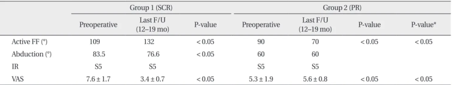
Results: The ROM of the shoulder joint was better in anterior elevation in the superior capsule reconstruction group than in the partial repair group at the final follow-up (active forward flexion, 132° vs. 70°; P < 0.05). The former group also had better VAS scores (3.4 ± 0.7 vs. 5.6 ± 0.8; P < 0.05). In terms of the patient’s subjective satisfaction, the proportion of “satis- fied” and above was higher in the superior capsule reconstruction group (53.3% vs. 27.3%). The radiologically measured AHDs were 5.98, 9.12, and 7.62 mm at presurgery, postsurgery, and final follow-up, respectively, in the superior capsule reconstruction group. In the partial repair group, the AHDs were 6.06, 7.26, and 4.94 mm, respectively.
Conclusion: Superior capsule reconstruction can be considered a useful surgical technique for patients with irreparable massive rotator cuff tear with no arthritis.
Keywords: Massive rotator cuff tear; Superior capsule reconstruction
Comparison of clinical outcome between superior capsule reconstruction and partial repair in irreparable massive rotator cuff tear
Jun Seok Kim, Hyung Kan Kim, Jae Yeon Kong, Jang Seok Choi
Department of Orthopedic Surgery, West Busan Centum Hospital, Busan, Korea
Copyright © 2019 Korean Orthopedic Society for Sports Medicine. All rights reserved.
CC This is an open-access article distributed under the terms of the Creative Commons Attribution Non-Commercial License (http://creativecommons.org/licenses/
by-nc/4.0) which permits unrestricted noncommercial use, distribution, and reproduction in any medium, provided the original work is properly cited.
Received January 24, 2019; Revised August 19, 2019; Accepted August 19, 2019 Correspondence to: Jun Seok Kim, https://orcid.org/0000-0002-8857-2796
Department of Orthopedic Surgery, West Busan Centum Hospital, 226 Saebyeok-ro, Sasang-gu, Busan 46973, Korea. Tel: +82-51-329- 3000, Fax: +82-51-329-3197, E-mail: [email protected]
Arthroscopy and Orthopedic Sports Medicine
AOSM
INTRODUCTION
Rotator cuff disease is the most common cause of chronic shoulder pain in adults. In some cases, massive rotator cuff tear is irreparable. Several surgical techniques for ir- reparable massive rotator cuff tear have been reported;
however, no definitive technique has been established.
For patients with irreparable massive rotator cuff tear and arthritis, reverse total shoulder arthroplasty has been known as the most effective technique. For patients with- out arthritis, several techniques have been introduced to date: surgical treatment on the biceps; conventional
treatments, such as rotator cuff debridement, rotator cuff reconstruction, superior capsule reconstruction (SCR), tendon transfer, and reverse total shoulder arthroplasty;
and combinations of the above. Among these, Burkhart [1] and Denard et al. [2] described the grounds for partial repair (PR) to restore the force couple, whereas Mihata et al. [3,4] argued for the use of SCR for irreparable massive rotator cuff tear through a biomechanical analysis after SCR for the stability of the superior capsule. However, no comparative study on the treatment effects of SCR and rotator cuff reconstruction on patients with irreparable massive rotator cuff tear has been conducted to date.
To address this, we comparatively analyze the treatment effects of the two surgical techniques. We hypothesize that SCR is a better surgical method than PR because the former includes rotator cuff reconstruction and SCR for better supracondylar stability of the humerus beyond the mere bridging effect.
METHODS
Subjects and data analysis
The subjects of this study were 26 patients to whom magnetic resonance imaging (MRI) and at least 1-year follow-up monitoring were available among patients with irreparable massive rotator cuff tear who received PR and SCR at the our hospital between January 2013 and November 2014. Of the 26 patients, 15 received SCR and 11 received PR. The following cases of irreparable rotator cuff tear, as assessed by MRI, were included in this study:
(1) tear of ≥ 5 cm, (2) at least two tendon tears, (3) severe fatty infiltration of the Goutallier stage 3 or above [5,6], (4) tendon involution of the glenoid level or above within the surgical findings [7], and (5) not being able to pull the scapula spine to the binding site even after debridement and relaxation [8]. The range of motion (ROM), visual analogue scale (VAS) score, patients’ subjective satisfac- tion (very satisfied, satisfied, rather the same, and dissat- isfied), and radiological acromiohumeral distance (AHD) were compared and analyzed between group 1 (SCR) and group 2 (PR) before and after surgery. The Mann–
Whitney U test was used to compare the sizes of the two groups, and Wilcoxon signed-rank test was used to com- pare the sizes within each group for statistical analysis.
IBM SPSS Statistics ver. 23.0 (IBM Corp., Armonk, NY, USA) was used as the statistical program. The statistically significant difference was recognized if the P-value was less than 0.05.
Surgical technique
The surgery was performed with the patient in the lateral decubitus position under general anesthesia. Any accom- panied lesion was evaluated using diagnostic arthros- copy. If the tear of the long head of the biceps tendon was at least 50% or accompanied by dislocation, tenodesis or tenotomy was performed. All procedures were performed by an experienced surgeon (J.S.K.) using rotator cuff su- tures.
Partial repair
After subscapularis observation, any tear that takes up at least one-fourth of the superior binding site was sutured with a suture anchor. The arthroscope was moved to the subacromial space, and debridement and bursectomy were performed in the same space. After separating the rotator interval and thoroughly detaching the adhesion around the supraspinatus, the size and quality of the tear on the superior rotator cuff were checked, and its non- suturability was reaffirmed. The surgical purpose of PR was to suture the subscapularis and posterior cuff to restore the balance of the force couple and restore the stable fulcrum of the shoulder joint. After preparations of the binding site at the infraspinatus, single-row repair was performed on the infraspinatus with a suture anchor.
Superior capsule reconstruction
Similarly, lesions along the long head of the biceps and subscapularis were treated, and the subacromial space was observed using an arthroscope to remove patho- logical cyst tissues, and decompression was performed in the subacromial space. A marker was used at the 45°
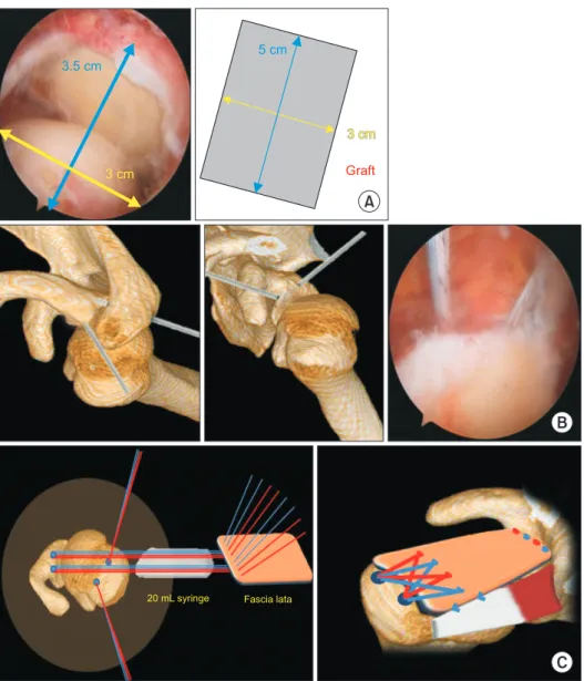
abduction position to measure the defect size on the superior capsule in the anterior, posterior, medial, and lateral directions. The size of the graft was considered the same as that of the defect site in the anterior and poste- rior directions and 1.5 cm longer in the medial and lateral directions, considering the labrum binding site (Fig. 1A).
The greater trochanter of the ipsilateral and lateral thigh was vertically incised, and the fascia lata was extracted at a length twice and thrice greater than the medial and lat- eral length of the graft and then folded in two or three lay- ers. The surroundings were sutured to prevent creasing.
The 4.5-mm-diameter suture anchor was inserted at the positions between 10 o’clock and 11 o’clock and between 12 o’clock and 1 o’clock in the superior glenoid fossa (be- tween 11 and 12 o’clock and between 1 and 2 o’clock of the left shoulder joint), respectively (Fig. 1B). The bind- ing site was prepared on the rotating cuff at the greater tuberosity, and a 4.5-mm suture anchor was inserted at the medial foot print for the upcoming suture bridge.
The lateral portal was extended to approximately 2 cm and then a 20 mL synringe was inserted. All glenoid an- chors thread were retrieved out through the synringe and each thread penetrated upward from the bottom in the medial part of the prepared graft. Subsequently, the graft penetrated through the syringe and was positioned at the subacromial space (Fig. 1C). Each thread positioned
in the joint pit was paired and then knotted to complete four horizontal stitches. The pre-inserted thread at the medial binding point at the greater tuberosity of the hu- merus penetrated the graft at the subacromial space, and a SwiveLock anchor (Arthrex, Naples, FL, USA) was used to complete the suture bridge at the lateral row. Side- to-side suture with No. 2 nonabsorbable fiber wire was performed between the last remaining infraspinatus and graft (Fig. 1D). If the medial, lateral, and posterior side sutures were completely rigid, the space between the subscapularis and graft was not sutured to prevent con- tracture after the surgery.
Rehabilitation
The patients were required to wear the shoulder abduc- tion brace for 6 weeks after the surgery and were initi-
ated on a stretching program from the 6th week. If their subscapularis recovered, external rotation was limited to 45°. Rotator cuff strengthening and scapular stabilizing exercises were initiated from the 12th week after surgery.
B
C
5 cm
3 cm Graft
20 mL syringe Fascia lata 3.5 cm
3 cm
A
Fig. 1. Superior capsule reconstruction procedure. (A) Graft preparation. (B) Fixation of 4.5-mm diameter suture an- chor. Below, arthroscopic image of ten- sor fascia graft position in subacromial space. (C) The diagram of graft suture using a 20-mL syringe. (D) Diagram of completed superior capsule reconstruc- tion using tensor fascia lata.
Table 1. Demographic data of patients
Group 1 (SCR) Group 2 (PR)
Sex (male/female) 11/4 5/6
Age (yr) 63.7 (57–71) 65.2 (54–79)
Dominant arm involved 13/15 6/11
Associated treatment
Subscapularis repair 2 2
Biceps tenotomy or tenodesis 1 3
Values are presented as number only or mean (range).
SCR, superior capsule reconstruction; PR, partial repair.
RESULTS
Demographic data
Table 1 shows the demographic data of the SCR and PR groups. In the SCR group, the average age was lower and the proportion of males was higher (Table 1).
Comparison of measurements of clinical findings (ROM, VAS score, and patient satisfaction)
The ROMs of the shoulder joint and VAS scores (measured for pain assessment) were better in the SCR group than in the PR group at the final follow-up (active forward flexion [FF], 132° vs. 70°, P < 0.05; 3.4 ± 0.7 vs. 5.6 ± 0.8, P
< 0.05) (Table 2). In the SCR group, anterior elevation and VAS scores improved but abduction decreased after the surgery. In the PR group, anterior elevation decreased af- ter the surgery. Internal rotation did not change in either group after the surgery (Table 2).
In terms of patient satisfaction, one patient was very satisfied and seven patents were satisfied in the SCR group; four patients reported rather the same, and three patients were dissatisfied. In the PR group, one patient was very satisfied, two patients were satisfied, three pa- tients felt rather the same, and five patients were dissatis- fied. The proportion of “satisfied” and above was higher in the SCR group than in the PR group (53.3% vs. 27.3%)
(Table 3).
Radiological comparison
In the SCR group, the radiologically measured AHD was 5.98 mm before surgery, 9.12 mm after surgery, and 7.62 mm at the final follow-up. In the PR group, AHD was 6.06, 7.26, and 4.94 mm, respectively. In the PR group, the AHD decreased at the final follow-up after surgery (Fig. 2). MRI was performed in 15 patients after SCR, and the status of the graft was good in 12 of the 15 patients (80.0%), which is consistent with the findings of Mihata et al. [9] (83.3%) (Fig. 3). Two of the three cases of graft failure occurred in women, whose MRI showed tear at the medial and lateral region of the greater tuberosity of the humerus after 2 years and 7 months and 3 years and 6 months, respectively, after surgery. The remaining one case was male, who showed a tear at the binding site of the labrum (Fig. 4). At the final follow-up, the group with graft failure Fig. 2. Comparisons of acromiohumeral distance (AHD) between the superior capsule reconstruction (SCR) group and partial repair (PR) group. The graphs show comparisons between the groups for AHD pre- operatively, postoperatively, and at the last follow-up (F/U). At the last follow-up, a lower value was obtained compared with the value before surgery.
SCR PR
Preoperative Postoperative Last F/U 10
8
6
4
2
0
5.98 9.12
7.62
6.06 7.26
(mm)AHD 4.94 Table 2. Comparisons of ROM and VAS score preoperatively and at the last follow-up
Group 1 (SCR) Group 2 (PR)
Preoperative Last F/U
(12–19 mo) P-value Preoperative Last F/U
(12–19 mo) P-value P-value*
Active FF (°) 109 132 < 0.05 90 70 < 0.05 < 0.05
Abduction (°) 83.5 76.6 < 0.05 60 60
IR S5 S5 S5 S5
VAS 7.6 ± 1.7 3.4 ± 0.7 < 0.05 5.3 ± 1.9 5.6 ± 0.8 < 0.05 < 0.05
Values are presented as mean only or mean ± standard deviation.
ROM, range of motion; VAS, visual analogue scale; SCR, superior capsule reconstruction; PR, partial repair; F/U, follow-up; FF, forward flexion; IR, inter- nal rotation; S, sacrum.
*P-value between the two groups.
Table 3. Patient-rated satisfaction
Group 1 (SCR) Group 2 (PR)
Very satisfied 1 (6.7) 1 (9.1)
Satisfied 7 (46.7) 2 (18.2)
Rather the same 4 (26.7) 3 (27.3)
Dissatisfied 3 (20.0) 5 (45.5)
Values are presented as number (%).
SCR, superior capsule reconstruction; PR, partial repair.
on MRI showed worse ROMs (mean active FF, 129.0° vs.
133.7°; abduction, 71.3° vs. 77.9°; internal rotation, S5 vs. S5), VAS scores (3.7 vs. 3.3), and AHDs (4.73 mm vs.
8.34 mm). Two patients reported dissatisfaction and one reported rather the same. As they recovered in terms of pain and functions after the surgery, their progress is be- ing monitored without re-surgery.
DISCUSSION
This study indicates that the results of SCR were better than those of PR in patients with irreparable massive ro- tator cuff tear. This study is significant because it reports the results of SCR and PR in patients with irreparable massive rotator cuff tear for the first time and explores the available appropriate treatment options prior to re-
placement arthroplasty.
The present study showed that the ROM of the shoulder joint (anterior elevation), pain, and patient satisfaction results were better with SCR than with PR for massive rotator cuff tear. This is presumably because SCR restores the stability of the fulcrum of the shoulder joint in pa- tients with irreparable massive rotator cuff tear. Mihata et al. [3,9] presented biomechanical evidence of the
“reverse trampoline effect” of SCR on the shoulder joint.
There have been a number of recent studies that present higher-quality evidence, whereas the systemic review article reported that SCR improves the clinical findings in functions, pain, ROM of the joint, and AHD [10]. SCR may show poor results in cases of re-surgery, women, and fatty degeneration of the infraspinatus [11]. In a previous study, even with graft failure on MRI, pain and functional Fig. 3. Radiologic assessment: postoperative graft consistency in the superior capsule reconstruction group based on magnetic resonance imaging findings. (A) Preoperative coronal magnetic resonance image. (B) Postoperative coronal magnetic resonance image. (C) Twenty-one months after ar- throscopic superior capsule reconstruction; 12 of 15 shoulders (80.0%) had no graft tears or no retears of the repaired rotator cuff tendon during the follow-up period (mean, 19 months after surgery). The results are similar to the results of Mihata’s data.
A B C
Graft
Graft
Fig. 4. Three patients (20.0%) showed graft tear on follow-up coronal magnetic resonance imaging. (A) Two tears at me- dial row failure with remnant tissue at greater tuberosity. (B) One tear at glenoid anchor pullout.
A B
results were good, with not much difference from the group with good graft [12]. As PR is performed based on the force couple theory, Shon et al. [13] and Wellmann et al. [14] commented that it is an effective technique for functional assessment and pain relief but pathologically reduced AHD does not recover. This is consistent with the findings of the present study that showed decreasing AHD in the follow-up monitoring. The cause of such a result is presumably insufficient restoration of the verti- cal force couple, in contrast to reinforcement of the hori- zontal force couple in PR. SCR reinforces the horizontal force couple by stabilizing the fulcrum in the shoulder joint, and stretching the AHD longer than the thickness of the implanted graft centralizes the humeral head, with the reconstructed superior capsule acting as the fixed ful- crum.
Two of the three SCR cases that were dissatisfactory were related to graft failure. The remaining case showed full recovery of the graft and motion of the shoulder joint, but the pain did not recede. We suggest that the cause of this problem was having attempted SCR while damaging the glenoid labrum when approaching it during surgery.
The superior glenoid labrum further deepens the shoul- der joint to play an extremely important role in increasing the stability of the joint [15], but the patient had a dam- aged superior labrum, which caused instability and sub- sequently increased the motion of the scapulohumeral joint, resulting in the patient’s dissatisfaction despite full recovery.
Unlike single tear, primary reconstruction of massive rotator cuff tear is mostly difficult, with a high likelihood of retear after reconstruction. Therefore, prognosis of sur- gical treatment for massive tear is poor, despite the small size of the tear. Nonsurgical treatment may be considered for such massive tear, but it can be an alternative for pa- tients with low demand or expectation of shoulder joint motion or with an accompanied condition for which sur- gical treatment is not possible [16]. Surgical techniques for massive tear include rotator cuff reconstruction (if possible) or reconstruction using load-sharing rip-stop stitch technique that can distribute the load if the tissue conditions are not good. However, reconstruction is not possible in approximately 5% of all cases of massive tear.
In this case, the following methods can be implemented:
reconstruction using a derma-based patch, tenotomy or fixation of the long head of the biceps, tendon transfer, partial suture, SCR, joint replacement, etc. Positive re- sults of the reconstruction using derma-based patch was
reported previously, but long-term follow-up is required [17]. The surgical method controlling the long head of the biceps is effective for pain relief [18], but the objective of this surgery is pain alleviation and not functional re- covery or treatment of rotator cuff tear arthrosis. Tendon transfer may be considered if the patient’s rotator cuff cannot be reconstructed without degenerative change in the glenohumeral joint, with the main symptoms being muscular weakness and pain. Its indication is extremely limited, and sufficient time and rehabilitation are re- quired for recovery. There is a recent trend of directly performing reverse total shoulder arthroplasty in patients with massive tear, whereas Guery et al. [19] reported at least one complication in 38% of patients with irrepara- ble massive rotator cuff tear in whom this procedure was performed, regardless of arthritis. Particularly, there is increasing concern for young and active patients. Guery et al. [19] reported positive results in the ROM and func- tions up to 2 years from surgery, but the survival rate of the joint replacement reduced to 91% after 5 years and 30% after 8 years. Sadoghi et al. [20] reported no adverse effect of previously performed rotator cuff reconstruction on the outcomes of total shoulder arthroplasty. Consid- ering these evidences, costs, and other complications, it is desirable to attempt reconstruction earlier than total shoulder arthroplasty in patients with massive rotator cuff tear without arthritis. In the present study, the SCR group showed better ROM of the shoulder joint (anterior elevation), VAS scores, and AHD changes compared with the PR group. Therefore, SCR is considered a more suit- able surgical method for patients with irreparable mas- sive rotator cuff tear without arthritis.
The present study has several limitations. First, the study group was small and the follow-up period was rela- tively short. Second, it did not adequately unify pain re- lief techniques during the postsurgical follow-up period, and the SCR group had a relatively lower average age and high frequency of males, which may have affected the results. Third, the results of some patients may have been affected by tendon fixation or tendonectomy on the long head of the biceps as well as subscapularis reconstruc- tion. Therefore, studies with long-term follow-up with larger patient groups are needed.
SCR stabilizes the superior shoulder joint and humerus.
Therefore, it can be considered a useful surgical method in patients with irreparable massive rotator cuff tear without arthritis.
CONFLICT OF INTEREST
No potential conflict of interest relevant to this article was
reported.
REFERENCES
1. Burkhart SS. Partial repair of massive rotator cuff tears: the evo- lution of a concept. Orthop Clin North Am 1997;28:125-32.
2. Denard PJ, Lädermann A, Jiwani AZ, Burkhart SS. Functional outcome after arthroscopic repair of massive rotator cuff tears in individuals with pseudoparalysis. Arthroscopy 2012;28:1214-9.
3. Mihata T, McGarry MH, Kahn T, Goldberg I, Neo M, Lee TQ. Bio- mechanical effects of acromioplasty on superior capsule recon- struction for irreparable supraspinatus tendon tears. Am J Sports Med 2016;44:191-7.
4. Mihata T, McGarry MH, Pirolo JM, Kinoshita M, Lee TQ. Superior capsule reconstruction to restore superior stability in irreparable rotator cuff tears: a biomechanical cadaveric study. Am J Sports Med 2012;40:2248-55.
5. Goutallier D, Postel JM, Bernageau J, Lavau L, Voisin MC. Fatty muscle degeneration in cuff ruptures. Pre- and postoperative evaluation by CT scan. Clin Orthop Relat Res 1994;304:78-83.
6. Goutallier D, Postel JM, Gleyze P, Leguilloux P, Van Driessche S.
Influence of cuff muscle fatty degeneration on anatomic and functional outcomes after simple suture of full-thickness tears. J Shoulder Elbow Surg 2003;12:550-4.
7. Patte D. Classification of rotator cuff lesions. Clin Orthop Relat Res 1990;254:81-6.
8. Bedi A, Dines J, Warren RF, Dines DM. Massive tears of the rota- tor cuff. J Bone Joint Surg Am 2010;92:1894-908.
9. Mihata T, Lee TQ, Watanabe C, et al. Clinical results of ar- throscopic superior capsule reconstruction for irreparable rotator cuff tears. Arthroscopy 2013;29:459-70.
10. Ekhtiari S, Adili AF, Memon M, et al. Sources, quality, and report- ed outcomes of superior capsular reconstruction: a systematic review. Curr Rev Musculoskelet Med 2019;12:173-80.
11. Woodmass JM, Wagner ER, Borque KA, Chang MJ, Welp KM, Warner JJP. Superior capsule reconstruction using dermal al-
lograft: early outcomes and survival. J Shoulder Elbow Surg 2019;28:S100-9.
12. Lim S, AlRamadhan H, Kwak JM, Hong H, Jeon IH. Graft tears af- ter arthroscopic superior capsule reconstruction (ASCR): pattern of failure and its correlation with clinical outcome. Arch Orthop Trauma Surg 2019;139:231-9.
13. Shon MS, Koh KH, Lim TK, Kim WJ, Kim KC, Yoo JC. Arthroscopic partial repair of irreparable rotator cuff tears: preoperative fac- tors associated with outcome deterioration over 2 years. Am J Sports Med 2015;43:1965-75.
14. Wellmann M, Lichtenberg S, da Silva G, Magosch P, Habermeyer P. Results of arthroscopic partial repair of large retracted rotator cuff tears. Arthroscopy 2013;29:1275-82.
15. Strauss EJ, Salata MJ, Sershon RA, et al. Role of the superior la- brum after biceps tenodesis in glenohumeral stability. J Shoulder Elbow Surg 2014;23:485-91.
16. Kim YS, Lee HJ. Massive tear of the rotator cuff. J Korean Orthop Assoc 2013;48:54-60.
17. Burkhart SS. Reconciling the paradox of rotator cuff repair ver- sus debridement: a unified biomechanical rationale for the treat- ment of rotator cuff tears. Arthroscopy 1994;10:4-19.
18. Boileau P, Baqué F, Valerio L, Ahrens P, Chuinard C, Trojani C.
Isolated arthroscopic biceps tenotomy or tenodesis improves symptoms in patients with massive irreparable rotator cuff tears.
J Bone Joint Surg Am 2007;89:747-57.
19. Guery J, Favard L, Sirveaux F, Oudet D, Mole D, Walch G. Reverse total shoulder arthroplasty. survivorship analysis of eighty re- placements followed for five to ten years. J Bone Joint Surg Am 2006;88:1742-7.
20. Sadoghi P, Vavken P, Leithner A, et al. Impact of previous rotator cuff repair on the outcome of reverse shoulder arthroplasty. J Shoulder Elbow Surg 2011;20:1138-46.