<접수일:2008년 5월 17일, 심사통과일:2008년 8월 7일>
※통신저자:이 지 현
부산시 중구 대청동4가 12번지 메리놀병원 류마티스내과
Tel:051) 461-2475 Fax:051) 441-6950, E-mail:[email protected]
강직척추염 환자의 대동맥 탄력성의 변화, 유병기간과 BASDAI 사이의 연관성
부산 메리놀병원 류마티스내과
김현정ㆍ이지현ㆍ허정은ㆍ최성원ㆍ최재원ㆍ박근모ㆍ조경임
= Abstract =
Aortic Elasticity was not Related with Duration, Bath Ankylosing Spondylitis Disease Activity Index
Hyun Jung Kim, Ji Hyun Lee, Jung Eun Huh, Seong Won Choi, Jae Won Choi, Keun Mo Park, Kyung Im Cho
Division of Rheumatology, Department of Internal Medicine, Maryknoll Medical Center, Busan, Korea
Objective: Cardiac manifestations are well recognized complication of ankylosing spondylitis (AS). They include aortic incompetence, conduction defects, mitral valve disease, pericarditis and cardiomyopathy. There was one study to evaluate the change of aortic elasticity in AS patient and the association between the aortic strain and duration of AS, Bath Ankylosing Spondylitis Disease Activity Index (BASDAI). We designed this study to determine whether aortic elasticity changes in Korean AS patients and is associated with the duration of AS or BASDAI.
Methods: 18 AS patients without cardiovascular involvement and 18 sex and age- matched healthy subjects were enrolled in the study. Aortic strain and distensibility was calculated from aortic diameters measured by echocardiography and blood pressure measured by sphygmo- manometry.
Results: The mean aortic strain and mean aortic distensibility in AS group indicated that there was not any correlation with those of control group, based on the statistical analysis. Moreover, there was no statistical correlation between the means of aortic strain, aortic distensibility and the duration or BASDAI of AS.
Conclusion: In patients with AS without cardiac involvement, the aortic elasticity was not
decreased than that of control group, and aortic strain and distensibility were not correlated with the duration or BASDAI of AS.
Key Words: Ankylosing spondylitis, Aortic elasticity, Aortic strain, Aortic distensibility index, Echocardiography
서 론
척추관절증은 human leukocyte antibody (HLA)-B27 과 천장관절염과 말초관절염을 동반하는 질환으로 강직척추염, 라이터 증후군, 반응성 관절염, 건선 관 절염, 염증성 장질환 관절염과 비분류성 척추관절염 등이 있으며, 발병기전은 확실치는 않지만 특징적인 임상양상, 방사선학적 소견 및 유전적인 특징들을 공유하므로 공통된 원인기전이 있을 것으로 생각되 고 있다 (1-3).
척추관절증은 관절 증상 이외에도 건부착부염, 포 도막염, 피부, 위장관 및 심장 침범 등의 증상이 동 반될 수 있는데 (4-8), 특히 강직척추염 환자의 심장 침범 중 대동맥판 폐쇄부전과 (9) 전도장애가 (10) 잘 알려져 있고, 이외에도 승모판 이상, 심근 질환 (11) 및 심낭염 등 다양한 증상들이 유병기간 및 침 범부위에 따라 다양하게 나타날 수 있다 (6,12).
비록 많은 연구에서 강직척추염의 심장 침범에 대해 다루어 왔지만 (4-11), 대동맥 탄력성에 대해서 는 오직 한 연구에서만 보고를 하였다 (13).
Demiralp 등은 35명의 심장질환이 없는 강직척추염 환자들을 대상으로 일반인에 비해 동맥 탄력성이 감소되어 있었으며, 강직척추염의 유병기간과 대동 맥 탄력성 감소 정도와는 연관관계가 없었다고 보 고하였다.
대동맥 탄력성의 감소는 심혈관계 질환의 유병률 및 사망률을 증가시킬 수 있는 위험인자로 알려져 있다 (14). 이에 저자들은 강직척추염 환자에서 대동 맥 탄력성의 변화를 알아보는 것이 심혈관계 질환에 의한 위험을 예측하는 데 도움이 될 것으로 생각하 고, 연구를 시행하였으며, 동시에 대동맥 탄력성의 변화와, 강직척추염의 유병기간과 bath ankylosing spondylitis disease activity index (BASDAI) 사이의 연 관 관계가 있는지에 대해서도 알아보았다.
대상 및 방법 1. 대상
2007년 1월부터 2007년 12월까지 본원 류마티스내 과를 방문한 환자 가운데, 1984년 개정된 modified New York criteria를 (15) 따라 진단된 강직척추염 환 자 18명을 대상으로 하였다. 대조군은 환자군의 나 이와 성별에 맞추어 무작위로 선택한 건강한 성인 18명을 대상으로 하였다. 갑상선 질환, 당뇨 그리고 고혈압, 협심증이나 혈관염 등의 심혈관 질환 환자 는 대상에서 제외하였다. 모든 환자 및 대조군에서 본 연구 참여에 동의하는 서명을 받았으며, 헬싱키 선언에 준수하여 시행하였다.
2. 방법
임상기록을 검토하여 성별, 나이, 키, 몸무게, 고혈 압을 포함한 심장질환 유무를 조사하였다. 연구 당 시, 심장내과 전문의가 대상군의 HLA-B27 여부나, 강직척추염의 유병기간 또는 BASDAI에 대해서 알 지 못하도록 한 상태에서 얻은 심초음파 결과를 통 해 대동맥 탄력성을 구하였다.
VIVID 7 (General Electric, Horten, Norway)의 1.5∼
3.6 MHz 탐촉자를 사용하여 미국 심장초음파학회에 서 권장한 방법으로 (16), 2차원 심장초음파검사 (two-dimensional echo-cardiography)와 도플러심장초음 파검사(Doppler echocardiography)를 시행하였다.
대상자가 안정, 공복상태에 있을 때 누운 자세로 5분 이상 쉬게 한 후, 좌심실 M형 심초 음파도의 기록은 strip chart recorder에 지속 50 mm/sec로 하였 고, 도플러 신호는 100 mm/sec로 기록하였다. 2차원 심장초음파검사로 M형 심초음파도를 통해 얻은 흉 골연 장축 단면도에서 좌심실이완기말지름(left ven- tricular end-diastolic diameter, LVEDD), 좌심실수축기 말지름(left ventricular end-systolic diameter, LVESD),
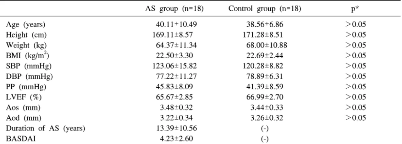
Table 1. Clinical and demographic characteristics of both groups and the comparisons
AS group (n=18) Control group (n=18) p*
Age (years) Height (cm) Weight (kg) BMI (kg/m2) SBP (mmHg) DBP (mmHg) PP (mmHg) LVEF (%) Aos (mm) Aod (mm)
Duration of AS (years) BASDAI
40.11±10.49 169.11±8.57 64.37±11.34
22.50±3.30 123.06±15.82 77.22±11.27 45.83±8.09 65.67±2.85 3.48±0.32 3.22±0.34 13.39±10.56
4.23±2.60
38.56±6.86 171.28±8.51 68.00±10.88
22.69±2.44 120.28±8.82 78.89±6.31 41.39±8.59 66.99±2.70 3.44±0.33 3.26±0.32
(-) (-)
>0.05
>0.05
>0.05
>0.05
>0.05
>0.05
>0.05
>0.05
>0.05
>0.05
*The Mann-Whiteny U test
BMI: body mass index, AS: ankylosing spondylitis, SBP: systolic blood pressure, DBP: diastolic blood pressure, PP:
pulse pressure, LVEF: left ventricular ejection fraction, Aos: aortic systolic diameter, Aod: aortic diastolic diameter, BASDAI: bath ankylosing spondylitis disease activity index
좌심실질량(left ventricle mass), 좌심실질량지수(left ventricle mass index), 좌심방지름(left atrium diameter, LAD), 상행대동맥 직경(aortic root diameter), 상행대 동맥 수축기 직경(systolic aortic diameter, Aos), 상행 대동맥 이완기 직경(diastolic aortic diameter, Aod) 등 을 측정하였다 (17). 상행대동맥 직경은 흉골연 장축 단면도에서 대동맥판막의 3 cm 상방에서 M형 심초 음파도를 이용하여 측정하였는데, Aos은 수축기 시 대동맥의 직경이 가장 커져있을 때 측정하였고, Aod 은 QRS complex의 첨부에서 대동맥 직경이 가장 작 을 때로 하였다. 심박출계수(ejection fraction, EF)는 다음 식을 이용하여 구하였다 (18).
EF (%)=(LVEDD2−LVESD2)/LVEDD2×100
검사 중 혈압을 3번 이상 혈압계(sphygmometer)로 수축기 혈압(systolic pressure)과 이완기 혈압(diastolic pressure)를 측정하였다. 대동맥 탄력 지수는 대동맥 긴장도(aortic strain) (19,20), 대동맥 팽창도 지수 (aortic distensibility index)를 (21) 사용하여 측정하였 는데, 다음과 같은 공식을 사용하여 계산하였다.
aortic strain (%)=100×(Aos−Aod)/Aod
aortic distensibility index (cm−2dyn−110−6)=2×aortic strain×pulse pressure
3. BASDAI scoring
BASDAI 점수는 임상의가 강직척추염 치료의 효 과를 판단하는데 이용하는 편리한 방법이다. 배 (22) 등이 한글로 번역하여 통계적으로 유의성이 입증된 설문지를 사용하였고, 강직성 척추염의 주된 증상 5 가지에 대한, 6개의 문항에 대해 환자가 점수로 답 하는 것으로 1∼10씩(불편함이 없으면 1점, 최대로 불편할 때 10점) 점수화하여 이에 대한 평균을 이용 하였다 (23).
4. 통계적 분석
측정 수치는 평균±편차로 나타내었고, 각 군의 평 균을 비교하였다. 통계처리는 SSPS (Version 12.0, Chicago, USA)를 이용하였다. 각 군의 지표 사이의 상관관계는 비모수 독립 2-표본 검정으로 Mann- Whitney 검정을 사용하였다. 강직척추염 환자군 내 의 대동맥 탄력성 지표들과 유병기간과 BASDAI 사 이의 상관관계는, 검정을 위한 검정통계량으로 Spearman의 순위상관계수(r)를 계산하여 분석 조사하 였다. 통계적 유의 수준은 p-value가 0.05 이하인 경 우에 유의한 것으로 간주하였다.
Table 2. The means of aortic strain, distensibility of groups and the comparisons between the groups Parameters AS group (n=18) Control group (n=18) p*
Aortic strain (%) Aortic distensibility index (cm−2dyn−110−6)
8.53±4.53 7.92±4.63
9.79±2.66 7.86±2.60
>0.05
>0.05
*The Mann-Whiteny U test
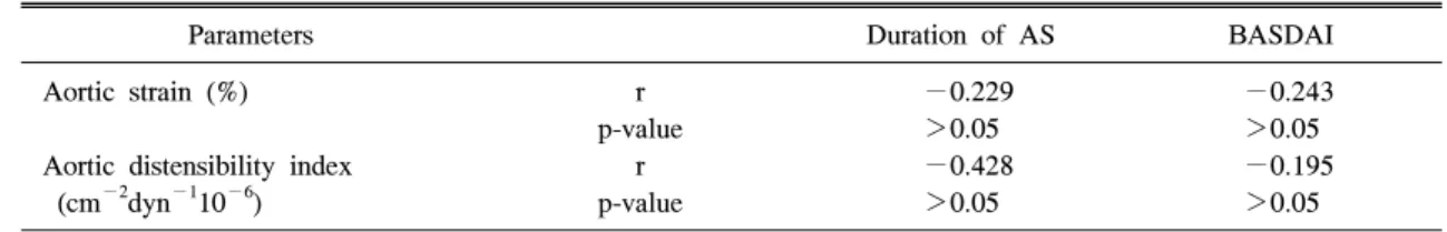
Table 3. Statistical correlations between the indexes of aortic elasticity and duration, BASDAI of AS patients
Parameters Duration of AS BASDAI
Aortic strain (%)
Aortic distensibility index (cm−2dyn−110−6)
r p-value
r p-value
−0.229
>0.05 −0.428
>0.05
−0.243
>0.05 −0.195
>0.05
*The Spearman rank test
결 과 1. 임상적 특징
강직척추염 환자의 평균 연령은 40.11±10.49세이 고, 남녀 비는 14:4로 남자가 많았다. 대조군의 평 균 연령은 38.56±6.86세이며 남녀 비는 동일하였다.
강직척추염 환자에서 유병기간은 13.39±10.56년(범 위 9∼45년)이고, BASDAI score는 4.23±2.61이였다.
신장은 169.11±8.57 cm, 체중은 64.39±11.34 kg, 신체 비만지수는 22.50±3.30 kg/m2였다.
2. 심장 기능 지표의 특징
강직척추염 환자의 수축기 혈압은 123.06±15.82 mmHg, 이완기 혈압은 77.22±11.27 mmHg이며, 대조 군의 수축기 혈압과 이완기 혈압은 각각 120.27±
8.82 mmHg, 78.89±6.31 mmHg이며, 양 군 사이의 유 의한 차이는 보이지 않았다.
강직척추염 환자에서 LVEF는 65.67±2.85%, Aos, Aod는 각각 3.48±0.32 mm, 3.22±0.34 mm이며, 대조 군의 값과 비교하면 역시 통계적으로 유의한 차이를 찾지 못하였다(표 1).
3. 대동맥 탄력성의 비교
강직척추염 환자의 대동맥 긴장도는 8.53±4.53%이 며, 9.43±2.66%의 대조군과 비교하여 통계적으로 유
의한 차이가 없음을 알 수 있었다. 대동맥 팽창도 지수는 강직척추염 환자에서 7.92±4.63 cm−2dyn−1 10−6 였으며, 이는 대조군의 7.86±2.60 cm−2dyn−110−6 와 비슷하였다. 이 역시 통계적으로 유의한 차이가 없 었다(표 2).
4. 대동맥 탄력성과 강직척추염의 유병기간과 BASDAI 사이의 연관성
강직척추염 환자에서 대동맥 긴장도와 팽창도 지 수를 각각 유병기간과 BASDAI 사이의 통계적 연관 성을 알아보았으나, 의의 있는 연관성을 얻지 못하 였다(표 3).
고 찰
강직척추염은 원인이 잘 알려지지 않은 HLA- B27 와 관련된 만성 염증성 질환이다 (24). 주로 초기에 천장관절에 침범하고, 후반기에 중추골격의 이상을 일으킨다. 말초 관절 침범 역시 중요한 양상을 보인 다. 이 질환은 급성 전방 포도막염, 대동맥판 폐쇄부 전, 심장 전도 이상, 폐 상엽의 섬유화, 신경학적 이 상 또는 이차적 신장 아밀로이드증 등과 같은 골격 계 외 질환과도 관련되기도 한다 (4-10,25,26).
강직척추염의 유병률은 남녀비가 2.5:1에서 5:1 정도로 남성에서 우세하게 나타난다 (27). 본 연구의
환자군의 남녀비는 14:4로 그 범주 내에 속하여, 연 구 대상의 남녀 성비는 적합하다고 할 수 있다.
강직척추염 환자에서 심혈관계 침범에 대한 많은 연구들이 있었다. 대동맥염, 판막염, 그리고 맥관벽 혈관염이 발생하였다는 보고가 있다 (28). 이러한 염 증반응은 대동맥근의 이완, 대동맥환과 대동맥첨판 의 근위부의 섬유화를 일으킨다. 이러한 병리학적인 변화는 히스속을 포함한 방실전도계까지 침범하여 완전심장차단을 일으킬 수 있다 (29). 대동맥활밑 섬 유화는 경우에 따라 경흉부 심초음파를 통해서 대동 맥환과 대동맥판막하 비대로 보여진다 (30).
우리는 이러한 염증성 변화가 대동맥벽에 일어나 면서, 대동맥 탄력성에 영향을 줄 수 있을 것이라고 예상하였다. 만약 염증 반응에 의하여 대동맥 탄력 성이 감소한다면, 좌심실의 후부하가 증가되어 좌심 실의 구조적 이상이 발생하게 된다. 즉, 심실의 비후 가 발생하는데 가장 중요한 요인은 수축기 말기에 심실벽에 가중되는 스트레스로 알려져 있는데 (31), 이는 심실의 구조적인 특성 및 대동맥의 경직도에 의해 영향을 받는다 (32). 증가된 수축기 말기의 스 트레스를 해소하기 이하여 심근의 수축기 및 이완기 경직도가 증가되게 되고 (33,34), 이렇게 증가된 심 근의 경직도는 초기에는 이완기 장애를 일으키고, 후기에는 수축기 장애를 유발시키게 된다. 이로 인 하여 좌심실 비대, 혈관 손상, 동맥경화에 의한 중심 동맥 경직 등의 합병증이 발생하고, 이것이 사망에 중요한 역할을 할 것으로 여겨지며 전향적으로 심혈 관병의 발생을 예측하는데 중요한 인자가 될 수 있 을 것이다 (35).
이러한 과정을 강직척추염 환자에서 관찰한다면, 질병 예후를 예상하는데 도움이 될 것이라 생각하 고, 본 연구를 시행하였다. 본 연구의 저자들은 강직 척추염 환자의 대동맥 탄력성이 정상인에 비해 감소 되어 있을 것으로 예상하였으나, 그 결과는 달랐다.
즉, 각각 18명의 강직척추염 환자군과 정상인 대조군 사이의 대동맥 탄력성 지표들의 통계적인 유의한 차 이가 없었던 것이다. 또한 대동맥 탄력성 지표들과 질병 유병기간, 그리고 BASDAI 사이의 연관성 역시 관찰하지 못하였다. 이는 Demiralp E 등이 심장질환 이 없는 강직척추염 환자, 35명을 대상으로 대동맥 탄력성을 조사하였고, 정상인에 비해 대동맥 탄력성
이 감소되었다는 결과와 상이한 것이다. 하지만 대 동맥 탄력성 지표들과 강직척추염의 유병기간과 통 계적인 연관성이 없었다는 결과는 동일하였다 (13).
본 연구에서 환자군과 대조군 사이에 대동맥 탄력 성이 유의한 차이가 없었던 이유는 다음과 같은 몇 가지로 생각된다. 첫째, 대동맥 긴장도와 팽창도 지 수를 구하기 위한 인자들이 환자군과 대조군 사이에 유의한 차이가 없었기 때문에 이러한 결과를 얻은 것으로 생각이 된다. 둘째, 환자군의 나이를 고려하 였을 때, 69세의 남환을 제외하면 대부분 3,40대이므 로(평균 36.28±11.8세, 유병기간 10.89±7.52년), 다른 강직척추염 환자들에 비해 상대적으로 짧은 유병기 간에 의해 대동맥의 염증성 변화 역시 적었을 것이 고, 대조군과 비교하였을 때, 통계학적으로 유의한 차이를 보이지 않았으리라 생각된다. 셋째, 경흉부 심초음파를 이용하여 대동맥의 유순도(compliance), 팽창도(distensibility), 또는 경직도(stiffness)를 구하는 방법은 관혈적으로 혈관 조영술을 통한 맥파전달속 도(pulse wave velocity)에 비하여 (34,36-38), 측정에 따른 오차 및 상완에서의 혈압이 대동맥의 혈압을 정확하게 반영하지 못하여 정확도 면에서 약점이 있 는데 이것 역시 영향을 주었을 것으로 생각된다 (36,40).
본 연구는 몇 가지 제한점을 가지고 있다. 첫째, 연구 대상군이 오직 심장 질환이 없는 강직성 척추 염 환자인 점이다. 이에 대상의 평균 연령이 낮고, 유병기간이 짧아서 정상인과 비교하였을 때, 대동맥 탄력성의 유의한 차이가 없었을 것이다. 둘째, 대상 군의 수가 너무 작아서, 본 연구의 결과를 확정하기 힘들다는 점이다. 따라서, 더 많은 강직척추염 환자 를 대상으로 대동맥 탄력성의 변화에 대한 연구가 필요하다고 본다.
결 론
본 연구는 18명의 심장질환이 없는 강직척추염 환 자와 18명의 심장질환이 없는 정상인을 대상으로 대 동맥 탄력성을 비교, 조사하였으며, 강직척추염 환자의 대동맥 탄력성이 정상인에 비해 감소되지 않았음을 관찰하였다. 대동맥 긴장성과 팽창도 지수와 같은 탄력성 지표들과 강직척추염의 유병기간과 BASDAI
사이의 연관성이 존재하지 않음을 발견하였다. 이런 관찰의 중요성은 전향적인 연구를 통해 평가되어야 할 것이다.
참고문헌
1) Harris E, Budd R, Genovese M, Firestein G, Sargent J, Sledge C. Kelly’s textbook of rheumatology. 7th ed. p. 1125-41, Philadelphia, Saunders, 2005.
2) Smith JA, Marker-Hermann E, Colbert RA.
Ankylosing spondylitis: Pathogenesis if ankylosing spondylitis: current concepts. Best Pract Res Clin Rheumatol 2006;20:571-91.
3) Sieper J, Rudwaleit M, Khab MA, Braoun J.
Ankylosing spondylitis: concepts and epidemiology of spondyloarthritis. Best Pract Res Clin Rheumatol 2006;20:401-17.
4) Bernstein L, Broch OJ. Cardiac complications in spondylitis ankylopoietica. Acta Med Scand 1949;
135:185-94.
5) Graham DC, Smythe HA. The carditis and aortitis of ankylosing spondylitis, Bull Rheum Dis 1958;9:171-4.
6) Davidson P, Baggenstoss AH, Slocumb DH, Daugherty GW. Cardiac and aortic lesions in rheumatoid spondylitis. Mayo Clin Proc 1963;36:
427-35.
7) Kinsella TD, Johnson LG, Sutherland RI. Cardio- vascular manifestations of ankylosing spondylitis. Can Med Assoc J 1974;111:1307-9.
8) O'Neill TW, Bresnihan B. The heart in ankylosing spondylitis. Ann Rheum Dis 1992;51:705-6.
9) Schilder DP, Harvey P, Hufnagel CA. Rheumatoid spondylitis and aortic insufficiency. N Engl J Med 1956;225:11-7.
10) Bergfeldt L, Edhag O, Vallin H. Cardiac conduction disturbances, an underestimated manifestation in ankylosing spondylitis. Acta Med Scand 1982;212:
212-7.
11) Brewerton DA, Goddard DH, Moore RB. The myocardium in ankylosing spondylitis. A clinical, echocardiographic, and histopathological study. Lancet 1987;1:995-8.
12) Lethinen K. 76 patients with ankylosing spondylitis seen after 30 years of disease. Scand J Rheumatol 1983;12:3-11.
13) Demiralp E, Kardesoglu E, Kiralp MZ, Cebeci BS, Keskin I, Ozmen N, et al. Arotic elasticity in patients with ankylosing spondylitis. Acta Cariol Dec 2004;
59:630-4.
14) Arnett DK, Evans GW, Riley WA. Arterial stiffness:
a new cardiovascular risk factor? Am J Epidemiol 1994;140:669-82.
15) Van der Linden S, Valkenburg HA, Cats A..
Evaluation of diagnostic criteria for ankylosing spondylitis: a proposal for modification of the New York criteria. Arthritis Rheum 1984;27:361-8.
16) Henry WL, DeMaria A, Gramiak MC, King DL, Kisslo JA, Popp RL, et al. Report of the American Society of Echocardiography Committee on Nomen- clature and Standards in two-dimensional echocar- diography. Circulation 1980;62:212-5.
17) Sahn DJ, DeMaria A, Kisslo J, Weyman A. Recom- mendations regarding quantification in M-mode echocardiography: results of a survey of echocar- diographic measurements. Circulation 1978;58:1072 -83.
18) Teichholz LE, Kreulen T, Herman MV. Probrems in echocardiographic volume determinations: Echocar- diographic-angiographic correlations in the presence or absence of asynergy. Am J Cardiol 1976;37:7-11.
19) Eren M, Gorgulu S, Uslu N, Celik S, Dagdeviren B, Tezel T. Relation between aortic stiffness and left ventricular diastolic function in patients with hyper- tension, diabetes, or both. Heart 2004;90:37-43.
20) Lacombe F, Dart A, Dewar E, Jennings G, Cameron J, Laufer E. Arterial elastic properties in man: a comparison of echo-Doppler indices of aortic stiffness. Eur Hear J 1992;13:1040-5.
21) Stefanadis C, Wooley CF, Bush CA, Kolibash AJ, Boudoulas H. Aortic distensibility in post-stenotic aortic dilatation: the effect of co-existing coronary artery disease. J Cardiol 1988;18:189-95.
22) Park HJ, Kim SH, Lee JE, Jun JB, Bae SC. Korean translation and validation of Bath ankylosing spondylitis disease activity index (BASDAI): a pilot test. Health Qual. Life Res 2003;12:817.
23) Calin A, Nakache JP, Gueguen A, Zeidler H, Mielants H, Dougados M. Defining disease activity in ankylosing spondylitis is a combination of variables (Bath Ankylosing Spondylitis Disease Activity Index) an appropriate instrument? Rheumatology 1999;38:
878-82.
24) Ruddy S, Harris ED, Sledge CB, Budd RC, Sergent TS. Kelley’s textbook of rheumatoloty. 6th ed. p. 1046, USA, Philadelphia, Pennsylvania, W.B. Saunders Co, 2001.
25) Arnett FC. Ankylosing spondylitis. Koopman WJ,
Arthritis and allied conditions: A textbook of rheu- matology. 12th ed. p. 1317, New York, Lippincott Williams and Wilkins, 2001.
26) Bulkley BH, Roberts WC. Ankylosing spondylitis and aortic regurgitation. Description of the characteristic cardiovascular lesion from a study of eight necropsy patients. Circulation 1973;48:1014-27.
27) Robert DI. The spondyloarthropathyies: Clinical subsets of the spondyloarthropathies: Ankylosing spondylitis: epidemiology. Cecil 23rd ed. p. 2017-19, New York, Saunders, 2008.
28) Ball GV, Hathaway B. Ankylosing spondylitis with widespread arteritis. Arthritis Rheum 1966;9:737-45.
29) Tucker CR, Fowles RE, Calin A, Popp RL. Aortitis in ankylosing spondylitis: Early detection of aortic root abnormalities with two dimensional echocar- diography. Am J Cardiol 1982;49:680-6.
30) Erol MK, Yilmaz M, Oztasyonar Y, Sevimli S, Senocak H. Aortic distensibility is increaseing in elite athletes. Am J Cardiol 2002;89:1002-4.
31) Grossman W, Jones D, McLaurin LP. Wall stress and patterns of hypertrophy in the hyman left ventricle. J Clin Invest 1975;56:56-64.
32) Bouthier JD, de Luca N, Safar ME, Simon AC.
Cardiac hypertrophy and arterial distensibility in essential hypertenstion. Am Heart J 1985;109:1345-52.
33) Weber KT. Cardiac interstitium in health and disease:
the fibrillar collagen network. J Am Coll Cardiol
1989;13:1637-52.
34) Rhee MY, Han SS, Lyu S, Lee MY, Kim YK, Yu SM. Short-term treatment with angiotensin II antago- nist in essential hypertension: Effects of Losartan on left ventricular diastolic function, left ventricular mass, and aortic stiffness. Korean Circ J 2000;30:
1341-9.
35) Boutouyrie P, Tropeano AI, Asmar R, Gautlier I, Benetos A, Lacolley P, et al. Aortic stiffness is an independent predictor of primary coronary events in hypertensive patients: a longitudinal study. Hyper- tension 2002;39:10-5.
36) Stefanadis C, Stratos C, Boudoulas H, Kourouklis C, Toutouzas P. Distensibility of the ascending aorta;
comparison of invasive and non-invasive techniques in healthy men and in men with coronary artery disease. Dur Heart J 1990;11:990-6.
37) Glasser SP, Arnett DK, McVeigh GE. Vascular compliance and cardiovascular disease: a risk factor or a marker? Am J Hypertens 1997;10:1175-89.
38) O’Rourke MF. Wave travel and reflection in the arterial system. J Hypertens 1999;17 Suppl 5:45-7S.
39) Park SM, Seo HS, Lim HE, Shin SH, Park CG, Oh DJ, et al. Assessment of the arterial stiffness index as a clinical parameter for atherosclerotic coronary artery disease. Korean Circ J 2004;34:677-83.
40) Jung HO. Strain and strain rate in echocardiography.
Korean Echo J 2002;10:2;13-7.