골다공증성 불안정 대퇴골 전자간 골절에서 내고정물의 종류에 따른 방사선학적 결과 비교
건양대학교 의과대학 정형외과학교실1, 아주대학교 의과대학 정형외과학교실2, 연세대학교 의과대학 정형외과학교실3
김광균1․원예연2․이우석3․조인호1
Comparison of the Radiologic Outcomes following the Kinds of Implants in Treating Unstable Osteoporotic Intertrochanteric Fracture
Kwang-Kyoun Kim1, Ye-Yeon Won2, Woo-Suk Lee3, In-Ho Jo1
1Department of Orthopedic Surgery, Konyang University, College of Medicine, Daejeon,
2Department of Orthopedic Surgery, Ajou University, College of Medicine, Suwon,
3Department of Orthopedic Surgery, Yonsei University, College of Medicine, Seoul, Korea
Objectives: To determine whether kinds of implants would influence on the radiologic outcomes in the treatment of unstable osteoporotic intertrochanteric fractures.
Materials and Methods: In this retrospective study, radiologic outcomes of 151 patients with unstable osteoporotic intertrochanteric fractures undergoing surgical treatments were compared based on the types of fixation implants as follows : PFNA (53 cases, group Ⅰ), gamma nail 3(31 cases, group Ⅱ), CHS with TSP (43 cases, group Ⅲ), and helical blade type LCP-DHS with TSP (24 cases, group Ⅳ). On the follow-up radiographs after operations, we assessed differences of bone union durations, neck-shaft ankle changes, lag screw or helical blade slippages, and varus alpha angle changes among the study groups.
Results: All the radiologic outcomes evaluated in this study were not significantly different among the study groups. The average bone union durations of the group Ⅰ, Ⅱ, Ⅲ and Ⅳ were 17.7, 18.0, 18.2, and 17.8 weeks, respectively (P=0.429). The average variation of neck-shaft angle of the group Ⅰ, Ⅱ, Ⅲ and
Ⅳ were 3.6o, 3.1o, 3.7o and 2.9o, respectively (P=0.273). The average lag screw or blade slippage of the group Ⅰ, Ⅱ, Ⅲ and Ⅳ were 5.1 mm, 3.3 mm, 3.6 mm and 2.7 mm, respectively (P=0.154). The average variation of varus alpha of the group Ⅰ, Ⅱ, Ⅲ and Ⅳ were 5.3o, 4.7o, 4.1o and 4.6º, respectively (P=0.894).
Conclusions: This study indicates that four typical types of fixation implants for treating unstable osteoporotic intertrochanteric fractures would not lead to differences in postoperative radiological outcomes.
Key Words: Unstable femoral intertrochanteric fracture, Proximal femoral nail antirotation, Compression hip screw, Gamma nail, Locking compression plate dynamic hip screw, Radiologic results
Received: November 19, 2013 Revised: January 1, 2013 Accepted: February 6, 2013
Corresponding Author: Ye-Yeon Won, Department of Orthopedic Surgery, Ajou University, College of Medicine, San 5 Wonchon-dong, Yeongtong-gu, Suwon 443-749, Korea
Tel: +82-31-219-5220, Fax: +82-31-219-5229, E-mail: [email protected]
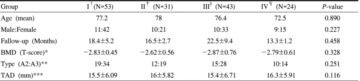
Table 1. Demographic data of the cases
Group I†(N=53) II‡ (N=31) III§ (N=43) IV¶ (N=24) P-value
Age (mean) 77.2 78 76.4 72.5 0.890
Male:Female 11:42 10:21 10:33 9:15 0.227
Fallow-up (Months) 18.4±5.2 16.5±2.7 22.5±9.4 13.3±1.2 0.458
BMD (T-score)* -2.83±0.45 -2.62±0.56 -2.87±0.76 -2.79±0.61 0.328
Type (A2:A3)** 19:34 12:19 15:28 10:14 0.251
TAD (mm)*** 15.5±6.09 16±5.82 15.4±6.71 16.3±5.91 0.116
*BMD: Bone mineral density, **Type: AO classification, ***TAD: Tip-apex distance
†PFNA (Proximal Femoral Nail Antirotation), ‡Gamma nail, §CHS (Compression Hip Screw) with TSP (Trochanteric Stabilizing Pate), ¶LCP (Locking Compression Plate) DHS (Dynamic Hip Screw) with TSP (Trochanteric Stabilizing Pate) 골다공증성 불안정 대퇴골 전자간 골절은 대부분
고령에서 발생하며 평균수명의 연장에 따라 발생 빈도가 증가하는 추세이며 장기간의 침상 안정 및 회복 후에도 일상생활 자립도에 영향을 미치어 사 회 경제적으로 매우 큰 영향력을 미치는 골절이 다.1-4 따라서, 수술의 목적은 견고한 고정으로 조기 에 체중부하를 시작하고 기능 회복을 하는 것이며, 이를 위해 다양한 내고정물이 개발되고 있다.5,6 특 히, 골다공증이 동반된 환자나,7 불안정성 전자 간 골절의 경우 골절 편의 과도한 전위나 지연나사의 대퇴 골두 천공, 과도한 내전 변형, 하지 단축 등으 로 인해 더욱 견고한 고정을 필요로 하며, 이에 따 라 다양한 내고정물이 고안되고 있다.8-10 항회전 근 위대퇴 골수정(Proximal Femoral Nail Antirotation, PFNAⓇ), 감마 골수정 3(Gamma Nail 3)과 압박고 나 사(Compression Hip Screw)와 전자부 안정화 금속판 (Trochanteric Stabilizing Plate)의 부가적 사용, 나선 칼날(Helical Blade)을 사용한 잠김 압박 금속판 나사 (Locking Compression Plate Dynamic Hip Screw)와 전 자부 안정화 금속판(Trochanteric Stabilizing Plate)의 부가적 사용이 불안정 골절에 있어 고정력을 증가 시킬 수 있다고 알려져 있다. 그러나, 각 각의 내고 정물에 따라 장 ․ 단점이 알려져 있으나, 골다공증을 동반한 불안정 전자간 골절에서 내고정물에 따른 방사선학적 결과의 비교는 드물었다.5-9 이에 저자들 은 골절의 고정력을 더욱 필요로 하는 골다공증을 동반한 대퇴골 불안정성 골절에 있어 내고정물의 선택이 방사선학적 결과에 영향을 미치는가에 대해
연구하고자 하였다.
대상 및 방법
1. 연구 대상
2005년 5월부터 2011년 5월까지 AO 분류상 A2.2 형부터 A3.3까지 해당되는 골다공증성 불안정성 대 퇴골 전자간 골절로 수술적 치료를 받은 환자 중 세 계보건기구에서 제시한 진단 기준(T score -1.0 이 상; 정상, -1.0에서 -2.5 사이; 골감소증, -2.5이하;
골다공증)으로 골다공증이 동반되어 있고, 최소 12 개월 이상 방사선학적 경과관찰이 가능하였던 환자 를 후향적으로 분석하였다. 기왕에 반대측 고관절에 골절이 있었던 18예, 동반된 정형외과 골절이 있었 던 19예는 제외하였다. 총 151명의 환자가 포함되었 고 남자가 40명, 여자가 111명이었으며 환자들의 연 령은 최소 45세에서 최대 93세로 평균 76.1세이었다.
모든 환자에서 각 군의 연령, 성별, 평균 추시 기간, BMD, AO/OTA 분류에 따른 골절 형태, 골밀도, 첨 단-정점거리(tip-apex distance)는 Table 1에 제시하였 다. 골밀도 측정값은 이중 에너지 X-선 흡수 방법을 이용하여, 반대측 고관절부와 요추체 전후면에서 측 정한 값을 이용하였다.
2. 내고정물에 따른 분류
수술에 사용된 내고정물에 따라 항회전 근위 대퇴 골수정(Proximal Femoral Nail Antirotation, PFNAⓇ, Synthes, Switzerland)을 사용한 53예(1군), 감마 골수
Fig. 1. A-D Radiographs of each groups. A: group Ⅰ fixed with PFNA (Proximal Femoral Nail Antirotation) B:
group II fixed with Gamma nail 3 C: group Ⅲ fixed with CHS (Compression Hip Screw) with TSP (Trochanteric Stabilizing Pate) D: group Ⅳ fixed with LCP (Locking Compression Plate) DHS (Dynamic Hip Screw) blade with TSP (Trochanteric Stabilizing Pate).
정 3(Gamma Nail 3, Stryker, Germany)을 사용한 31예 (2군), 압박고 나사(Compression Hip Screw, Aesculap, Germany)에 부가적으로 전자부 안정화 금속판(Tro- chanteric Stabilizing Plate)을 사용한 43예(3군), 나선 칼 날을 사용한 잠김 압박 금속판 압박고 나사(Locking Compression Plate Dynamic Hip Screw, Synthes, Switzerland)에 전자부 안정화 금속판(TSP)을 부가적 으로 사용한 24예로 분류하였다(Fig. 1).
3. 방사선학적 평가
방사선학적 평가로 골유합은 전후면 및 측면 사진 에서 피질골 가골교(cortical callus bridge)가 3개 이상 보이며, 골절선이 보이지 않는 경우로 하였다. 정복 정도를 측정하기 위해 수술 직후와 최종 추시에서 대퇴 경간각의 내반 변화(varization of neck shaft angle), 골절부의 함몰 정도를 측정하기 위해 지연나 사 또는 나선형 칼날의 활강 정도(screw or helical blade sliding), 대퇴 골두내에서 지연나사 또는 나선 형 칼날의 전위 정도를 측정하기 위해 내반 붕괴 알 파 각11(varus collapse alpha angle)을 측정하였다(Fig.
2). 내반 붕괴 알파 각은 수술 직후 방사선사진에서 대퇴골 경부의 축과 내고정물 사이의 각도와 6개월 이후 추시에서 같은 방식으로 측정한 각도의 차이로
계산하였다.
4. 통계분석
각 군 간의 통게학적 분석은 SPSS (13.0 for window) 통계프로그램을 이용하였다. 각 군에 따른 각각의 방사선학적 계측치를 one-way ANOVA test로 통계검 정을 시행하였으며, 유의 수준은 P 값이 0.05 미만인 경우를 통계학적으로 유의한 것으로 하였다.
결 과
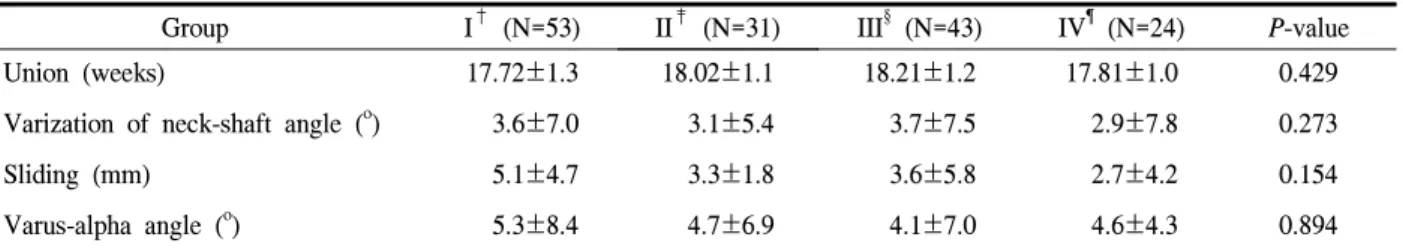
골 유합 기간은 1군에서는 평균 17.7주이었으며, 2 군에서는 평균 18.0주, 3군에서는 평균 18.2주, 4군에 서는 평균 17.8주로 각 군 간에 유의한 차이를 보이 지 않았다(P=0.429). 대퇴 경간각의 내반 변화는 1군 에서는 평균 3.6도 내반 되었고, 2군에서는 평균 3.1 도, 3군에서는 평균 3.7도, 4군에서는 2.9도로 각 군 간에 유의한 차이를 보이지 않았다(P=0.273). 지연나 사 또는 나선형 칼날의 활강 정도는 1군에서는 평균 5.1 mm, 2군에서는 3.3 mm, 3군에서는 3.6 mm, 4구 에서는 2.7 mm로 각 군 간에 유의한 차이를 보이지 않았다(P=0.154). 내반 붕괴 알파 각의 변화는 1군에 서는 5.3도, 2군에서는 4.7도, 3군에서는 평균 4.1도,
Table 2. Comparative results of four groups
Group I† (N=53) II‡ (N=31) III§ (N=43) IV¶ (N=24) P-value
Union (weeks) 17.72±1.3 18.02±1.1 18.21±1.2 17.81±1.0 0.429
Varization of neck-shaft angle (o) 3.6±7.0 3.1±5.4 3.7±7.5 2.9±7.8 0.273
Sliding (mm) 5.1±4.7 3.3±1.8 3.6±5.8 2.7±4.2 0.154
Varus-alpha angle (o) 5.3±8.4 4.7±6.9 4.1±7.0 4.6±4.3 0.894
†PFNA (Proximal Femoral Nail Antirotation), ‡Gamma nail, §CHS (Compression Hip Screw) with TSP (Trochanteric Stabilizing Pate), ¶LCP (Locking Compression Plate) DHS (Dynamic Hip Screw) blade with TSP (Trochanteric Stabilizing Pate)
Fig. 2. A-C. Measurement of parameters using radiographs A: Varization of neck-shaft angle(o) (a) Immediate postoperative radiograph. a: neck shaft angle (b) a’:neck shaft angle at last follow up, Varization neck shaft angle=a-a’. B: Sliding(mm) (a) Immediate postope- rative radiograph. a: Screw length at immediate post- operative, b: Screw sliding at Immediate postoperative.
(b) Follow up radiograph. a’: Screw length at the last follow up. b’ Screw sliding at at the last follow up, Corrected follow up Sliding= b’/Correcting factor – b. (*correction factor=Screw length at immediate postoperative(a) / Screw length at the last follow up(a’)) C: Varus-collapse angle (o) α: angle between neck axis and screw axis (a) Immediate postoperative radiograph. a: varus collapse angle (b) a’: varus collapse angle α at last follow up, change of varus-collapse angle α =a-a’.
4군에서는 4.6도로 각 군 간에 유의한 차이를 보이 지 않았다(P=0.894)(Table 2). 수술 후 1군에서 골두 천공이 2예, 3군에서 금속판의 뽑힘(pull-out)이 1예,
부가적으로 사용한 전자부 안정화 금속판의 파손이 1예에서 발생하였다(Fig. 3).
Fig. 3. Radiograph shows. A: perforation of the helical blade into the acetabulum B: pull out from the proximal femur of the CHS with TSP.
고 찰
고령에서 발생하는 골다공증성 불안정 대퇴골 전 자간 골절은 골절에 따른 유병률과 사망률이 높아 해부학적 정복과 내고정을 시행하여 조기 보행을 유 도하는 것이 치료의 원칙이다. 특히, 골다공증성 불 안정 대퇴골 전자간 골절에서는 부적절한 정복 및 내고정물의 사용에 따라 과도한 활강, 지연 나사의 관절내 돌출, 금속판의 파손 등의 문제가 유발될 수 있다.1-3 이러한 문제를 조절하기 위해 불안정성 골 절의 치료를 위한 많은 내고정 기구들이 개발되었 고, 이들은 각 각의 장점을 가지고 있다. 골절의 내 고정에 이전부터 널리 사용된 압박고 나사는 골절부 의 압박을 유도하여 골유합을 유도하나 과도한 활강 은 내고정 실패로 이어질 수 있다. 이에 따라 압박고 나사에 전자부 안정화 금속판을 추가하여 지나친 감 입에 의한 원위 정복의 소실, 지연 나사의 골두 천공 등을 줄일 수 있었다.5-9 그러나, 절개부위의 확장으 로 수술 후 출혈 및 수술 시간의 연장 등에 대한 단 점이 있다. 이에 비해 절개 부위의 축소와 수술 시간 을 단축할 수 있는 골수내 금속 정이 개발 되었고, 이들은 짧은 지렛대로 인해 골절을 생역학적으로 보 다 안정적으로 고정할 수 있는 이론적인 장점이 있
다.12-14 그러나, 1980년대 소개된 감마정의 경우 근위
부 직경이 두껍고 10도 외반각으로 인해, 금속정 원 위부 대퇴골 골절, 대전자부 골절의 합병증이 보고
되었다.6,7,9 근위 대퇴정의 경우 Z-effect로 인한 골두
천공 등의 합병증의 비율이 높게 보고된 바 있다.7 이러한 단점들을 극복하기 위해 제품의 형태에 대한 변화를 주었고, 본 연구에서 사용한 골수내 금속정 은 과거 감마정의 단점을 보완한 3세대 감마정 및 항회전 근위 대퇴정이었고, 술 중 이나 술 후 골절의 합병증은 없었다. 항회전 근위대퇴 골수정의 경우 나선형 칼날의 사용으로 인해 칼날의 홈 사이로 해 면골이 압착되는 효과로 인해 골절부 안정성을 높여 주는 장점 및 골 소실을 줄이고 골편의 회전을 줄인 다는 장점이 있다.15,16 저자들의 경우 항회전 근위 대 퇴 골수정군에서 골두 천공이 2예가 있었는데, 이는 항회전 근위 대퇴정이 골절편의 활강을 과도하게 억 제하여, 상대적으로 날카로운 면을 가진 나선형 칼 날이 골두에 대해 상대적으로 외반전위 되면서 골두 를 천공하거나, 삽입할 나선형 칼날의 길이 측정의 오류로 실제 삽입할 깊이보다 확공을 길게 하였거나 삽입된 나선형 칼날을 지지하는 뼈의 미세구조가 약 해져 고정력이 감소하는 것이 영향을 미쳤을 것으로 생각된다.
나선형 칼날과 지연나사간의 고정력에 대한 비교 연구에서 Windolf 등14은 압박고 금속판을 이용한 고 정에서 나선형 칼날이 생역학적으로 대퇴골두에 고 정력이 강하다고 보고하였다. 또한 Strauss 등15은 골 수내정을 이용한 연구에서 나선형 칼날과 지연나사 간에 최종 항복 응력에 차이가 없으나 나선형칼날이 골두의 전위 및 골절부의 확공정도가 적어 압박고 금속판에 비해 생역학적으로 더 우수하다고 보고하 였다. 저자들의 경우는 방사선학적 결과에서 의미 있는 차이는 없었고, 나사의 대퇴 골두에 대한 고정 력이 골두 천공이나 내반 붕괴 각의 유지에 영향을 미치지만, 실제 체내에서 반복되는 하중의 방향 및 양과 각 개체의 골질이 영향을 미치기 때문에, 체외 에서 시행한 생역학 실험의 결과를 직접 임상에 적 용하는 것은 한계가 있다고 생각된다.
최근 골절 치료에 널리 유용되는 잠김 압박 금속 판은 기존의 금속판에 비해 뽑힘 저항력이 커서 골 다공증 환자에서 고정력을 증가시키는 것으로 보고
되었다.17,18 최근에는 압박 고 나사 금속판에도 금속 판 부위를 잠김 압박 금속판으로 제작하여 뼈에 고 정력을 증가시킨 제품이 소개되었다. 그러나, 저자 들이 검색을 통한 문헌 고찰에서 나선 칼날을 사용 한 잠김 압박 금속판의 임상적 또는 방사선학적 결 과에 관한 문헌 보고는 찾을 수 없었다. 저자들의 결 과에서는 일반 압박고 나사와 방사선학적 결과에 차 이는 없었으나, 압박고 나사에서 발생한 금속판 뽑 힘(Fig. 3B)과 같은 합병증을 감소시키는 데 도움이 될 것으로 판단된다.
본 연구의 제한점은 첫째, 방사선학적 계측의 결 과에 대한 비교 연구로 임상 결과에 직접 적용하는 데 한계가 있고 생각된다. 골다공증성 근위 대퇴부 골절이 술전 다양한 질환을 동반한 혈역동학적으로 불안정한 고령 환자에서 발생하며 조기 체중 부하 등 보행 능력이 술 후 폐렴, 욕창 등 합병증의 예방 에 중요한 점을 고려할 때, 방사선학적인 계측치의 차이에 대한 연구도 필요하지만 임상적으로 출혈량, 수술 시간, 수술후 보행 능력 및 사망률 등을 포함한 합병증이 중요하며 향후 이에 대한 연구가 함께 진 행되어야 할 것으로 생각된다. 둘째, 본 연구는 장기 간의 방사선 자료에 대한 후향적 연구로 방사선 촬 영자의 교체 및 촬영 방법에 따른 오차가 발생 가능 성이 있다는 점이다.
결 론
골다공증성 불안정 대퇴골 전자간 골절의 치료에 서 4가지의 전형적인 내고정물의 선택은 방사선학 적 결과에 영향을 미치지 않았다. 따라서, 환자의 대 부분이 혈역동학적으로 불안정한 고령이며, 조기 체 중 부하가 임상 결과에 중요한 점을 고려하여 시술 자의 경험에 따른 숙련도에 따라 내고정물을 선택하 여야 할 것으로 사료된다.
참 고 문 헌
1. Nam WD, Park IH, Han KY. Treatment of inter- trochanteric fracture using proximal femoral nail for patients over 90 years old. J Korean Hip Soc
2009;21:339-44.
2. Parker MJ, Palmer CR. A new mobility score for predicting mortality after hip fracture. J Bone Joint Surg Br 1993;75:797-8.
3. Kim YS, Kwon SY, Lee YM, Han SK. Treatment of Intertrochanteric Fractures in Patients with Severe Osteoporosis with Dynamic Compression Hip Screws. J Korean Hip Soc 2009;21:29-34.
4. Rho JY, Kim SB, Heo YM, Cho SJ, Chae DS, Lee WS. Proximal Femoral Nail Antirotation versus Compression Hip Screw with Trochanter Stabilizing Plate for Unstable Intertrochanteric Hip Fractures.
J Korean Fract Soc 2010;23;161-6
5. Aune AK, Ekeland A, Odegaard B, Grøgaard B, Alho A. Gamma nail vs compression screw for trochanteric femoral fractures. 15 reoperation in a prospective, randomized study of 378 patients. Acta Orthop Scand 1994;65:127-30.
6. Babst R, Renner N, Biedermann M, Rosso R, Heberer M, Harder F, et al. Clinical results using the trochanter stabilizing plate (TSP): the modular extension of the dynamic hip screw (DHS) for internal fixation of selected unstable intertro- chanteric fractures. J Orthop Trauma 1998;12:
392-9.
7. Lorich DG, Geller DS, Nielson JH. Osteoporotic pertrochanteric hip fractures: management and current controversies. Instr Course Lect 2004;53:441-54.
8. Buciuto R, Uhlin B, Hammerby S, Hammer R.
RAB-plate vs Richards CHS plate for unstable trochanteric hip fractures. A randomized study of 233 patients with 1-year follow-up. Acta Orthop Scand 1998;69:25-8.
9. Madsen JE, Naess L, Aune AK, Alho A, Ekeland A, Strømsøe K. Dynamic hip screw with tro- chanteric stabilizing plate in the treatment of unstable proximal femoral fractures: a comparative study with the Gamma nail and compression hip screw. J Orthop Trauma 1998;12:241-8.
10. Su ET, DeWal H, Kummer FJ, Koval KJ. The
effect of an attachable lateral support plate on the stability of intertrochanteric fracture fixation with a sliding hip screw. J Trauma 2003;55:504-8.
11. Sommers MB, Roth C, Hall H, Kam BC, Ehmke LW, Krieg JC, et al. A laboratory model to evaluate cutout resistance of implants for pertro- chanteric fracture fixation. J Orthop Trauma 2004;
18:361-8.
12. Swiontkowski MF, Harrington RM, Keller TS, Van Patten PK. Torsion and bending analysis of internal fixation techniques for femoral neck fractures: the role of implant design and bone density. J Orthop Res 1987;5:433-44.
13. Simmermacher RK, Bosch AM, Van der Werken C. The AO/ASIF-proximal femoral nail (PFN): a new device for the treatment of unstable proximal femoral fractures. Injury 1999;30:327-32.
14. Windolf, M, Braunstein V, Dutoit C, Schwieger K.
Is a helical shaped implant a superior alternative to
the Dynamic Hip Screw for unstable femoral neck fractures? A biomechanical investigation. Clin Biomech 2009;24:59-64.
15. Strauss E, Frank J, Lee J, Kummer FJ, Tejwani N.
Helical blade versus sliding hip screw for treatment of unstable intertrochanteric hip fractures: a Bio- mechanical evaluation. Injury 2006;37:984-9.
16. Ahrengart L, Törnkvist H, Fornander P, Thorngren KG, Pasanen L, Wahlström P, et al. A randomized study of the compression hip screw and Gamma nail in 426 fractures. Clin Orthop Relat Res 2002;
401:209-22.
17. Niemeyer P, Südkamp NP. Principles and clinical application of the locking compression plate (LCP).
Acta Chir Orthop Traumatol Cech 2006;73:221-8.
18. Jewell DP, Gheduzzi S, Mitchell MS, Miles AW.
Locking plates increase the strength of dynamic hip screws. Injury 2008;39:209-12.