ISSN: 2233-601X (Print) ISSN: 2093-6516 (Online)
Received: August 23, 2018, Revised: November 7, 2018, Accepted: November 8, 2018, Published online: June 5, 2019
Corresponding author: In Kyu Park, Department of Thoracic and Cardiovascular Surgery, Seoul National University Hospital, Seoul National University College of Medicine, 101 Daehak-ro, Jongno-gu, Seoul 03080, Korea
(Tel) 82-2-2072-2342 (Fax) 82-2-764-3664 (E-mail) [email protected]
© The Korean Society for Thoracic and Cardiovascular Surgery. 2019. All right reserved.
This is an open access article distributed under the terms of the Creative Commons Attribution Non-Commercial License (http://creativecommons.org/
licenses/by-nc/4.0) which permits unrestricted non-commercial use, distribution, and reproduction in any medium, provided the original work is properly cited.
Clinical Outcomes of Surgical Treatment for Primary Chest Wall Soft Tissue Sarcoma
Seung Hwan Yoon, M.D., Joon Chul Jung, M.D., In Kyu Park, M.D., Ph.D., Samina Park, M.D., Chang Hyun Kang, M.D., Ph.D., Young Tae Kim, M.D., Ph.D.
Department of Thoracic and Cardiovascular Surgery, Seoul National University Hospital, Seoul National University College of Medicine, Seoul, Korea
Background: This study investigated the clinical outcomes of surgical treatment of primary chest wall soft tis- sue sarcoma (CW-STS). Methods: Thirty-one patients who underwent surgery for CW-STS between 2000 and 2015 were retrospectively reviewed. The disease-free and overall survival rates were estimated using the Kaplan-Meier method, and prognostic factors were analyzed using a Cox proportional hazards model. Results:
The median follow-up duration was 65.6 months. The most common histologic type of tumor was malignant fibrous histiocytoma (29%). The resection extended to the soft tissue in 14 patients, while it reached full thickness in 17 patients. Complete resection was achieved in 27 patients (87.1%). There were 5 cases of lo- cal recurrence, 3 cases of distant metastasis, and 5 cases of combined recurrence. The 5-year disease-free rate was 49%. Univariate analysis indicated that incomplete resection (p<0.001) and stage (p=0.062) were possible risk factors for recurrence. Multivariate analysis determined that incomplete resection (p=0.013) and stage (p=0.05) were significantly associated with recurrence. The overall 5- and 10-year survival rates were 86.8% and 64.3%, respectively. No prognostic factor for survival was identified. Conclusion: Long-term pri- mary CW-STS surgery outcomes were found to be favorable. Incomplete microscopic resection and stage were risk factors for recurrence.
Key words: 1. Chest wall 2. Sarcoma 3. Recurrence
Introduction
The chest wall can contain various kinds of mass- es, such as traumatic hematomas, benign tumors, ma- lignant tumors, and primary soft tissue sarcomas (STS). Primary chest wall tumors are relatively rare and constitute only a small fraction of human malig- nant tumors, with an incidence of less than 2% of the population [1,2], representing approximately 5%
of all thoracic neoplasms [3]. Primary chest wall tu-
mors can be classified as either osteogenic or soft-tissue tumors based on their tissue of origin, and as either benign or malignant [4]. Approximately 50%–80% of chest wall tumors are malignant [3,5]
and roughly 45% originate in soft tissue [6]. The crude incidence rate of STS in South Korea was 2.2 per 100,000 people in 2015 [7]. STS arises in the body’s non-epithelial extra-skeletal tissue, excluding the reticuloendothelial system. STS is highly hetero- geneous and consists of various histologic types;
https://doi.org/10.5090/kjtcs.2019.52.3.148
therefore, therapeutic strategies and clinical outcomes differ according to the origin of the mass. For this reason, it is important to accurately evaluate and di- agnose such masses. The implementation of proper surgical strategies for treating chest wall neoplasms could decrease the risk of recurrence and improve patient survival. Primary chest wall STS (CW-STS) is relatively rare, and few experiences of treating CW-STS have been described in the literature. This study investigated the clinical outcomes of surgical treatment of primary CW-STS, with the goal of iden- tifying prognostic factors.
Methods
In this study, patients who underwent surgical treatment for primary CW-STS between 2000 and 2015 were reviewed retrospectively. Demographic and clinicopathological data were obtained by re- viewing medical records. The data consisted of sex, age, symptoms, tumor location, tumor pathology type, surgical procedures performed, the extent of chest wall defects, reconstruction techniques, postoperative complications, adjuvant treatments received, instances of recurrence, and patients’ survival status. The 2002 World Health Organization criteria were used for his- topathologic classification [8], and the ‘Fédération Nationale des Centres de Lutte Contre le Cancer’ sys- tem was used for histologic grading [9]. The seventh edition of American Joint Committee on Cancer stag- ing system for STS of the extremities or trunk was used for the staging classification [10]. Desmoid tu- mors were excluded from the analysis.
All patients received contrast-enhanced chest com- puted tomography (CT) scans. Magnetic resonance imaging (MRI) was performed when tumors invaded other structures, such as the spine, the sternum, in- tra-thoracic organs, or intra-abdominal organs. Bone scans were performed if the tumor was suspected of having invaded bone. Positron emission tomography (PET) scans were conducted to rule out distant metastasis.
In most cases, preoperative histologic diagnoses were made by core needle biopsy. If the result of an incisional biopsy was non-diagnostic, an incisional or excisional biopsy was performed, depending on the size of the tumor.
The primary surgical strategy in these circum-
stances is the en bloc wide excision of tumors with 2- to 4-cm resection margins. Full-thickness chest wall resection was considered in cases of skeletal invasion. If there was evidence of subcutaneous tissue invasion, the overlying skin was also excised. The re- construction method was selected based on the ex- tent of the chest wall defect. For small defects, only soft tissue reconstruction was performed, with or without synthetic patches. For larger defects, soft tis- sue reconstruction was performed with myocutaneous flaps, with or without synthetic patches. Skeletal re- construction with rigid artificial materials was re- stricted to patients who might experience respiratory impairment due to paradoxical movement. Adjuvant chemotherapy or radiation therapy was offered to pa- tients at high risk of recurrence or who had received incomplete resection. Routine check-ups and contrast CT scans were conducted every 6 months after treat- ment for 5 years, and routine check-ups were con- ducted every 12 months for 10 years after treatment.
Survival duration was defined as the length of time from when a patient underwent surgery to his or her death or most recent follow-up. The disease-free in- terval (DFI) was defined as the length of time from when a patient underwent surgery to when re- currence was clinically or radiologically detected. The DFI and overall survival rates were estimated using the Kaplan-Meier method. The prognostic effects of sex, age (<60 years or ≥60 years), initial diagnostic method (incisional biopsy or another method), tumor diameter (≤5 cm or >5 cm), depth of invasion (soft tissue or bone), pathologic stage (I or II/III), com- pleteness of resection (R0 or R1/2), and whether the patient received either adjuvant chemotherapy or ad- juvant radiotherapy were tested using the log-rank test. Parameters with p-values ≤0.2 in univariate analyses were included in the multivariate analysis conducted using a Cox proportional hazards model.
All p-values <0.05 were considered to indicate stat- istical significance. Statistical analyses were performed using the IBM SPSS software package ver. 23.0 (IBM Corp., Armonk, NY, USA).
This study was approved by the Institutional Review Board of Seoul National University Hospital (IRB approval no., H-1601-011-730) and it complied with the Declaration of Helsinki. Informed consents of the patients were waived by Institutional Review Board.
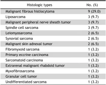
Table 2. Distribution of histologic types (n=31)
Histologic types No. (%)
Malignant fibrous histiocytoma 9 (29.0)
Liposarcoma 3 (9.7)
Malignant peripheral nerve sheath tumor 3 (9.7)
Spindle cell sarcoma 3 (9.7)
Leiomyosarcoma 2 (6.5)
Synovial sarcoma 2 (6.5)
Malignant skin adnexal tumor 2 (6.5)
Fibromyxoid sarcoma 1 (3.2)
Primary eccrine carcinoma 1 (3.2)
Sarcomatoid carcinoma 1 (3.2)
Extrarenal malignant rhabdoid tumor 1 (3.2)
Myxofibrosarcoma 1 (3.2)
Granular cell tumor 1 (3.2)
Undifferentiated sarcoma 1 (3.2)
Table 1. Patients’ characteristics
Characteristic Value
Age (yr) 54.1±16.4
Sex
Male 18 (58.1)
Female 13 (41.9)
Diagnostic method
No biopsy 3 (9.7)
Needle biopsy 15 (48.4)
Excisional biopsy 8 (25.8)
Incisional biopsy 5 (16.1)
Tumor size (cm)
<5 13 (41.9)
≥5 18 (58.1)
Depth of invasion
Soft tissue only 22 (71.0)
Bone invasion 9 (29.0)
Stage
I 15 (48.4)
II/III 16 (51.6)
Neoadjuvant chemotherapy 4 (12.9)
Neoadjuvant radiotherapy 1 (3.2)
Complete resection
R0 27 (87.1)
R1 4 (12.9)
Adjuvant chemotherapy 5 (16.1)
Adjuvant radiotherapy 11 (35.5)
Values are presented as mean±standard deviation or number (%).
Results
Data from 31 patients were analyzed in this study.
The patients’ characteristics are described in Table 1.
Of the patients, 18 were male and 13 were female.
Their mean age was 54.1 years (range, 13–81 years).
The initial presenting symptoms were non-painful masses in 22 patients (71%), painful masses in 3 pa- tients (9.7%), pain without a palpable mass in 4 pa- tients (12.96%), and lymphadenopathy in 1 patient (3.2%). Only 1 patient (3.2%) was asymptomatic.
Contrast-enhanced CT scans were performed as the initial work-up of 29 patients (93.5%), while the ini- tial work-up of 2 patients (6.5%) involved MRI. Four patients (12.9%) received MRI as an additional work-up to evaluate the extent to which adjacent structures had been invaded. Bone scans were per- formed on 6 patients (19.4%), and PET scans were performed on 15 patients (48.4%) to rule out distant metastasis. The preoperative histologic diagnosis was
made by needle biopsy in 15 patients, by incisional biopsy in 5 patients, and by excisional biopsy in 8 patients, while 3 patients underwent curative re- section without a preoperative histologic diagnosis.
In 9 cases (29%), the histologic type of the tumors was malignant fibrous histiocytoma, followed by lip- osarcoma, malignant peripheral nerve sheath tumor, spindle cell sarcoma, leiomyosarcoma, malignant skin adnexal tumor, and synovial sarcoma (Table 2). The histologic grade of the tumor was grade I in 3 pa- tients (9.6%), grade II in 10 patients (32.3%), grade III in 6 patients (19.3%), and unknown in 12 pa- tients (38.7%). The median tumor size was 5.4 cm (range, 1.3–17 cm). The depth of invasion of the growths was to soft tissue in 22 patients (71%) and to bone in 9 patients (29%). Four patients received neoadjuvant chemotherapy and 1 patient received neoadjuvant radiotherapy. The extent of resection was soft tissue in 14 patients and en bloc rib re- section in 17 patients. Complete resection was ach- ieved in 27 patients (87.1%), and 4 patients (12.9%) received microscopically incomplete (R1) resection.
One patient initially underwent marginal resection due to a misdiagnosis of tuberculosis and underwent a second operation. Another patient had R1 resection because the tumor was close to the brachial plexus.
The reason for incomplete resection in the other 2 patients could not be documented, because they un- derwent surgery at another hospital. Chest wall re- construction was necessary in 21 patients (67.7%), including 17 patients who underwent full-thickness
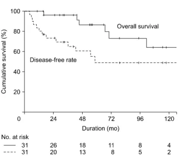
Fig. 1. Kaplan-Meier curves for overall survival and disease-free rates.
resection. Chest wall reconstruction was performed with a skin rotation flap in 1 patient, with a my- ocutaneous flap in 4 patients, with a polytetrafluoro- ethylene (PTFE) patch in 8 patients, and with a my- ocutaneous flap combined with a PTFE patch in 7 patients. One patient underwent skeletal re- construction using polymethyl methacrylate bone cement. The muscle harvested for the myocutaneous flap came from the latissimus dorsi in 7 patients, the pectoralis major in 2 patients, and the rectus abdom- inis in 2 patients.
Two patients required postoperative mechanical ventilator support for 1 and 2 days, respectively.
None of the patients experienced postoperative respi- ratory complications or death. Of the 15 patients who underwent chest wall reconstructions with a PTFE patch, 4 (26.7%) suffered from wound in- fections, which were treated by removing the PTFE patch. Three patients, including 1 patient who re- ceived preoperative radiotherapy, suffered from wound infection within 4 weeks after the operation.
In those patients, conservative treatment, such as drainage and antibiotic therapy, was initially ad- ministered to gain time for fibrous tissues to develop around the graft. The PTFE patch was then removed after the fibrous tissues had matured. The other pa- tient experienced a delayed infection 7 months after surgery, and the patch was removed promptly.
Adjuvant chemotherapy and radiotherapy was pro- vided to 5 and 11 patients, respectively.
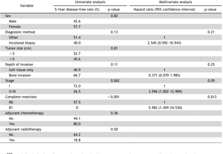
The median follow-up duration was 65.6 months (range, 10.2–191.5 months). There were 5 cases of local recurrence, 3 cases of distant metastasis, and 5 cases of combined recurrence. All cases of recurrence occurred within 5 years after surgery. Among the 10 patients with local recurrence, 3 had undergone R1 resection and 7 had undergone R0 resection. Four of the patients who had local recurrence, but had un- dergone R0 resection, had a close resection margin (less than 1 cm). The 5-year disease-free rate was 49% (Fig. 1). Incomplete resection was a significant prognostic factor for recurrence, and stage was shown to be a marginal prognostic factor in the uni- variate analysis. The multivariate analysis showed that incomplete resection and stage were significantly associated with recurrence (Table 3). Incomplete re- section was shown to be the only significant prog- nostic factor for local recurrence in both the uni-
variate and multivariate analyses (hazard ratio, 4.670;
95% confidence interval, 1.074–20.313; p=0.04).
Of the 13 patients who experienced recurrence, 7 died of CW-STS, 3 patients were alive with disease at the end of the study period, and 3 patients were alive without disease after treatment for recurrence.
There were no other deaths due to CW-STS. The 5- and 10-year overall survival rates were 86.8% and 64.3%, respectively (Fig. 1). No prognostic factor for the overall likelihood of survival was identified. The survival rate of patients who experienced recurrence was significantly worse than that of patients who had not experienced recurrence (10-year recurrence survival rate, 18.5%; 10-year no-recurrence survival rate, 100.0%; p=0.003) (Fig. 2).
Discussion
The present study showed that complete resection was the factor that most strongly influenced the re- currence of CW-STS, and that stage was a meaningful predictor of the prognosis of CW-STS. Although in- complete resection was not shown to be a statisti- cally significant prognostic factor for overall survival, it is nevertheless logical that incomplete resection must be a poor prognostic factor for survival, since the prognosis of patients who experienced recurrence was significantly worse than that of those who did not. Malignant chest wall tumors are relatively rare,
Table 3. Univariate and multivariate analyses of risk factors for recurrence
Variable Univariate analysis Multivariate analysis
5-Year disease-free rate (%) p-value Hazard ratio (95% confidence interval) p-value
Sex 0.82
Male 42.6
Female 57.7
Diagnostic method 0.13 0.21
Other 51.4 1
Incisional biopsy 40.0 2.545 (0.592–10.943)
Tumor size (cm) 0.81
<5 52.7
≥5 45.6
Depth of invasion 0.11 0.25
Soft tissue only 40.9 1
Bone invasion 66.7 0.371 (0.070–1.985)
Stage 0.062 0.05
I 72.0 1
II/III 26.5 3.596 (1.002–12.909)
Complete resection <0.001 0.013
R0 57.5 1
R1 0 5.982 (1.459–24.530)
Adjuvant chemotherapy 0.36
No 44.1
Yes 80.0
Adjuvant radiotherapy 0.50
No 64.2
Yes 18.8
Fig. 2. Kaplan-Meier curves for overall survival of the recurrence- free group and the recurrence group.
representing roughly 5% of all thoracic neoplasms and 1%–2% of all primary tumors [11]. Malignant fi- brous histiocytoma is the most common histologic
type of CW-STS overall, and was also the most com- mon histologic type in the present study [12]. The reported 5-year survival rate after receiving treat- ment for CW-STS was 60%–80% [4]. The 5- and 10-year survival rates in the present study were 86.8% and 64.3%, respectively, which are similar to the results of previous studies. Only a few studies of prognostic factors for CW-STS patient survival have been conducted, and those studies have produced in- consistent results because of the rarity of the disease. A multicenter study of the Anatolian Society of Medical Oncology suggested that patients with re- sectable and low-grade tumors might have better prognostic outcomes [13]. McMillan et al. [14] re- ported that tumor grade was a significant factor in- fluencing the survival rate and recurrence patterns in patients with CW-STS. Gross et al. [15] identified tu- mor grade and diameter as the most important fac- tors influencing the survival rate of patients with CW-STS. Duranti et al. [16] evaluated 337 patients with resected thoracic STS and found that those with
high-grade tumors, with pulmonary and mediastinal STS, and who had undergone R1 resection were more likely to experience poor outcomes than those with low-grade tumors, with CW-STS, and who had undergone R0 resection. Unal et al. [13] reported that adjuvant chemotherapy increased the survival rate. In the present study, complete resection and stage were prognostic factors for predicting recurrence. There were no instances of macroscopi- cally incomplete resection in this study. However, lo- cal recurrence developed in 4 cases of R0 resection with a narrow margin. Therefore, the resection mar- gins of large, high-stage tumors must be examined more comprehensively using a microscopic evaluation of resection margins under frozen examination, and such tumors must be subject to more extended re- section to reduce the possibility of local recurrence because STS spreads laterally along the fascial plane [4,17]. No risk factors influencing the survival rates of patients with STS were found in this study.
However, given that the prognosis of patients experi- encing recurrence was significantly worse than that of patients who did not experience recurrence and that incomplete resection was a poor prognostic fac- tor for recurrence, incomplete resection and resect- ability are likely to be poor prognostic factors for survival [17]. Galetta et al. [18] reported that in- complete tumor resection, not receiving adjuvant therapy, and a tumor diameter greater than 10 cm adversely affected the survival rate.
Wide surgical resection with 2- to 4-cm resection margins is the preferred treatment for CW-STS [4].
From a practical perspective, reconstruction after re- section is the most important procedure. Advancements in reconstructive surgical techniques have led sur- geons to be more aggressive and enabled them to perform more extensive resections, resulting in re- duced morbidity and mortality rates [19]. After large, full-thickness resections, it is important to re- construct the thoracic cage to increase structural sta- bility and to restore soft tissue coverage. PTFE patch- es are the most frequently used type of synthetic material [19-21]. In this study, PTFE patches were used in a majority of cases and produced favorable thoracic cage stability without respiratory complica- tions. Skeletal reconstruction with rigid materials has been strictly limited to use in patients with extensive resections and impaired pulmonary function. Rigid
materials were used in only 1 patient in this study.
Tsukushi et al. [22] reported that non-rigid re- construction with flap transfer and careful respira- tory management were effective even after extensive full-thickness chest wall resections.
Complications after chest wall resection are com- mon and occur in 46%–69% of patients [3,6]. Wound complications, such as infection, dehiscence, and hematoma, have been reported to occur in 8%–20%
of patients [3,6,23]. Complications arise more fre- quently when synthetic materials are irradiated, con- taminated or infected, or laid directly over viscera [24,25]. Deschamps et al. [26] reported that 3% (4 of 133) of wounds became infected after using PTFE patches and no patches were removed. Weyant et al.
[27] reported that the 90-day wound complication rate was 7.2% and the 90-day prosthesis removal rate was 4.1% in patients who received synthetic patches. In the present study, early wound complica- tions were observed in 3 patients (9.7%), and con- servative care, such as drainage and antibiotic treat- ment, was administered to gain enough time for fi- brotic tissues to grow around the defect, after which the infected prostheses were removed.
This retrospective study had several limitations.
Due to the rarity of CW-STS, the subject population was small, the patients were heterogeneous, and the study covered a long period of time. Thus, the reli- ability of the statistical analyses presented in this study may not be high, and surgical resection and re- construction strategies were not standardized throughout study period. Another limitation is in- sufficient data about the tumor grade, which is likely an important prognostic factor. We could not per- form a meaningful analysis of the effect of tumor grade on patients’ prognoses. An organized pro- spective multi-institutional registry must be built to overcome the innate limitations of studies of CW-STS.
In conclusion, the short- and long-term survival of patients who were surgically treated for CW-STS were favorable. Microscopic incomplete resection was the most important prognostic factor for recurrence.
The resection margin should be comprehensively evaluated intraoperatively.
Conflict of interest
No potential conflict of interest relevant to this ar-
ticle was reported.
ORCID
Seung Hwan Yoon: https://orcid.org/0000-0001-7290-3776 Joon Chul Jung: https://orcid.org/0000-0003-4555-5782 In Kyu Park: https://orcid.org/0000-0003-3550-5554 Samina Park: https://orcid.org/0000-0001-9625-2672 Chang Hyun Kang: https://orcid.org/0000-0002-1612-1937 Young Tae Kim: https://orcid.org/0000-0001-9006-4881
References
1. Faber LP. Chest wall tumors: introduction. Semin Thorac Cardiovasc Surg 1999;11:250.
2. Incarbone M, Pastorino U. Surgical treatment of chest wall tumors. World J Surg 2001;25:218-30.
3. Hsu PK, Hsu HS, Lee HC, et al. Management of primary chest wall tumors: 14 years’ clinical experience. J Chin Med Assoc 2006;69:377-82.
4. Shah AA, D’Amico TA. Primary chest wall tumors. J Am Coll Surg 2010;210:360-6.
5. King RM, Pairolero PC, Trastek VF, Piehler JM, Payne WS, Bernatz PE. Primary chest wall tumors: factors affecting survival. Ann Thorac Surg 1986;41:597-601.
6. Burt M. Primary malignant tumors of the chest wall: the Memorial Sloan-Kettering Cancer Center experience.
Chest Surg Clin N Am 1994;4:137-54.
7. Korea Central Cancer Registry, Ministry of Health and Welfare. Annual report of cancer statistics in Korea in 2015 [Internet]. Goyang: National Cancer Information Center; 2018 [cited 2018 Jul 2]. Available from: https://
www.cancer.go.kr/lay1/program/S1T211C223/cancer/
view.do?cancer_seq=4397&menu_seq=4402.
8. Jo VY, Fletcher CD. WHO classification of soft tissue tu- mours: an update based on the 2013 (4th) edition.
Pathology 2014;46:95-104.
9. Trojani M, Contesso G, Coindre JM, et al. Soft-tissue sarco- mas of adults; study of pathological prognostic variables and definition of a histopathological grading system. Int J Cancer 1984;33:37-42.
10. Coindre JM, Terrier P, Bui NB, et al. Prognostic factors in adult patients with locally controlled soft tissue sarcoma:
a study of 546 patients from the French Federation of Cancer Centers Sarcoma Group. J Clin Oncol 1996;14:
869-77.
11. Kroll SS, Walsh G, Ryan B, King RC. Risks and benefits of using Marlex mesh in chest wall reconstruction. Ann Plast Surg 1993;31:303-6.
12. Fang ZW, Chen J, Teng S, Chen Y, Xue RF. Analysis of soft tissue sarcomas in 1118 cases. Chin Med J (Engl) 2009;122:
51-3.
13. Unal OU, Oztop I, Yasar N, et al. Clinicopathologic charac- teristics, treatment outcomes, and prognostic factors of primary thoracic soft tissue sarcoma: a multicenter study of the Anatolian Society of Medical Oncology (ASMO).
Thorac Cancer 2015;6:85-90.
14. McMillan RR, Sima CS, Moraco NH, Rusch VW, Huang J.
Recurrence patterns after resection of soft tissue sarco- mas of the chest wall. Ann Thorac Surg 2013;96:1223-8.
15. Gross JL, Younes RN, Haddad FJ, Deheinzelin D, Pinto CA, Costa ML. Soft-tissue sarcomas of the chest wall: prog- nostic factors. Chest 2005;127:902-8.
16. Duranti L, Gronchi A, Stacchiotti S, et al. Localised thora- cic sarcomas: outcome improvement over time at a single institution. Eur J Cancer 2013;49:2689-97.
17. Tsukushi S, Nishida Y, Sugiura H, Nakashima H, Ishiguro N.
Soft tissue sarcomas of the chest wall. J Thorac Oncol 2009;4:834-7.
18. Galetta D, Pelosi G, Leo F, et al. Primary thoracic synovial sarcoma: factors affecting long-term survival. J Thorac Cardiovasc Surg 2007;134:808-9.
19. Kilic D, Gungor A, Kavukcu S, et al. Comparison of mersi- lene mesh-methyl metacrylate sandwich and polytetra- fluoroethylene grafts for chest wall reconstruction. J Invest Surg 2006;19:353-60.
20. Eschapasse H, Gaillard J, Fournial G, et al. Use of acrylic prosthesis for the repair of large defects of the chest wall (author’s transl). Acta Chir Belg 1977;76:281-5.
21. Mansour KA, Thourani VH, Losken A, et al. Chest wall re- sections and reconstruction: a 25-year experience. Ann Thorac Surg 2002;73:1720-5.
22. Tsukushi S, Nishida Y, Sugiura H, et al. Non-rigid re- construction of chest wall defects after resection of mus- culoskeletal tumors. Surg Today 2015;45:150-5.
23. Chapelier A, Macchiarini P, Rietjens M, et al. Chest wall reconstruction following resection of large primary malig- nant tumors. Eur J Cardiothorac Surg 1994;8:351-6.
24. Althubaiti G, Butler CE. Abdominal wall and chest wall reconstruction. Plast Reconstr Surg 2014;133:688e-701e.
25. Butler CE, Langstein HN, Kronowitz SJ. Pelvic, abdominal, and chest wall reconstruction with AlloDerm in patients at increased risk for mesh-related complications. Plast Reconstr Surg 2005;116:1263-75.
26. Deschamps C, Tirnaksiz BM, Darbandi R, et al. Early and long-term results of prosthetic chest wall reconstruction.
J Thorac Cardiovasc Surg 1999;117:588-91.
27. Weyant MJ, Bains MS, Venkatraman E, et al. Results of chest wall resection and reconstruction with and without rigid prosthesis. Ann Thorac Surg 2006;81:279-85.