INTRODUCTION
Fibrodysplasia ossificans progressiva (FOP; OMIM 135100) is a rare genetic disorder characterized by the progressive development of ectopic ossification of the skeletal muscles and subsequent joint stiffness. The worldwide prevalence of FOP is estimated to be approximately 1/2,000,000 (1). The majority of FOP cases are sporadic, but in familial cases, inheritance is autosomal-dominant with variable expression (2). Children with FOP appear normal at birth except con- genital malformations of the great toes or phalanges (3, 4).
However, in general, sporadic episodes of painful soft tissue swellings (flare-ups) occur during the first decade of life (3).
Even minimal trauma such as minor soft tissue injuries, mus- cle overstretching, overexertion and fatigue, intramuscular injections, falls, or influenza-like illnesses may lead to episod- ic flare-ups (1, 4). These soft tissue nodules rarely regress spontaneously, and usually they rapidly mature through an endochondral ossification to form normal lamellar bone (3, 5). Heterotopic ossification in FOP is not random but pro- ceeds in a direction that is axial to appendicular, cranial to caudad, and proximal to distal (3). The diaphragm, extraoc- ular, cardiac, and smooth muscles are characteristically spared from ossification (3, 6).
FOP is diagnosed based on clinical and radiographic find- ings. When established ectopic ossification has been confirmed
in a FOP patient, no remedies are available to improve func- tional capability. Thus, early diagnosis and the avoidance of provoking events is essential to delay the onset of catastroph- ic restriction of motion. Nonetheless, the rates of diagnostic errors and of inappropriate invasive medical procedures are astonishing, which is probably caused by a lack of physician awareness (7).
Recently, FOP was found to be caused by a heterozygous point mutation of c.617G>A; p.R206H in the gene coding activin A type I receptor (ACVR1) on chromosome 2q23- 24 (8). This mutation is reported to be recurrent regardless of races (8-10). To the best of our knowledge, the c.617G>A mutation is the only one that has been associated with FOP to date.
Korean familial FOP patients were included in the previ- ous multi-center linkage analysis study to locate this disease locus to ACVR1 gene (8). In the present study, we conduct- ed mutation analysis of c.617G>A in ACVR1 in sporadic Korean patients who were clinically and radiologically diag- nosed or suspected to have FOP.
MATERIALS AND METHODS
Twelve patients were included in this study. The pheno- types of the patients are summarized in Table 1. Ten patients
433
Dong Yeon Lee, Tae-Joon Cho, Hye Ran Lee, Moon Seok Park, Won Joon Yoo, Chin Youb Chung, and In Ho Choi
Department of Orthopaedic Surgery, Seoul National University College of Medicine, Seoul, Korea
Address for correspondence Tae-Joon Cho, M.D.
Department of Orthopaedic Surgery, Seoul National University Children’s Hospital, 28 Yeongeon-dong, Jongno-gu, Seoul 110-744, Korea
Tel : +82.2-2072-2878, Fax : +82.2-765-3367 E-mail : [email protected]
*This work was supported by Seoul National University Research Grant (SNU-2006-041-E00208).
DOI: 10.3346/jkms.2009.24.3.433
ACVR1 Gene Mutation in Sporadic Korean Patients with Fibrodysplasia Ossificans Progressiva
Fibrodysplasia ossificans progressiva (FOP; OMIM 135100) is a rare but extreme- ly disabling genetic disorder of the skeletal system, and is characterized by the pro- gressive development of ectopic ossification of skeletal muscles and subsequent joint ankylosis. The c.617G>A; p.R206H point mutation in the activin A type I recep- tor (ACVR1) gene has been reported to be a causative mutation of FOP. In the pre- sent study, mutation analysis of the ACVR1 gene was performed in 12 patients diag- nosed or suspected to have FOP. All patients tested had a de novo heterozygous point mutation of c.617G>A; p.R206H in ACVR1. Mutation analysis confirmed a diagnosis of FOP in patients with ambiguous features, and thus, could be used for diagnostic purposes. Early confirmation through mutation analysis would allow med- ical professionals to advise on the avoidance of provoking events to delay catas- trophic flare-ups of ectopic ossifications.
Key Words : Myositis Ossificans; Mutation Analysis; ACVR1 Gene
Received : 29 December 2007 Accepted : 11 July 2008
had definite clinical manifestations of FOP, i.e., progressive ectopic ossification with resultant joint ankylosis (Fig. 1).
Detailed clinical manifestations in some patients (case 7, 8, and 10) have been reported previously (11). Two patients (cases 1 & 2) showed ambiguous clinical features. An 8-yr old boy (case 1) was referred under a diagnosis of hereditary mul- tiple exostosis. He did not have any restriction of joint motion except for limitation of terminal flexion in both elbow joints.
No soft tissue mass or ectopic ossification was observed in this patient except for an osteochondroma-like bony spur on the right distal humerus. However, he was suspected to have FOP due to big toe anomalies. A 15-yr-old girl (case 2) vis- ited complaining of in-toeing gait and calf pain after exer- cise. Although having mild flexion contractures on both hip and knee joints, she was athletically active. No ectopic ossification observed except for osteochondroma-like bony spurs on both distal femora (Fig. 2A). Big toe anomalies (Fig.
2B) and the 5th finger symphalangism lead us to suspect FOP. The parents recalled that a subcutaneous painless migrat- ing scalp nodule had been detected when she was 6 months old, which spontaneously resolved in 2 weeks. Mutation analysis revealed heterozygous c.617G>A; p.R206H muta- tion in ACVR1, and she started to experience series of flare- up at age of 16 yr.
Mutation analysis
Peripheral blood was obtained from all patients and, if pos- sible, from their parents (case 1, 2, and 6) after obtaining in- formed consent. Genomic DNA was extracted from circu- lating leukocytes using standard procedures. A portion of genomic DNA encompassing exon 4 of ACVR1 was ampli- fied by polymerase chain reaction (PCR) using specific pri- mers (5′-CCAGTCCTTCTTCCTTCTTCC-3′, sense and 5′-AGCAGATTTTCCAAGTTCCATC-3′, antisense) (8).
PCR products were sequenced directly using an ABI Prism 3700 automated sequencer (Applied Biosystems, Foster City, CA, U.S.A.). Results were further verified by restriction en- donuclease digestion of PCR products using Cac8I (New England Biolabs, Beverly, MA, U.S.A.) and HphI (New Eng- land Biolabs).
RESULTS
DNA sequence analysis demonstrated the invariable pres- ence of a heterozygous point mutation of c.617G>A in all ten patients with obvious clinical manifestations (Fig. 3A).
The mutation was not detected in any of their family mem-
*Previous diagnosis when the patients were referred to the authors.
FOP, fibrodysplasia.
Age (yr) of parents at birth of the patient Current Previous Great toe History of Age (yr) of
Caseage (yr) Sex
diagnosis* abnor- migrating onset of Ankylosis Current status
mality nodule ossification Father Mother
1 10 M Hereditary multiple + - Not yet None Normal life 34 31
exostosis
2 16 F Congenital foot + + 16 Neck Community ambulator 30 27
deformity
3 33 F FOP + + 10 Whole body Bed ridden >45 44
involved
4 15 F FOP + - 6 Neck, shoulder, Wheel-chair bound 30 31
back, hip, knee
5 19 M FOP + + 5 Neck, shoulder, Community ambulator 30 27
back
6 6 M Infantile + - 5 Neck, shoulder, Community ambulator 31 29
fibromatosis back
7 33 F Myositis ossificans + + 2 Whole body Bed ridden 32 29
involved
8 19 F FOP + + 2 Neck, shoulder, Wheel-chair bound 34 32
back, hip, knee
9 7 F Myositis ossificans + + 5 Neck, shoulder, Community ambulator 33 31
back
10 14 F FOP + + 2 Neck, shoulder, Wheel-chair bound 35 28
back, hip, knee
11 21 F Myositis ossificans + + 11 Neck, shoulder, Community ambulator 32 31
back, Rt knee
12 12 M FOP + + 9 Neck, back, Community ambulator 36 32
Rt ankle Table 1.Pertinent data of the patients
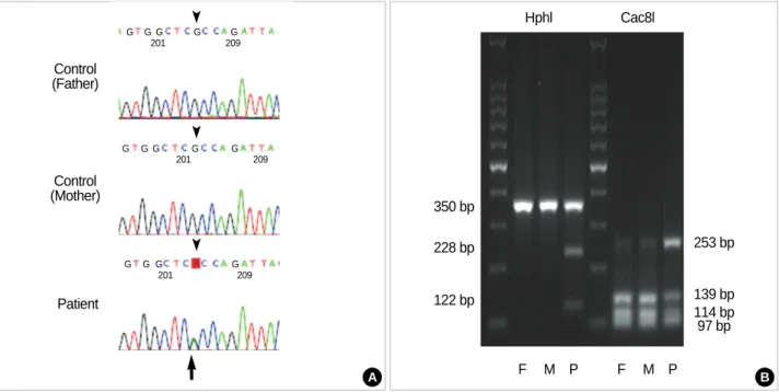
bers tested. In addition to direct DNA sequence analysis, the presence of c.617G>A mutation was also verified by restric- tion endonuclease digestion (Fig. 3B). The c.617G>A ACVR1 mutation eliminates a Cac8I site and forms a new HphI site.
The PCR product of 350 bp from the G allele (control-par-
ents) was digested by Cac8I and produced three bands (139, 114, and 97 bp), whereas the A allele (FOP patients) appeared as two bands (253 and 97 bp). For HphI, PCR products of controls were not digested (one band) whereas bands of 228 and 122 bp, corresponding to the A allele, were detected for
Fig. 2.Radiographic findings suggestive of FOP in a patient with ambiguous clinical features (case 2). (A) This patient showed no ectopic ossification but an osteochondroma-like bony spurs were observed on both distal femora, (B) Big toe abnormalities in this patient showed a slanting of metatarso-phalangeal joint (hallux valgus deformity).
Fig. 1.Radiographic findings of FOP patients with unambiguous clinical features. Infiltration of paraspinal muscles mimicking a tumorous condition (A), ossification around the neck (B), thoracolumbar spine (C), and left thigh, which caused permanent loss of motion (D).
A B
A
B C D
all FOP patients (8).
In two patients (case 1 & 2) with ambiguous clinical fea- tures, DNA sequence analysis demonstrated and restriction endonuclease digestion analysis confirmed the presence of a heterozygous de novo c.617G>A mutation.
DISCUSSION
This is the first report on mutation analysis conducted in sporadic Korean FOP patients. It shows that all 12 spora- dic FOP patients had invariable heterozygous mutation of c.617G>A in ACVR1.
FOP is an extremely rare disorder and most cases are spo- radic (1). Clinical manifestations of typical FOP are consid- erably uniform, i.e., usually ectopic ossifications occur and progress during the first decade of life and most patients are wheelchair bound by the end of the second decade (1, 3, 6).
Delayed onset of ossification after age 15 yr is quite rare, al- though reports have been issued concerning mild cases with a late onset of ossification and unusually slow progression that remained ambulatory till their mid-forties; however, these cases were not confirmed by mutational analysis (12).
This diverse clinical course causes diagnostic and counseling difficulties in patients with atypically mild FOP patients.
However, diagnosis of FOP can be erratic before the onset of established ectopic ossification (7, 11). Diagnostic errors and inappropriate medical procedures, for example, attempts
to remove the heterotopic bone, may lead to explosive new bone formations and can aggravate the natural history of FOP (7). Thus, early diagnosis and confirmation of FOP is essential if such iatrogenic hazards are to be avoided.
To achieve early diagnosis before the flare-ups of ectopic ossification, great toe abnormalities and a history of migrat- ing pre-osseous soft tissue mass on the scalp, neck or back during infancy or early childhood may be informative (11, 13).
In particular, great toe abnormality is one of the most strin- gent and unambiguous features of FOP patients, and usual- ly presents as short, malformed great toes with or without valgus deviation (6). All patients in the present study also showed great toe anomaly. However, these clinical manifes- tations without ectopic ossification only suggest diagnosis of FOP and cannot confirm it.
The variable clinical manifestation of FOP argues against the homogeneity of the FOP mutation (9). Shore et al. (8) reported that the c.617G>A ACVR1 mutation was not iden- tified in a family who showed ambiguous FOP features. How- ever, in the present study, mutational analysis confirmed the diagnosis even in those with exceptionally mild phenotype, e.g., in case 2. In a study using in-silico modeling of wild- type and mutant ACVR1, substitution with histidine (p.R 207H), and only histidine, created a pH-sensitive switch with- in the activation domain of the receptor that lead to ligand- indepenent activation of ACVR1 in FOP (1). The mecha- nism affecting the severity of disease progression remains to be elucidated. However, our findings demonstrate that muta-
Fig. 3.Mutation analysis of the ACVR1 in FOP patients with definite clinical manifestations. (A) Direct sequencing of the PCR products of the ACVR1 showed the presence of the c.617G>A mutation. R=adenine or guanine, (B) Restriction endonuclease digestion of the PCR prod- uct (350 bp). The G allele (control) was digested by Cac8I to produce three bands, whereas the A allele appeared as two bands. Because FOP patients were heterozygous for this mutation, the 139 bp and 114 bp bands were also presented. The PCR product not digested with HphI corresponds to the G allele (control) in contrast to digested products corresponding to the A allele (FOP).
Control (Mother)
Patient Control (Father)
Hphl
350 bp
253 bp
139 bp 114 bp 97 bp F M P F M P 228 bp
122 bp
Cac8l
G
G
G G G G
G G G G
G G 201
201
201 209
209 209
G G
A B
tional analysis of the ACVR1 is helpful for confirming or ex- cluding a diagnosis of FOP in clinically ambiguous patients.
Furthermore, we recommend mutation analysis in young patients with the early stigma of FOP, e.g., great toe anoma- ly and a migrating mass on the scalp, neck or back during infancy or early childhood, to allow patients to avoid provok- ing events during earlier life.
The incidence of human spontaneous mutation increases according to parental age, especially paternal age (14). In the present study, the mean paternal and maternal age was 34 and 31, respectively. Although paternal ages were over thir- ties in all cases, we could not determine the effect of parental age on ACVR1 mutation.
In conclusion, the present study shows that the de novo c.617G>A; p.R206H heterozygous point mutation in the ACVR1 is present in all sporadic Korean FOP patients ex- amined. Moreover, mutation analysis confirms a diagnosis of FOP in patients with ambiguous features, which enable the medical personnel to give early appropriate medical advice concerning the prevention of provoking events with hope for delay or prevention of catastrophic flare-ups of ectopic ossifications.
REFERENCES
1. Groppe JC, Shore EM, Kaplan FS. Functional modeling of the ACVR1 (R206H) mutation in FOP. Clin Orthop Relat Res 2007; 462: 87-92.
2. Shore EM, Feldman GJ, Xu M, Kaplan FS. The genetics of fibrodys- plasia ossificans progressiva. Clin Rev Bone Miner Metab 2005; 3:
201-4.
3. Cohen RB, Hahn GV, Tabas JA, Peeper J, Levitz CL, Sando A, Sando N, Zasloff M, Kaplan FS. The natural history of heterotopic ossifi- cation in patients who have fibrodysplasia ossificans progressiva. A study of forty-four patients. J Bone Joint Surg Am 1993; 75: 215-9.
4. Kaplan FS, Glaser DL, Shore EM, Deirmengian GK, Gupta R, Delai P, Morhart R, Smith R, Le Merrer M, Rogers JG, Connor JM, Kit-
terman JA. The phenotype of fibrodysplasia ossificans progressiva.
Clin Rev Bone Miner Metab 2005; 3: 183-8.
5. Kaplan FS, Tabas JA, Gannon FH, Finkel G, Hahn GV, Zasloff MA.
The histopathology of fibrodysplasia ossificans progressiva. An endo- chondral process. J Bone Joint Surg Am 1993; 75: 220-30.
6. Connor JM, Evans DA. Fibrodysplasia ossificans progressiva. The clinical features and natural history of 34 patients. J Bone Joint Surg Br 1982; 64: 76-83.
7. Kitterman JA, Kantanie S, Rocke DM, Kaplan FS. Iatrogenic harm caused by diagnostic errors in fibrodysplasia ossificans progressiva.
Pediatrics 2005; 116: e654-61.
8. Shore EM, Xu M, Feldman GJ, Fenstermacher DA, Cho TJ, Choi IH, Connor JM, Delai P, Glaser DL, LeMerrer M, Morhart R, Rogers JG, Smith R, Triffitt JT, Urtizberea JA, Zasloff M, Brown MA, Kaplan FS. A recurrent mutation in the BMP type I receptor ACVR1 causes inherited and sporadic fibrodysplasia ossificans progressiva. Nat Genet 2006; 38: 525-7.
9. Nakajima M, Haga N, Takikawa K, Manabe N, Nishimura G, Ikegawa S. The ACVR1 617G>A mutation is also recurrent in three Japanese patients with fibrodysplasia ossificans progressiva. J Hum Genet 2007; 52: 473-5.
10. Lin GT, Chang HW, Liu CS, Huang PJ, Wang HC, Cheng YM. De novo 617G-A nucleotide mutation in the ACVR1 gene in a Taiwanese patient with fibrodysplasia ossificans progressiva. J Hum Genet 2006; 51: 1083-6.
11. Choi IH, Chung CY, Cho TJ, Lee DY, Suk SI, Kim WJ, Cho HO, Lee CS, Yoo HW, Yun YH. Fibrodysplasia Ossificans Progressi- va. J Korean Orthop Assoc 1998; 33: 1069-75.
12. Janoff HB, Tabas JA, Shore EM, Muenke M, Dalinka MK, Schle- singer S, Zasloff MA, Kaplan FS. Mild expression of fibrodysplasia ossificans progressiva: a report of 3 cases. J Rheumatol 1995; 22:
976-8.
13. Kaplan FS, Smith RM. Fibrodysplasia ossificans progressiva (FOP).
J Bone Miner Res 1997; 12: 855.
14. Crow JF. The origins, patterns and implications of human sponta- neous mutation. Nat Rev Genet 2000; 1: 40-7.