Aldosterone Modulates Cell Proliferation and Apoptosis in the Neonatal Rat Heart
In the present study, we investigated whether and how the mineralocorticoid receptor antagonist spironolactone affects cardiac growth and development through apoptosis and cell proliferation in the neonatal rat heart. Newborn rat pups were treated with
spironolactone (200 mg/kg/d) for 7 days. The cell proliferation was studied by PCNA immunostaining. The treatment with spironolactone decreased proliferating myocytes by 32% (P<0.05), and reduced myocytes apoptosis by 29% (P<0.05). Immunoblot and immunohistochemistry for the expression of p38, p53, clusterin, TGF-β2, and extracellular signal-regulated kinase were performed. In the spironolactone group, p38, p53, clusterin, and TGF-β2 protein expression was significantly decreased (P<0.05). These results indicate that aldosterone inhibition in the developing rat heart induces cardiac growth impairment by decreasing proliferation and apoptosis of myocytes.
Key Words: Aldosterone; Muscle Cells; Apoptosis; Cell Proliferation Hyung Joo Sohn, Kee Hwan Yoo,
Gi Young Jang, Jang Hoon Lee, Byung Min Choi, Jung Hwa Lee, In Sun Bae, Hyung Eun Yim, Chang Sung Son, and Joo Won Lee Department of Pediatrics, Korea University College of Medicine, Korea University Hospital, Seoul, Korea Received: October 1 2009
Accepted: March 9 2010 Address for Correspondence:
Gi Young Jang, M.D.
Department of Pediatrics, Korea University Hospital, 123 Jeokgeum-ro, Danwon-gu, Ansan 425-707, Korea Tel: +82.31-412-5096, Fax: +82.31-405-8591 E-mail: [email protected]
This study was supported by the Korea University Grant (K0619631) and by the Myung-Moon Pediatrics Research Foundation.
DOI: 10.3346/jkms.2010.25.9.1296 • J Korean Med Sci 2010; 25: 1296-1304
INTRODUCTION
Our previous studies demonstrated that alterations in the renin angiotensin aldosterone system (RAAS), of the developing rat, affect cardiac and renal growth and development (1). To date, many studies have reported that RAAS plays a major role in the pathophysiology of the cardiovascular system (2, 3). Aldoste- rone, the final product of the RAAS, has been highlighted as a potentially important component of the RAAS. In the cardiovas- cular system, aldosterone modulates vascular tone by increas- ing the pressor response to catecholamines or by upregulation of angiotensin II receptors (4), and it provides a homeostastic response to hypovolemia (5). However, aldosterone excess causes collagen deposition and remodeling in vessels (5). Therefore, excessive aldosterone secretion promotes perivascular and car- diac fibrosis, myocyte hypertrophy and diastolic dysfunction that is independent of the other components of the RAAS by a variety of mechanisms (6).
The mineralocorticoid receptor antagonists, spironolactone and epelerenone, can prevent many of these deleterious effects, on the cardiovascular system. The mineralocorticoid receptor antagonist, spironolactone has been used to reverse these ad- verse effects on the cardiovascular system in many patients with chronic heart disease such as congestive heart failure or myo- cardial hypertrophy. Although several studies have investigated the role of aldosterone in the adult cardiovascular system (2, 3),
relatively little attention has been focused on the role of aldo- sterone in modulating cardiac growth during the perinatal pe- riod. Aldosterone contributes to cardiomyocyte apoptosis both in vivo and in isolated cells (7). Apoptosis and cell proliferation are essential for normal cardiac development; dysregulation of this process can lead to a variety of cardiac diseases during post- natal development.
As a final product of the RAAS, aldosterone is now recognized as an important modulator of myocyte apoptosis (8). Among the mitogen-activated protein kinase (MAPK) family members, p38 is activated by hemodynamic stress and cellular stress; it is thought to inhibit cell growth and induce apoptosis. However, the extracellular signal-regulated kinase (ERK) promotes cell proliferation and differentiation (9). Clusterin and p53 are apop- tosis related molecules. Clusterin plays a major role in prevent- ing apoptosis and p53 may enhance the renin-angiotensin sys- tem and activates apoptosis (10). The transforming growth fac- tor (TGF)-β is known as a multifunctional growth factor; it is in- volved in modulating cell proliferation, differentiation and ex- tracellular matrix formation (11).
The intracellular molecular mechanisms involved in the con- trol of cardiac growth and development, by aldosterone in vivo, have not been elucidated. Therefore, the present study was de- signed to investigate the role of endogenous aldosterone in car- diac growth and development, through apoptosis and cell pro- liferation, in the neonatal rat heart.
MATERIALS AND METHODS Animal preparation
Forty-five neonatal rat pups from five pregnant Sprague Dawley rats were breastfed by their own mothers throughout the study.
The body weights were measured daily from birth. The rat pups were given a dose of 200 mg/kg of spironolactone (Sigma Chem- ical Co., St. Louis, MO, USA) (S group) or normal saline (control group) through an orogastric tube. The dose of 200 mg/kg of spironolactone has been shown to block the effects on aldoste- rone (7).
This dose of spironolactone is known to protect cardiovascu- lar system against the harmful effects of hemodynamic overload (12). The rats were sacrificed at 7 days of age. Their hearts were removed, weighed and harvested for this study. RNA analysis, protein assays, detection of apoptosis, and immunohistochem- istry were performed. The Animal Care Committee of Korea Uni- versity Guro Hospital approved the experimental protocol.
Immunohistochemical staining
For assessment of the expression of p38, p53, clusterin, and TGF β-2, the harvested hearts were prepared in 10% formalin solu- tion (Sigma Chemical Co.) and embedded in paraffin. The samples were then cut into 4-μm sections and the sections were deparaffinized with xylene, followed by rehydration in a descend- ing series of ethanol concentrations. The endogenous peroxidase was quenched by 3% hydrogen peroxide in methanol for 15 min.
The sections were incubated with primary antibodies against p38 (Santa Cruz Biotechnology, Santa Cruz, CA, USA), p53 (Cal- biochem, Cambridge, USA), clusterin (Upstate Biotechnology, Incorporated, NY, USA), and TGF β-2 (Promega, Madison, WI, USA). After the incubation, the sections were washed twice for 5 min in phosphate-buffered saline (PBS). The sections were then incubated for 30 min with secondary antibodies, washed in PBS and incubated for 50 min with Vectastain ABC reagent (Burlingame, CA, USA). The bound antibodies were detected using 3,3´-diaminobenzidine, which produces a brown color.
The sections were counterstained with a 0.5% methyl green so- lution (Trevigen, Gaithersburg, MD, USA), then the samples were dehydrated and mounted. After dehydration, we evaluat- ed the specimens under the light microscope.
Detection of cell proliferation
To assess cell proliferation, the harvested hearts were prepared in 10% formalin or Bouin solution (Sigma Chemical Co.), embed- ded in paraffin, then immunohistochemical staining for the proliferating cell nuclear antigen (PCNA) was carried out. The PCNA-positive cells were detected using the avidin-biotin im- munoperoxidase method (Vectastain ABC kit). The primary an- tibodies were the monoclonal anti-mouse PCNA antibody (1:100 dilutions, DAKO, Glostrup, Denmark) in the study group and
PBS in the negative control group. Each slide was cross-stained with 0.5% methyl green solution (Trevigen). After dehydration, we compared the slides under the light microscope. The PCNA positive cells were evaluated by counting 50 areas (25×25 μm) and the average was calculated. The counts were carried out randomly throughout all the observed fields.
Detection of apoptosis
To determine whether apoptosis is involved in the developing rat heart, we performed terminal deoxynucleotide transferase- mediated nick-end labeling (TUNEL) staining for apoptotic cells in the heart of the control and spironolactone-treated group.
Apoptotic nuclei were labeled using the TACS TM 2 TdT In situ Apoptosis Detection Kit (Trevigen, Gaithersburg, MD, USA).
Tissues were fixed in 4% neutral buffered formalin for 4 hr at 4°C, dehydrated in graded alcohols and embedded in paraffin. The samples were then cut into 4 μm sections and dried onto silicon- ized slides (Sigma Chemical Co.). The slides were deparaffinized and digested for 10 min with proteinase K (20 μg/mL) and the endogenous peroxidase activity was quenched by 2% hydrogen peroxide for 5 min. Samples were equilibrated using the provid- ed buffer for 2 min and labeled with biotin dNTP mixtures, TdT, CoCl2, and a labeling buffer according to the manufacturer’s in- structions. The samples were rinsed in PBS for 2 min, and treat- ed with the provided Streptavidin-Horseradish Peroxidase De- tection solution for 10 min and finally washed 2 times in PBS for 2 min each wash. The color was developed using 0.05% 3,3´- diaminobenzidine and 0.05% hydrogen peroxidase in PBS for 5 min. The sections were rinsed in water and counter-stained in methyl green for 5 min, then the samples were dehydrated and mounted. After dehydration, we compared them under the light microscope. The positive apoptotic cells were counted in 50 ar- eas (25×25 μm) and the average was calculated.
Isolation of RNA and analysis of mRNA
Five hearts from the control group and the spironolactone-treat- ed group were selected for RNA analysis and protein assays. Af- ter removal from the rats, the cardiac tissue was frozen in liquid nitrogen and stored at -70°C. The total cellular RNA was isolat- ed using the TRI-reagent (Molecular Research Center, Cincin- nati, OH, USA) and homogenized with a tissue tearor (Model 985-370, Biospec products, Bartlesville, OK, USA). Next, 37%
chloroform (200 μL/mL TRI reagent) was added to the homog- enates and centrifuged at 12,000 rpm for 15 min at 4°C separat- ing the sample into three layers: RNA, DNA and protein materi- al. The colorless clear upper layer was transferred into another Eppendorf tube, isopropanol was added, then left at room tem- perature for 15 min centrifuged at 12,000 rpm for 10 min at 4°C until a white-colored cellular RNA pellet was isolated. The pellet was dried at room temperature for 5 min after washing in 75%
ethanol and was dissolved in 25 μL Forma zol (Molecular Re-
search Center) at 55°C in a heating block for 10 min and then stored at -70°C. The RNA was quantified spectrophotometrical- ly at an absorbance of 260 nm.
cDNA synthesis by reverse transcription (RT) and PCR A cDNA Synthesis Kit (Boehringer Mannheim Corp., Indianap- olis, IN, USA) was used to obtain 1 μg of oligo dT primed first strand cDNA from the RNA template. AMV reverse transcriptase was used for synthesis of the first strand cDNA for use in subse- quent amplification reactions. The PCR reaction had a forward primer 5´-AATGCATCCTGCACCACCAA-3´ and a reverse prim- er 5´-GTAGCCATATTCATTGTCATA-3´ for the GAPDH designed based on the DNA template from rats. In addition, 5´-CGGGTA–
CCGACAATGAGCTCCA-3´, 5´-GGCCGCGGCCACTTCTGCA–
GAC-3´ for clusterin, 5´-ATGTCTCAGGAGAGGCCCACGTT–
CT-3´, 5´-TCAGGAGTCCATTTCTTCTTGGTC-3´ for p38, 5´-CT- GAGGTCGGCTCCGACTATACCACTATCC-3´, 5´-CTGATTCA–
GCTCTCGGAACATCTCGAAGCG-3´ for p53, 5´-CCTAGCCA–
GGGACGTTTTTC-3´, 5´-TAGACAGACTGAGCGCCACC-3´ for TGF β-2, all five 515, 310, 1080, 360, and 230 base pairs of the PCR products for each gene were obtained. The PCR reaction was carried out at different times and temperatures for each re- action period using the Perkin Elmer Cetus DNA Thermal Cy- cler (Model 2400, Foster City, CA, USA). These amplified PCR products were visible as a fluorescent band under UV rays, after agarose gel electrophoresis, at different time intervals and with ethidium bromide staining. Polaroid photographs were scanned using Epson GT-9500 (Seiko Corp, Nagano, Japan) and quanti- tated by densitometry (Image PC alpha 9, NIH, Bethesda, MD, USA) and the values were revised based on the GAPDH.
Protein extraction
Alcohol (100%) was added to the interface and the organic phase remained from the RNA separation; then the DNA portion was precipitated after the sample was centrifuged at 5,000 rpm for 5 min. Isopropanol was added to the remaining ethanol upper layer, which was centrifuged and dissolved in 0.3 M guanidine hydrochloride in 95% ethanol. Next, this layer was washed three times and a protein pellet was obtained. The extracted protein was dissolved in 1% SDS solution and preserved at -20°C. The Bradford method was used for the quantification of the protein.
Western blotting
To precipitate the DNA, the interface and the organic phase re- maining after the RNA extraction were centrifuged at 5,000 rpm for 5 min at 4°C after treatment with 100% alcohol. A residual phenol-ethanol upper layer was added with isopropanol, cen- trifuged and treated with 0.3 M guanidine hydrochloride dis- solved in 95% ethanol. This layer was centrifuged at 10,000 rpm for 10 min at 4°C after incubation at room temperature for 15 min, and this process was repeated three times. The sample was
then washed to obtain a protein pellet. The extracted proteins were solubilized in 5×SDS loading buffer for 5 min at 95°C and separated by electrophoresis on 10% SDS-polyacrylamide gels under reducing conditions. Subsequently, the proteins were transferred to nitrocellulose membranes (Amersham Life Sci- ence, Buckinghamshire, England). The nitrocellulose membranes were blocked in 5% skim milk with TBS-T (0.05% Tween 20 in 50 mM Tris, 150 mM NaCl, 0.05% NaN3, pH 7.4) at room tem- perature for 1 hr. The membranes were then incubated for 18 hr at 4°C with the respective primary antibodies against clusterin (1:100 dilution), p38 (1:1,000 dilution), p53 (1;200 dilution), TGF β-2 (1:200 dilution), ERK 1/2 (1:1,000 dilution), and JNK-2 (1:500 dilution). Thereafter, the membranes were washed two times with TBS-T and incubated for 40 min with an anti-rabbit IgG (Amersham Life Science) at room temperature. After wash- ing with TBS-T four times, the secondary antibody bound to the nitrocellulose was detected by incubation for 1 min with a detec- tion reagent (Amersham Life Science) and then exposed to medi- cal X-ray film (Agfa, Mortsel, Belgium) for 1 min. The film was developed by a FPM-3500 Fuji X-ray Film Processor (Fuji, Ota- wara, Japan). The developed X-rays were scanned using the Ep- son GT-9500 (Seiko Corp) and quantitated by densitometry (Im- age PC α 9).
Statistical analysis
The data are presented as mean±SEM. The differences between the two groups were analyzed by the Student-Newman-Keuls test; a value of P<0.05 was considered statistically significant.
RESULTS
On day 7, the average body weight was 13.1±1.4 g in the S group and 15.8±1.1 g in the control group. The heart weight was 0.08±
0.01 g in the S group and 0.11±0.01 g in the control group. The heart weight/body weight ratio was 0.005±0.0009 in the S group and 0.007±0.0008 in the control group. The body weights, heart weights and heart weight/body weight ratio of the S group were significantly reduced compared to the control group (P<0.05).
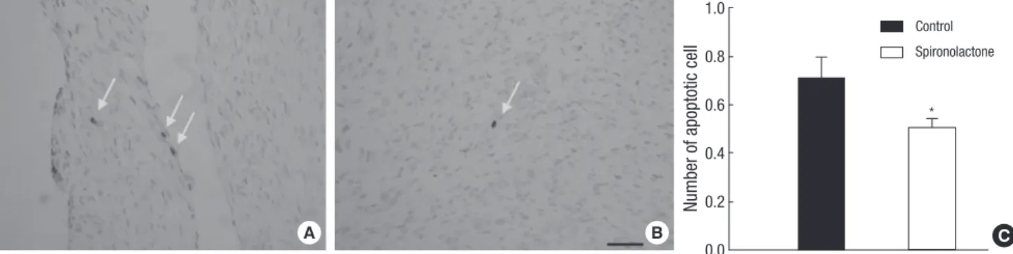
Proliferating cell nuclear antigen (PCNA) and apoptosis In PCNA immunostaining of the proliferating myocytes was lower in the myocardium of the neonatal rat heart in the S group compared to the control group. The PCNA-positive cells were 28.1±6.61 in the control group and 19.18±2.09 in the S group in each of the 25×25 μm fields (P<0.05, Fig. 1). Myocyte apoptosis was detected using the toatal TUNEL technique. The myocyte apoptosis was lower in the myocardium of neonatal rat heart in the S group compared to the control group. Apoptotic nuclei were present in 0.71±0.18 of the control group and in 0.50±0.08 of the S group for each 25×25 μm field (P<0.05, Fig. 2). Spironolactone treatment decreased proliferating myocytes by 32% (P<0.05) and
reduced myocyte apoptosis by 30% (P<0.05). However, apoptot- ic nuclei accounted for only 2.5% of the relative fraction of cells undergoing proliferation in the present study. Finally, a net re- duction of 32%, in cell proliferation, was observed in the S group compared to the control group.
Expression of p38
The semiquantitative RT-PCR showed that p38/GAPDH mRNA expression was 0.77±0.1 in the control group and 0.87±0.12 in
the S group (P=0.18; Fig. 3A). Immunoblot analysis revealed that the p38/tubulin protein expression was significantly decreased in the S group (1.04±0.15) compared to the control group (1.33±
0.19; P<0.05; Fig. 3B). Immnohistochemically, there was no dif- ference between control and spironolactone group.
Expression of p53
The semiquantitative RT-PCR of p53/GAPDH mRNA expres- sion was 1.02±0.11 in the control group and 1.13±0.08 in the S
Fig. 3. Representative expressions of p38 mRNA and protein. (A) In immunoblot analysis, p38 mRNA amount is similar between the control and S groups. (B) p38/tubulin protein expression is decreased significantly in the S group compared with the control group, *P<0.05.
p38 p38
Control Spironolactone Control Spironolactone
1,080 bp 38 kDa
GAPDH 515 bp Tubulin 57 kDa
p38/GAPDH mRNA expression p38/Tubulin protein expression
1.2 1.0 0.8 0.6 0.4 0.2 0.0
1.8 1.6 1.4 1.2 1.0 0.8 0.6 0.4 0.2 0.0
*
n=5 n=5
n=5 n=5
A B
Control Spironolactone
Control Spironolactone
Number of apoptotic cell
1.0
0.8
0.6
0.4
0.2
A B 0.0 C
*
Fig. 2. Distribution of apoptotic cells in the myocardium. (A, B) Apoptotic cells (arrows) in the S group (B) are less than those in control group (A) (bar=25 µm). (C) Number of apoptotic cells in control group (black bar) is reduced in spironolactone group (white bar), measured by counting 50 areas, 25×25 µm, *P<0.05.
Control Spironolactone
Number of PCNA positive cell
50
40
30
20
10
A B 0 C
Fig. 1. PCNA positive proliferating cardiac myocytes in control and spironolactone group. (A, B) Immunohistochemical staining for PCNA in both groups demonstrates that proliferating myocytes (arrows) in the S group (B) are less than those in control group (A) (bar=25 µm). (C) Number of PCNA positive proliferating cells in control group (black bar) is reduced in spironolactone group (white bar), measured by counting 50 areas, 25×25 µm, *P<0.05.
* Control Spironolactone
group (P=0.1; Fig. 4A). The immunoblot analysis revealed that the p53/tubulin protein expression was significantly decreased in the S group (0.71±0.11) compared to the control group (1.05±
0.11; P<0.05; Fig. 4B).
Expression of clusterin
The semiquantitative RT-PCR showed that the clusterin/GAP- DH mRNA expression was 0.90±0.22 in the control group and 0.93±0.17 in the S group (P=0.83; Fig. 5A). Immunoblot analysis
Fig. 4. Representative expressions of p53 mRNA and protein. (A) In immunoblot analysis, p53 mRNA amount is similar between the control and S groups. (B) p53/tubulin protein expression is decreased significantly in the S group compared with the control group, *P<0.05.
p38 p38
Control Spironolactone Control Spironolactone
360 bp 53 kDa
GAPDH 515 bp Tubulin 57 kDa
p38/GAPDH mRNA expression p38/Tubulin protein expression
1.4 1.2 1.0 0.8 0.6 0.4 0.2 0.0
1.4 1.2 1.0 0.8 0.6 0.4 0.2 0.0
*
n=5 n=5 n=5
n=5
A B
Control Spironolactone
Control Spironolactone
Fig. 5. Representative expressions of clusterin mRNA and protein. (A) In immunoblot analysis, clusterin mRNA amount is similar between the control and S groups. (B) clusterin/
tubulin protein expression is decreased significantly in the S group compared with the control group, *P<0.05.
Clusterin Clusterin
Control Spironolactone Control Spironolactone
310 bp 35 kDa
GAPDH 515 bp Tubulin 57 kDa
Clusterin/GAPDH mRNA expression Clusterin/Tubulin protein expression
1.4 1.2 1.0 0.8 0.6 0.4 0.2 0.0
1.4 1.2 1.0 0.8 0.6 0.4 0.2 0.0
*
n=5
n=5 n=5 n=5
A B
Control Spironolactone
Control Spironolactone
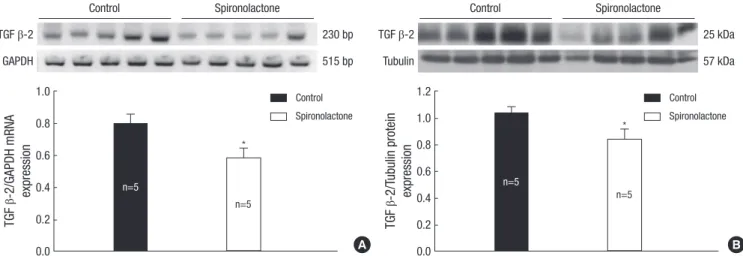
Fig. 6. Representative expressions of TGF β-2 mRNA (A) and protein (B) are reduced in the developing rat heart of the control and S groups, *P<0.05.
TGF β-2 TGF β-2
Control Spironolactone Control Spironolactone
230 bp 25 kDa
GAPDH 515 bp Tubulin 57 kDa
TGF β-2/GAPDH mRNA expression TGF β-2/Tubulin protein expression
1.0 0.8 0.6 0.4 0.2 0.0
1.2 1.0 0.8 0.6 0.4 0.2 0.0
*
*
n=5 n=5
n=5 n=5
A B
Control Spironolactone
Control Spironolactone
revealed that the clusterin/tubulin protein expression was sig- nificantly decreased in the S group (0.91±0.14) compared to the control group (1.12±0.07; P<0.05; Fig. 5B). Immunohistochemi- cally, there was no difference between control and spironolac- tone group.
Expression of TGF β
The semiquantitative RT-PCR showed that the TGF β-2/GA–
PDH mRNA expression was 0.79±0.12 in the control group and 0.58±0.13 in the S group (P<0.05; Fig. 6). Immunoblot analysis revealed that the TGF β-2/tubulin protein expression was sig- nificantly decreased in the S group (0.83±0.15) compared to the control group (1.03±0.09; P<0.05: Fig. 6). Immunohistoche–mi- cally, there was no difference between control and spironolac- tone group (Fig. 6). Immunoblot analysis showed that the TGF β-1/tubulin protein expression was 0.96±0.02 in the control group and 0.92±0.06 in the S group. There was no detectable difference between the two groups (Fig. 7).
Expression of ERK
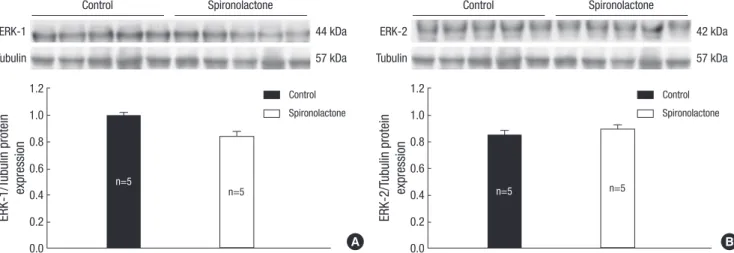
The immunoblot analysis showed that the ERK-1/tubulin pro- tein expression was 0.99±0.05 in the control group and 0.83±0.16 in the S group (Fig. 8A), and the ERK-2/tubulin protein expres- sion was 0.85±0.07 in the control group and 0.89±0.07 in the S group (Fig. 8B). There was no detectable difference between the two groups.
Expression of JNK
Immunoblot analysis showed that the JNK-2/tubulin protein expression was 0.90±0.05 in the control group and 0.94±0.08 in the S group (Fig. 9). There was no detectable difference between the two groups.
DISCUSSION
The renin-angiotensin-aldosterone system (RAAS) plays an im- portant role in regulating the cardiovascular system and is im- portant in regulating the growth of the myocardium (13). An- giotensin II, a product of the RAAS, causes an increase in blood
Fig. 7. TGF β-1 protein expression in the developing rat heart. There is no difference between the control and S groups.
TGF β-1
Control Spironolactone
25 kDa
Tubulin 57 kDa
TGF β-1/Tubulin protein expression 1.2 1.0 0.8 0.6 0.4 0.2 0.0
n=5 n=5
Control Spironolactone
Fig. 9. JNK-2 protein expression in the developing rat heart. There is no difference between the control and S groups.
JNK-2
Control Spironolactone
54 kDa
Tubulin 57 kDa
JNK-2/Tubulin protein expression 1.2 1.0 0.8 0.6 0.4 0.2 0.0
n=5 n=5
Control Spironolactone
Fig. 8. Representative expressions of ERK-1 (A) and ERK-2 proteins (B) are similar in the developing rat heart of the control and S groups.
ERK-1 ERK-2
Control Spironolactone Control Spironolactone
44 kDa 42 kDa
Tubulin 57 kDa Tubulin 57 kDa
ERK-1/Tubulin protein expression ERK-2/Tubulin protein expression
1.2 1.0 0.8 0.6 0.4 0.2 0.0
1.2 1.0 0.8 0.6 0.4 0.2 0.0
n=5 n=5 n=5 n=5
A B
Control Spironolactone
Control Spironolactone
pressure through its vasoconstrictive effect. In addition, angio- tensin II promotes cell growth via angiotensin II type I (AT1) re- ceptors in cardiac myocytes, which cause inotropic and chro- notropic changes and induce apoptosis in myocytes (14). A num- ber of studies have demonstrated that alterations in the RAAS of developing rats affect cardiac growth and development (1, 15).
A recent new wave of interest in aldosterone physiology has been generated by its role in cardiovascular homeostasis including blood-pressure homeostasis, myocardial fibrosis and remodel- ing of blood vessels (6). In addition, recent studies have shown that aldosterone induces cardiomyocyte apoptosis (6, 14). Ac- cording to Mano et al. (8), there are plasma membrane recep- tors that are specific for aldosterone in myocytes. Activation of aldosterone receptors stimulates myocyte apoptosis through a cacineurin dependent mitochondrial death-signaling pathway.
Chronic aldosterone receptor activation promotes vascular in- flammation, vascular stiffness and perivascular collagen forma- tion (16). The loss of myocytes by apoptosis in the dysfunctional heart results in reduced cardiac reserve capacity; this is one of the most important components of the pathogenesis of various cardiac diseases. Therefore, aldosterone excess has been shown to have a deleterious effect on the cardiovascular system (17).
However, there are few reports regarding the relationship be- tween aldosterone and cardiac myocyte apoptosis and cell pro- liferation in the developing heart, especially in the neonate. In this study, both cell proliferation and apoptosis were decreased in the spironolactone treated group. Blocking of aldosterone re- duced the number of proliferating myocytes by 32% and de- creased myocyte apoptosis by 29%. The decrease in cell prolif- eration was counterbalanced by a reduction in myocyte apop- tosis. However, the cells undergoing apoptosis accounted for only a minor fraction of the proliferating cells. Therefore, a net reduction of cell proliferation, in the cardiac myocytes, resulted from the blocking of aldosterone.
Although cardiomyocytes constitute only 30% of the ventric- ular cellular population in the mature rodent, they may occupy up to 85% of the total volume of the ventricular myocardium (18). Therefore, the net reduction of cell proliferation in myo- cytes, with aldosterone inhibition, may have contributed to the decrease in heart weight and heart weight to body weight ratio.
The present in vivo animal study showed that aldosterone, the final product of RAAS, might play a role in cardiac growth in de- veloping heart. The apoptotic and cell proliferation activity, in the cardiac myocytes, is mediated by several growth factors as- sociated with apoptosis and cell proliferation. Among the MAPK signaling cascade, the p38 MAPK signaling pathway is activated in cardiac cells by hemodynamic stress or ischemia; it is thought to induce myocyte apoptosis, hypertrophy and fibroblast prolif- eration. In some animal models using transgenic mice, p38 ac- tivation resulted in interstitial fibrosis, cardiac hypertrophy and myocardial dysfunction (19). In the present study, p38 protein
expression was significantly decreased by spironolactone; myo- cyte apoptosis was lower in the myocardium of the spironolac- tone treated group. These results suggest that p38 may be in- volved in the aldosterone-related intracellular signaling path- ways of myocyte apoptosis in the developing heart.
p53 is a tumor suppressor protein; it has been reported to up- regulate the cellular renin-angiotensin system, potentiating the synthesis and release of the octapeptide angiotensin II, which induces myocyte apoptosis via the activation of surface AT1 re- ceptors (20). In addition, p53 is a transcriptional regulator of the proapoptotic gene product Bax and antiapoptotic gene product Bcl-2 (21), the activation of p53 may upregulate Bax and down- regulate Bcl-2 in myocytes (22). The decrease in the Bcl-2: Bax ratio and upregulation of the cellular renin-angiotensin system, by the activation of p53, is thought to trigger myocyte apoptosis (23). The results of this study showed that p53 protein expression was significantly decreased. However, the p53 mRNA expres- sion was not significantly different in the spironolactone treated group compared to the control group. These findings suggest that p53 is involved in the intracellular signaling pathways of myocyte apoptosis in the developing rat heart.
Clusterin is a disulfide-linked heterodimeric glycoprotein, first isolated from ram rete testis fluid in 1983, it has been re- ported to have a wide array of functions such as lipid transport, tissue remodeling, sperm maturation, and complement regula- tion (24). Recent studies have demonstrated that clusterin has both anti-apoptotic and anti-proliferative activity (25). The role of clusterin may vary, but it may be anti-proliferative or anti- apoptotic. If an anti-proliferative effect of clusterin dominates, this would render the cells susceptible to apoptosis. In this study, while there was no significant effect of spironolactone on myo- cardial expression of clusterin mRNA, clusterin protein expres- sion was significantly decreased by treatment with spironolac- tone. These observations suggest that clusterin may participate in aldosterone-modulated intracellular signaling processes in myocyte apoptosis and proliferation.
The protein expressions of p38, p53, clusterin were found to be decreased significantly in the spironolactone group, however, their mRNA expression levels showed no significant difference compared with the control group. We suspected that mRNA ex- pressions of p38, p53, culsterin were slightly increased –although there was no statistical significance- as a secondary response to the decreased cellular proliferation and decreased protein ex- pressions.
Transforming growth factor-β (TGF-β) is a multifunctional growth factor molecule; it regulates cell proliferation and differ- entiation in many different cell types (26). Recent studies have demonstrated that TGF-β promotes cardiac myocyte differenti- ation from embryonic stem cells and upregulates cardiac tran- scription factors (27). The TGF-β family of peptides has three iso- forms of TGF-β (TGF–β1, -β2, and -β3). Among these isoforms
of TGF-β, TGF-β2 participates in cardiac myocyte differentiation.
TGF-β2-knockout mice have severe cardiovascular malforma- tions (28). In the present study, we found that the TGF-β2 pro- tein and mRNA expression were significantly decreased in the spironolactone-treated group compared to the control group.
These results suggest that TGF-β2 is involved in aldosterone-re- lated intracellular signaling pathways of cardiac growth and myocyte proliferation.
It is known that ERK and JNK are important regulators of car- diac myocyte hypertrophic growth (29). ERK 1/2 activation is occurred to protect cardiomyocytes from apoptotic stimuli.
However, there are no significant difference of ERK 1/2 expres- sion between two groups in our experiment. This suggest that ERK singnaling pathway may not be associated with the action of spironolactone.
The activation of JNK signaling pathway is associated with car- diac hypertrophic response (30). However, there are no signifi- cant difference of JNK expression between two groups in our experiment. This suggest that JNK singnaling pathway may not be associated with the action of spironolactone.
In conclusion, the results of this study show an influential role of the aldosterone for the normal physiologic growth and devel- opment of the rat heart. The blocking of aldosterone in the de- veloping heart causes cardiac growth impairment by decreasing cell proliferation and apoptosis of cardiac myocytes. In addition, the expression of p38, p53, clusterin, and TGF-β2 proteins is de- creased. These findings suggest that aldosterone is important for normal cardiac growth, and that p38, p53, clusterin, and TGF-β2 may play an important role in the impairment of cardiac growth.
In addition, aldosterone may post-transcriptionally modulate the expression of p38, p53, clusterin, and TGF-β2 during cardi- ac development. Further studies are needed to investigate the intracellular signaling mechanisms involved in a variety of met- abolic pathways responsible for reduction of cell proliferation and apoptosis by aldosterone inhibition in the developing heart.
REFERENCES
1. Choi JH, Yoo KH, Cheon HW, Kim KB, Hong YS, Lee JW, Kim SK, Kim CH. Angiotensin converting enzyme inhibition decreases cell turnover in the neonatal rat heart. Pediatr Res 2002; 52: 325-32.
2. Burniston JG, Saini A, Tan LB, Goldspink DF. Aldosterone induces myo- cyte apoptosis in the heart and skeletal muscles of rats in vivo. J Mol Cell Cardiol 2005; 39: 395-9.
3. Veliotes DG, Woodiwiss A, Deftereos DA, Gray D, Osadchii O, Norton GR. Aldosterone receptor blockade prevents the transition to cardiac pump dysfunction induced by beta-adrenoreceptor activation. Hyper- tension 2005; 45: 914-20.
4. Wang W, McClain JM, Zucker IH. Aldosterone reduces baroreceptor dis- charge in the dog. Hypertension 1992; 19: 270-7.
5. Connell JM, Davies E. The new biology of aldosterone. J Endocrinol 2005; 186: 1-20.
6. Rocha R, Stier CT Jr, Kifor I, Ochoa-Maya MR, Rennke HG, Williams GH, Adler GK. Aldosterone: a mediator of myocardial necrosis and renal ar- teriopathy. Endocrinology 2000; 141: 3871-8.
7. Haunstetter A, Izumo S. Apoptosis: basic mechanisms and implications for cardiovascular disease. Circ Res 1998; 82: 1111-29.
8. Mano A, Tatsumi T, Shiraishi J, Keira N, Nomura T, Takeda M, Nishika- wa S, Yamanaka S, Matoba S, Kobara M, Tanaka H, Shirayama T, Taka- matsu T, Nozawa Y, Matsubara H. Aldosterone directly induces myocyte apoptosis through calcineurin-dependent pathways. Circulation 2004;
110: 317-23.
9. Xia Z, Dickens M, Raingeaud J, Davis RJ, Greenberg ME. Opposing ef- fects of ERK and JNK-p38 MAP kinases on apoptosis. Science 1995; 270:
1326-31.
10. Leri A, Fiordaliso F, Setoguchi M, Limana F, Bishopric NH, Kajstura J, Webster K, Anversa P. Inhibition of p53 function prevents renin-angio- tensin system activation and stretch-mediated myocyte apoptosis. Am J Pathol 2000; 157: 843-57.
11. Moses HL, Yang EY, Pietenpol JA. TGF- stimulation and inhibition of cell proliferation: new mechanistic insights. Cell 1990; 63: 245-7.
12. Gallego M, Espina L, Vegas L, Echevarria E, Iriarte MM, Casis O. Spi- ronolactone and captopril attenuates isoproterenol-induced cardiac re- modelling in rats. Pharmacol Res 2001; 44: 311-5.
13. Barlucchi L, Leri A, Dostal DE, Fiordaliso F, Tada H, Hintze TH, Kajstu- ra J, Nadal-Ginard B, Anversa P. Canine ventricular myocytes possess a renin-angiotensin system that is upregulated with heart failure. Circ Res 2001; 88: 298-304.
14. De Angelis N, Fiordaliso F, Latini R, Calvillo L, Funicello M, Gobbi M, Mennini T, Masson S. Appraisal of the role of angiotensin II and aldoste- rone in ventricular myocyte apoptosis in adult normotensive rat. J Mol Cell Cardiol 2002; 34: 1655-65.
15. Beinlich CJ, White GJ, Baker KM, Morgan HE. Angiotensin II and left ventricular growth in newborn pig heart. J Mol Cell Cardiol 1991; 23:
1031-8.
16. Struthers AD. Aldosterone-induced vasculopathy. Mol Cell Endocrinol 2004; 217: 239-41.
17. Packer M. The neurohormonal hypothesis: a theory to explain the mech- anism of disease progression in heart failure. J Am Coll Cardiol 1992; 20:
248-54.
18. Engelmann GL, Vitullo JC, Gerrity RG. Morphometric analysis of cardiac hypertrophy during development, maturation, and senescence in spon- taneously hypertensive rats. Circ Res 1987; 60: 487-94.
19. Liao P, Georgakopoulos D, Kovacs A, Zheng M, Lerner D, Pu H, Saffitz J, Chien K, Xiao RP, Kass DA, Wang Y. The in vivo role of p38 MAP kinases in cardiac remodeling and restrictive cardiomyopathy. Proc Natl Acad Sci USA 2001; 98: 12283-8.
20. Kajstura J, Cigola E, Malhotra A, Li P, Cheng W, Meggs LG, Anversa P.
Angiotensin II induces apoptosis of adult ventricular myocytes in vitro. J Mol Cell Cardiol 1997; 29: 859-70.
21. Miyashita T, Reed JC. Tumor suppressor p53 is a direct transcriptional activator of the human bax gene. Cell 1995; 80: 293-9.
22. Vogelstein B, Kinzler KW. p53 function and dysfunction. Cell 1992; 70:
523-6.
23. Pierzchalski P, Reiss K, Cheng W, Cirielli C, Kajstura J, Nitahara JA, Rizk M, Capogrossi MC, Anversa P. p53 induces myocyte apoptosis via the activation of the renin-angiotensin system. Exp Cell Res 1997; 234: 57-65.
24. Hochgrebe TT, Humphreys D, Wilson MR, Easterbrook-Smith SB. A re- examination of the role of clusterin as a complement regulator. Exp Cell Res 1999; 249: 13-21.
25. Zhou W, Janulis L, Park II, Lee C. Novel anti-proliferative property of clusterin in prostate cancer cells. Life Sci 2002; 72: 11-21.
26. Boyer AS, Ayerinskas II, Vincent EB, McKinney LA, Weeks DL, Runyan RB. TGF beta2 and TGF beta3 have separate and sequential activities during epithelial-mesenchymal cell transformation in the embryonic heart. Dev Biol 1999; 208: 530-45.
27. Singla DK, Sun B. Transforming growth factor-beta2 enhances differen- tiation of cardiac myocytes from embryonic stem cells. Biochem Biophys Res Commun 2005; 332: 135-41.
28. Bartram U, Molin DG, Wisse LJ, Mohamad A, Sanford LP, Doetschman T, Speer CP, Poelmann RE, Gittenberger-de Groot AC. Double-outlet right ventricle and overriding tricuspid valve reflect disturbances of loop- ing, myocardialization, endocardial cushion differentiation, and apop- tosis in TGF-beta(2)-knockout mice. Circulation 2001; 103: 2745-52.
29. Bueno OF, De Windt LJ, Tymitz KM, Witt SA, Kimball TR, Klevitsky R, Hewett TE, Jones SP, Lefer DJ, Peng CF, Kitsis RN, Molkentin JD. The MEK1-ERK1/2 signaling pathway promotes compensated cardiac hy- pertrophy in transgenic mice. EMBO J 2000; 19: 6341-50.
30. Liang Q, Molkentin JD. Redefining the roles of p38 and JNK signaling in cardiac hypertrophy: dichotomy between cultured myocytes and animal models. J Mol Cell Cardiol 2003; 35: 1385-94.