INTRODUCTION
Telomerase activity is frequently detected in most malig- nant tumors, but not in premalignant lesions or in normal tissues. Therefore, most of telomerase studies have used the telomerase assay as a complementary method to distinguish between benign and malignant tumors, and telomerase activ- ity is widely accepted as a potential marker of tumor biolo- gy (1, 2). Detection of telomerase activity can be used for early diagnosis of cancer, in combination with the tradition- al cytology (3), and also has been proposed as a predictor of poor clinical outcome of cancer patients (4). In addition, telomerase activity was used as a marker for residual or re- currence of tumor (5). Recently, there is increasing evidence that telomerase activity is associated with cell proliferation without malignancy (6-9). Skin samples that had low levels of telomerase activity and uroepithelial cells that had no detectable telomerase activity were shown to have greatly elevated levels of telomerase activity when these cells were cultured (10, 11). So, it has been suggested that telomerase activity is a biomarker of cell proliferation, not of malignant transformation (11). At present, however, there is little in- formation about the telomerase activity in nonmalignant, chronic hyperproliferative skin diseases and its relationship with cell proliferation.
Psoriasis is a pathologic condition in which hyperprolifer- ation of keratinocytes with an increase in stem cells and transient amplifying cells is one of the characteristic features (12), and retinoic acid (RA) is a commonly used antiprolif- erative agent for the treatment of this disease. Therefore, using telomeric repeat amplification protocol (TRAP) assay, the current study was designed to investigate telomerase activity in lesional skin of psoriasis and correlation between telomerase activity and Ki-67 expression, a cellular prolifer- ation marker. The effect of RA on the expression of telom- erase activity in HaCaT cells, which are commonly used as a model for highly proliferative epidermis, was also investi- gated. In the present study we demonstrated that telom- erase activity was increased in lesional skin of psoriasis in association with increased Ki-67 labelling index, represent- ing another example showing telomerase activity correlates with cellular proliferation. Telomerase activity in HaCaT cells was suppressed by RA.
MATERIALS AND METHODS Tissue specimens
Lesional and nonlesional skin samples, which were sepa- Ho-Sun Jang, Chang-Keun Oh, Ju-Hyun Jo, Yu-Sun Kim*, Kyung-Sool Kwon
Department of Dermatology, College of Medicine, Pusan National University, Pusan; Medical Research Institute*, Pusan National University Hospital, Pusan, Korea
Received : 24 May 2001 Accepted : 18 June 2001
Address for correspondence Ho-Sun Jang, M.D.
Department of Dermatology, Pusan National University Hospital, 1-10, Ami-dong, Seo-ku, Pusan 602-739, Korea
Tel : +82.51-240-7337, Fax : +82.51-245-9467 E-mail : hsjang@hyowon.cc.pusan.ac.kr
*This work was supported by Korea Research Foundation Grant (KRF-99-003-F00186).
623
Detection of Telomerase Activity in Psoriasis Lesional Skin and Correlaton with Ki-67 Expression and Suppression by Retinoic Acid
Telomerase activity is usually detected in most tumor tissues but not in normal tissues. Recently, there is increasing evidence that telomerase activity is associ- ated with cell proliferation without malignancy, whereas there is little information about telomerase activity and its relationship with cell proliferation in chronic hyperproliferative skin diseases. Thus, we studied telomerase activity in skins from 10 patients with psoriasis and compared telomerase activity with the expression of Ki-67, a proliferation marker, using immunohistochemical staining.
The effect of retinoic acid on the telomerase activity in HaCaT cells was also evaluated. Telomerase activity was detected in 7 (70%) of 10 lesional skins of psoriasis and none of the nonlesional skin. Telomerase activity in lesional skin was significantly associated with Ki-67 labelling index. Retinoic acid treatment on HaCaT cells inhibited telomerase activity, which correlated with inhibition of cell proliferation by the agent. The results of our study represent another example that shows telomerase activity correlates with cellular proliferation. Further stud- ies on the regulation of the telomerase are needed to understand the cellular fac- tors involved in controlling telomerase activity.
Key Words : Cell proliferation; HaCaT Cells; Ki-67 Antigen; Psoriasis; Tretinoin; Telomerase
rated at least 10 cm from each other and located on sun-pro- tected area, were obtained from 10 psoriasis patients with a 4-mm punch biopsy. Lesional skin samples were obtained in duplicate, with one submitted for routine histology and immunohistochemistry and the other placed into a 1.5-mL microcentrifuge tube for assay of telomerase activity. None of the patients received systemic therapy before the collec- tion of skin samples. Specimens were stored at -80℃until the analysis of telomerase activity.
Cell line and culture conditions
HaCaT cells were kindly provided by Dr. NE Fusenig (DKFZ, Heidelberg, Germany) and cultured on the Dulbec- co’s modified Eagles medium (DMEM) supplemented with 10% fetal bovine serum (FBS), 100 units/mL penicillin, and 200 g/mL streptomycin at 37℃with 5% CO2in humidified air. All-trans RA (Sigma Co., St. Louis, U.S.A.) was diluted in dimethyl sulfoxide (DMSO) to a stock of 10-4 M. The stock was diluted in a culture medium to prepare various concentrations of RA. When cells were treated with RA, they were protected from light during the incubation period. HaCaT cells were seeded at a density of 1×10⁶cells/
mL in DMEM in six-well culture plates. After 24 hr, the medium was replaced by culture medium supplemented with 10-6, 10-8, and 10-10M RA or without RA supplemen- tation. After 5 days, cells were then harvested from the cul- ture plates by trypsin/EDTA treatment and the cell num- bers were determined by using hemocytometer. The cells were aliquoted (105cells), centrifuged, placed in 1.5-mL microcentrifuge tubes, and stored as pellets at -80℃until analyzed by TRAP assay.
Preparation of tissue and cells for telomerase activity analysis
Tissue specimens were cut into slices and these slices (40- 100 mg) were transferred to a sterile 1.5-mL microcentrifuge tubes containing 200 L of 1×CHAPS lysis buffer (10 mM Tris-HCl, pH 7.5, 1 mM MgCl2, 1 mM EGTA, 0.1 mM benzamidine, 5 mM mercaptoethanol, 0.5% CHAPS, 10%
glycerol) for telomerase extraction. Then these mixtures were dispersed by homogenization with disposable pestles attached to a Pellet Pestle Motor (Kontes Co., U.S.A.) for 10 sec. Pellets of HaCaT cells (105 cells) were also resuspended in 200 L of 1×CHAPS lysis buffer and homogenized.
Homogenized tissues and cells were then left on ice for 30 min and centrifuged at 12,000 g for 20 min at 4℃.Super- natants were collected and the protein concentrations were measured using BCA protein assay kit (Biorad Co., Her- cules, U.S.A.). When performing the TRAP assay, 2 g of protein was analyzed according to the manufacturer’s instruction.
Telomerase activity assay
Telomerase activity was measured using TRAP assay with TRAPEZE Telomerase Detection Kit (Oncor, Geithersburg, U.S.A.). Before polymerase chain reaction (PCR), 5′-end- labeling of the TS primer was done in a total of 20 L mix- ture containing 2.5 L of -32P-ATP (3000 Ci/mmol, 10 mCi/mL), 10 L of TS primer (5′-AATCCGTCGAGCAG AGTT-3′), 2 L of 10×Kinase buffer, 0.5 L of T4 Polynu- cleotide kinase (10 U/ L), and 5 L of distilled water. This mixture was incubated for 30 min at 37℃, and then 5 min at 85℃. In 0.2 mL tubes, an extract containing 2 L (1 g/
L) of protein was assayed in 50 L of reaction mixture (5 L 10×TRAP buffer [200 mM Tris-HCl, pH 8.3, 630 mM KCl, 15 mM MgCl2, 1 mM EGTA, 0.5% Tween 20], 1 L 50×dNTPs mix [25 mM each dATP, dTTP, dGTP and dCTP], 2 L32P-TS primer, 1 L TRAP primer mix [RP primer, K1 primer, TSK1 template], 0.4 L Taq polymerase [5 U/ L], and 38.6 L distilled water). The reaction mixture was incubated at room temperature for 30 min to allow telomerase to extend TS primer and heated to 94℃for 3 min to inactivate telomerase, and then followed by 30 cycles (94℃for 30 sec and 59℃for 30 sec) of PCR amplification of the telomeric products with GeneAmp Thermal Cycler Model 2400 (Perkin-Elmer, Foster, U.S.A.). Analyses of 25 L of the PCR products were performed on 12.5% nonde- naturing polyacrylamide gels. Gels were exposed to phos- phor screens overnight, and were visualized on a Phospho- Imager using MicroAnalyst software (Biorad Co.). Hu-man embryonic kidney 293 cells were used as positive control.
For negative control, samples were incubated at 85℃for 10 min prior to the TRAP assay to inactivate telomerase or water instead of extracts used as a template for PCR. All TRAP assays for every conditions were performed at least twice, with comparable results between trials.
Immunohistochemistry
Paraffin-embedded blocks of 10 skin lesions of psoriasis were used for immunohistochemistry to detect Ki-67 anti- gen. Ki-67 immunohistochemistry using the monoclonal antibody MIB1 (DAKO, Carpinteria, U.S.A.) and LSAB (labelled streptavidin biotin) kit (DAKO) was performed according to the manufacturer’s instructions following anti- gen retrieval with microwaves. Cut sections were then im- munostained with MIB1 (diluted 1:500) for 1 hr at room temperature, incubated with biotinylated anti-mouse sec- ondary antibody for 30 min, followed by incubation with streptavidin peroxidase and staining with AEC chromogen.
Labelling index (LI) was expressed as the number of positive cells/total number of epidermal cells (basal and suprabasal)
×100%. Five high power fields (×400 magnification) were counted in each sample. The skin lesions of psoriasis were divided into telomerase-positive and telomerase-nega-
tive lesions and the association between telomerase activity and Ki-67 labelling index was analyzed using paired t-test.
Values were expressed as mean percentage±standard error of the mean (SEM) and p value<0.05 was considered signif- icant.
MTT assay
Cellular growth in the presence or absence of RA was determined using the 3-[4,5-dimethylthiazol-2-yl]-2,5- diphenyltetrazolium bromide (MTT) assay. HaCaT cells were inoculated at the concentration of 1×10⁴ cells into 96-well microtiter plates. After overnight culture, RA was applied at a final concentration of 10-6, 10-8, and 10-10M in triplicate culture wells, and cultures were incubated for 5 days at 37℃.On the 5th day after the application of RA, 100 L of MTT (1 mg/mL) was added to each well. After 4 hr incubation at 37℃, the reaction was terminated by removing the supernatant and the dye was dissolved by adding 100 L DMSO, followed by a thorough mixing.
The plates were read at 570 nm on an ELISA reader (BIO- RAD, U.S.A.). The mean and standard deviations were derived from six replicates
Quantitation of telomerase activity
Individual bands of TRAP assay performed on HaCaT cells treated with RA were quantitated by PhosphoImager using MicroAnalyst software (BIORAD Co.). To compare relative amounts of telomerase activity between samples, the TRAP assay signals of the telomerase ladder were nor- malized to that of the internal standard. The signal intensity of the bands from RA-treated samples was compared with that from the untreated, control sample and expressed as a percentage of the telomerase activity detected in untreated cells. All results were expressed as mean±SEM. The statis- tical significance of differences between the measured telom- erase activity after the addition of the RA was determined by means of the Kruskal-Wallis test. The p value<0.05 was considered statistically significant.
RESULTS
Telomerase activity was detected in lesional skin but not in nonlesional skin of psoriasis
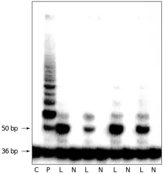
To demonstrate whether telomerase activity is involved in psoriasis skin, we assayed the telomerase activity in lesional and nonlesional skins of psoriasis. Using TRAP assay, telom- erase activity was detected in 7 (70%) out of 10 lesional skins of psoriasis. None of the nonlesional skin had detectable telomerase activity. Telomerase activity of lesional skin was clearly increased compared with that of nonlesional skin,
but TRAP assay revealed only a few ladders of telomerase products in lesional skin of psoriasis, indicating a relatively low level of telomerase activity or dilution of telomerase activity by telomerase-negative cells (Fig. 1). As controls, 2 normal skins obtained from patients undergoing plastic surgery were tested for telomerase activity and telomerase activity was not detected.
Correlation between telomerase activity and Ki-67 expression in lesional skin of psoriasis
As we found that most psoriasis lesional skins had increased telomerase activity and some lesional skins did not show telomerase activity, we performed immunohistochemical staining using monoclonal antibody MIB1 in telomerase- positive and -negative lesional skins to determine whether telomerase activity correlates with the proliferative state of the lesional epidermal cells. Ki-67-positive cells were dis- tributed in the basal and a few suprabasal cells mostly along the lower parts of the rete ridges of the lesional epidermis (Fig. 2). Mean LI of Ki-67 expression was calculated as 19.9
±5.7% in telomerase-positive psoriasis lesions (n=7) and 10.1±2.8% in telomerase-negative psoriasis lesions (n=3).
LI of Ki-67 expression in telomerase-positive lesions was significantly higher than that in telomerase-negative lesions (p<0.05), suggesting a significant association between telomerase activity and Ki-67 expression in psoriasis lesions (Fig. 3).
50 bp
36 bp
Fig. 1. Telomerase activity is detected in lesional skin of psoria- sis, but nonlesional skin has barely detectable telomerase activi- ty. Bottom line represents 36 bp internal control (S-IC) and lad- ders represent 6 bp repeats starting with 50 bp. C, negative control; P, positive control; L, lesional skin of psoriasis; N, non- lesional skin of psoriasis.
C P L N L N L N L N
Suppression of telomerase activity in HaCaT cells by RA
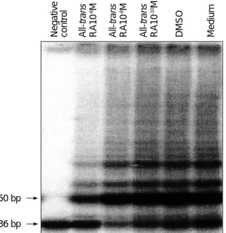
To reveal whether RA can inhibit telomerase activity, HaCaT cells which are known to express high levels of telomerase activity (13), were treated with RA and assessed for telomerase activity. Telomerase activity was markedly down-regulated in HaCaT cells treated with varying con- centrations of RA for 5 days (Fig. 4). A decline of telom-
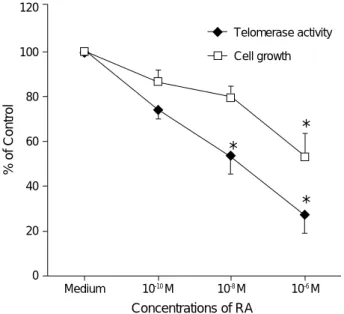
erase activity in RA-treated versus untreated cells was observed in a dose-dependent manner as follows: relative telomerase activity (mean±SD) was 74.1±3.2% at 10-10M of RA, 54.1±9.2% at 10-8M of RA, and 27.2±7.2% at 10-6M of RA compared to the untreated cells (Fig. 5). Sig- nificant suppression of telomerase activity in HaCaT cells
A B
Fig. 2.Telomerase activity-positive lesional skin of psoriasis (A) reveals increased expression of Ki-67, keratinocyte proliferation mark- er, compared to telomerase activity-negative lesional skin of psoriasis (B). Note that Ki-67-positive cells are predominantly localized in the basal and first few suprabasal layers (immunohistochemical stain,×200).
Telomerase Activity
Fig. 3. Ki-67 labelling index is significantly higher in telomerase activity-positive psoriasis lesional skin than in telomerase acti- vity-negative psoriasis lesional skin (*p<0.05).
Ki-67 Labelling Index (%)
30
25
20
15
10
5
0
Positive
*
Negative 50 bp
36 bp
Fig. 4. Suppression of telomerase activity in HaCaT cells treated with varying concentrations of retinoic acid as measured by the TRAP method. Telomerase activity declines in a dose-dependent manner. Bottom line represents 36 bp internal control (S-IC).
Negative control All-trans RA10-6M All-trans RA10-8M All-trans RA10-10M DMSO Medium
was observed at 10-8M and 10-6M of RA (p<0.05).
Correlation between inhibition of telomerase activity and cell proliferation by RA
To investigate whether the decreased telomerase activity observed during the treatment with RA correlates with in- hibition of cell proliferation by RA, MTT assay was per- formed. MTT assay showed a dose-response relationship for inhibition of HaCaT cells proliferation by RA concentrations of 10-10, 10-8, and 10-6M. HaCaT cells showed a decrease of proliferation during the 5 days of exposure to RA, and rela- tive proliferative activity (mean±SD) was 87.5±5.4% at 10-10M of RA, 80.3±5.6% at 10-8M of RA, and 54.3± 8.9% at 10-6M of RA compared to the untreated cells (Fig.
5). Significant inhibition of proliferation was attained at 10-6M of RA (p<0.05). The lesser degree of decline in telom- erase activity at the lowest RA dose for the inhibition of proliferation and the higher degree of decline at the highest dose of RA suggest a link between the proliferation and telomerase regulation.
DISCUSSION
Telomerase activity has been shown to be specifically ex- pressed in most human cancers and immortalized cell lines (1, 14) but is lacking in normal somatic cells except for
germline cells of testis or ovary, and hemopoietic stem cells (6, 15), which have been shown to express low levels of telomerase activity. Besides these cells, telomerase activity was demonstrated in proliferative basal layer of epidermis (10), endometrial tissue during the proliferative phase of the menstrual cycle (16), and highly proliferative normal oral mucosa (17). Telomerase activity was also detected in normal proliferating human uroepithelial, mammary, and prostate epithelial cultures, but not in uncultured or senes- cent cells (11). Detection of telomerase activity in these nor- mal proliferating somatic cells suggests that telomerase activity is associated not only with tumorigenic transforma- tion but also with the proliferation state of the cells (11). In addition, telomerase activity is increased in blood lympho- cytes from atopic dermatitis patients and correlates with cellular proliferation (9). In normal tissues like fibroblasts in which proliferation is generally absent, telomerase activity to maintain the tissue integrity is not required (10). Recent studies also strongly suggest that telomerase activation may be responsible in part for some of nonmalignant prolifera- tive skin diseases (18-20). The level of telomerase activity in nonmalignant skin diseases was much lower than that of malignant skin tumors (20). This can be interpreted as that, unlike malignant tumors in which most of cells express telomerase activity, only a subset of cells in nonmalignant proliferative diseases express telomerase activity, because the level of telomerase activity can be influenced by the propor- tion of telomerase-positive cells in the tissues, namely the dilution effect by the telomerase-negative cells or stroma (10). Thus, it may be speculated that the detection of the low level of telomerase activity in lesional skin of psoriasis, but not in normal skin in the present study results from selective expansion of a subset of cells expressing telomerase activity.
For cell proliferation in the epidermis, the transition from quiescent (G0/early G1 state) stem cell to proliferative tran- sient amplifying cell is required (7). In psoriasis, six- to seven-fold more stem cells than in normal epidermis have emerged from its normal G0 state into active cell cycle state to become transient amplifying cells (12). In addition, it has been suggested that telomerase is not an epidermal stem cell marker and the more actively proliferating transient amplifying cells exhibit more telomerase activity than epi- dermal stem cells (21). Therefore, telomerase activity in psoriasis observed in this study may result not only from stem cells but also from selective expansion of telomerase- positive cells such as expansion of transient amplifying cells.
In situ hybridization study to detect human telomerase RNA (hTR) also revealed moderate to strong levels of hTR in cells of the expanded rete ridges of active psoriasis and it was suggested that the increased hTR expression in supra- basal epidermal cells could be due to the migration of expanded proliferating keratinocytes into the suprabasal compartment (19). Another possible candidate responsible
Concentrations of RA
Fig. 5. Relative telomerase activity and cell proliferation by MTT assay in varying doses of retinoic acid (RA)-treated and control HaCaT cells. Inhibition of telomerase activity in HaCaT cells treated with RA for 5 days is in parallel with inhibition of cell pro- liferation (*p<0.05). Relative telomerase activity during RA treat- ment is expressed as a percentage of the telomerase activity detected in untreated cells.
% of Control
120
100
80
60
40
20
0
Medium 10-10 M 10-8 M 10-6 M
*
*
*
Telomerase activity Cell growth
for telomerase activity in psoriasis lesion is leukocyte infil- trates in the skin. Leukocytes have been shown to constitu- tively express low levels of telomerase activity (6), which can be increased with antigen stimulation (6, 20), and are known to play a critical pathogenic role in psoriasis. There- fore, the increased level of telomerase activity observed in psoriasis may be in part due to telomerase activity expressed by leukocytes themselves (19, 20). However, the levels of telomerase activity in lesional skin of psoriasis did not cor- relate with the degree of inflammatory cell infiltrate (20) and hTR was not expressed in infiltrating lymphocytes in active psoriasis (19).
Immunohistochemistry using monoclonal antibody to Ki-67 nuclear antigen is a useful method for the detection of proliferating cells in tissue samples (22). As Ki-67 expres- sion correlates well with growth fraction of cells and rapidly proliferating cells require telomerase in order to maintain chromosomal stability, telomerase expression might corre- late with Ki-67 expression. Mokbel et al. (23) reported that telomerase activity was significantly associated with Ki-67 expression in breast cancer, suggesting an association with cellular proliferation. Poremba et al. (24) also reported a positive correlation between the mean value of MIB1 immu- noreactivity and telomerase levels. The MIB1 proliferation index ranged from 12% for low-telomerase tumors to 63%
for high-telomerase tumors. Malignant melanomas also showed high telomerase activities correlated with cellular proliferation as measured by Ki-67 index (2, 25). In addi- tion, the distribution of hTR and Ki-67 was also similar in newborn foreskins, basal cell carcinomas, and squamous cell carcinomas, suggesting that the expression of hTR correlates with the proliferative state (19). It is known that telomerase activity (26) and expression of Ki-67 antigen (22) are high in both S and M phases of cell cycle. Therefore, in telom- erase-negative psoriasis lesion with a low level of Ki-67 expression shown in the present study, there is a possibility that epidermal cells bearing proliferative potential might be mainly in resting G0/G1 phase at the time of examination, and thus might exhibits little telomerase activity and not express Ki-67 antigen.
RA is a known modulator of cell proliferation and differ- entiation. In several cell lines, RA inhibited telomerase activi- ty by inducing cellular differentiation and inhibiting cell proliferation (27, 28). In case of HaCaT cells, treatment with higher doses (above 10-7to 10-6M) of RA significantly reduced cellular growth by increasing premature shedding of cells into the culture medium and by inducing an inhibi- tion of differentiation (29, 30). This resulted from a marked reduction in the number of desmosomes with complete dis- appearance of their ultrastructual components (30). These results suggest that inhibition of telomerase activity in Ha CaT cells by RA, unlike in other immortal cell lines in which RA-induced differentiation resulted in a decrease of telomerase activity (27, 28), be related to the inhibition of
proliferation but not to the induction of differentiation. In the present study, to further analyze the relationship between cell proliferation and telomerase activity, HaCaT cells grown with RA were tested by MTT assay. The inhibition of pro- liferation by RA closely paralleled the inhibition of telom- erase activity by this agent, further suggesting that telom- erase expression be linked to cell proliferation. Therefore, it may be speculated that one possible mechanism of epider- mal hyperplasia in psoriasis might be the increased telom- erase activity in psoriatic epidermis, and RA exerts its anti- proliferative activity by suppressing telomerase activity.
The exact biological significance of telomerase activity in psoriasis remains to be determined. Further studies on the regulation of the telomerase are needed to understand the cellular factors involved in controlling telomerase activity.
A better understanding of these factors will be important to provide a new insight on the therapy of this chronically an- noying disease.
REFERENCES
1. Shay JW, Bacchetti S. A survey of telomerase activity in human cancer. Eur J Cancer 1997; 33: 787-91.
2. Rudolph P, Schubert C, Tamm S, Heidorn K, Hauschild A, Michal- ska I, Majewski S, Krupp G, Jablonska S, Parwaresch R. Telom- erase activity in melanocytic lesions; a potential marker of tumor biology. Am J Pathol 2000; 156: 1425-32.
3. Yoshida K, Sugino T, Tahara H, Woodman A, Bolodeoku J, Nar- gund V, Fellows G, Goodison S, Tahara E, Tarin D. Telomerase activity in bladder carcinoma and its implications for noninvasive diagnosis by detection of exfoliated cancer cells in urine. Cancer 1997; 79: 362-9.
4. Hiyama E, Hiyama K, Yokoyama T, Matsuura Y, Piatyszek MA, Shay JW. Correlation of telomerase activity level with human neu- roblastoma outcome. Nature Med 1995; 1: 249-55.
5. Dalbagni G, Han W, Zhang ZF, Cordon-Cardo C, Saigo P, Fair WR, Herr H, Kim N, Moore MA. Evaluation of the telomeric repeat amplification protocol (TRAP) assay for telomerase as a diagnostic modality in recurrent bladder cancer. Clin Cancer Res 1997; 3: 1593-8.
6. Hiyama K, Hirai Y, Kyoizumi S, Akiyama M, Hiyama E, Piatyszek MA, Shay JW, Ishioka S, Yamakido M. Activation of telomerase in human lymphocytes and hematopoietic progenitor cells. J Immunol 1995; 155: 3711-5.
7. Greider CW. Telomerase activity, cell proliferation, and cancer.
Proc Natl Acad Sci USA 1998; 95: 90-2.
8. Holt SE, Wright WE, Shay JW. Regulation of telomerase activity in immortal cell lines. Mol Cell Biol 1996; 16: 2932-9.
9. Wu K, Volke A, Lund M, Bang K, Thestrup-Pederson K. Telom- erase activity is spontaneously increased in lymphocytes from patients with atopic dermatitis and correlates with cellular proliferation. J Dermatol Sci 1999; 22: 24-30.
10. Harle-Bachor C, Boukamp P. Telomerase activity in the regenera-
tive basal layer of the epidermis in human skin and in immortal and carcinoma-derived skin keratinocytes. Proc Natl Acad Sci USA 1996;
93: 6476-81.
11. Belair CD, Yeager TR, Lopez PM, Reznikoff CA. Telomerase activity: a biomarker of cell proliferation, not malignant transfor- mation. Proc Natl Acad Sci USA 1997; 94: 13677-82.
12. Bata-Csorgo Z, Hammerberg C, Voorhees JJ, Cooper KD. Flow cytometric identification of proliferative subpopulations within nor- mal human epidermis and the localization of the primary hyperpro- liferative population in psoriasis. J Exp Med 1993; 178: 1271-81.
13. Kallassy M, Martel N, Damour O, Yamasaki H, Nakazawa H.
Growth arrest of immortalized human keratinocytes and suppres- sion of telomerase activity by p21WAF1 gene expression. Mol Car- cinog 1998; 21: 26-36.
14. Kim NW, Piatyszek MA, Prowse KR, Harley CB, West MD, Ho PL, Coviello GM, Wright WE, Weinrich SL, Shay JW. Specific association of human telomerase acitivity with immortal cells and cancer. Science 1994; 266: 2011-5.
15. Wright W, Piatyszek M, Rainey W, Byrd W, Shay JW. Telomerase activity in human germline and embryonic tissues and cells. Dev Genet 1996; 18: 173-9.
16. Kyo S, Takakura M, Kohama T, Inoue M. Telomerase activity in human endometrium. Cancer Res 1997; 57; 610-4.
17. Kannan S, Tahara H, Yokozaki H, Mathew B, Nalinakumari KR, Nair MK, Tahara E. Telomerase activity in premalignant and malig- nant lesions of human oral mucosa. Cancer Epiderm Biomarkers Prevent 1997; 6: 413-20.
18. Parris CN, Jezzard S, Silver A, MacKie R, McGregor JM, Newbold RF. Telomerase activity in melanoma and non-melanoma skin can- cer. Br J Cancer 1999; 79: 47-53.
19. Ogoshi M, Le T, Shay JW, Taylor RS. In situ hybridization analysis of the expression of human telomerase RNA in normal and patho- logic conditions of the skin. J Invest Dermatol 1998; 110: 818-23.
20. Taylor RS, Ramirez RD, Ogoshi M, Chaffins M, Piatyszek MA, Shay JW. Detection of telomerase activity in malignant and nonma- lignant skin conditions. J Invest Dermatol 1996; 106: 759-65.
21. Bickenbach JR, Vormawald-Dogan V, Bachor C, Bleuel K, Schanapp G, Boukamp P. Telomerase is not an epidermal stem cell marker
and is downregulated by calcium. J Inves Dermatol 1998; 111: 1045- 52.
22. Cattoretti G, Becker MH, Key G, Duchrow M, Schluter C, Galle J, Gerdes J. Monoclonal antibodies against recombinant parts of the Ki-67 antigen (MIB1 and MIB3) detect proliferating cells in mic- rowave-processed formalin-fixed paraffin sections. J Pathol 1992;
168: 357-63.
23. Mokbel K, Parris CN, Ghilchik M, Williams G, Newbold RF. The association between telomerase, histopathological parameters, and Ki-67 expression in breast cancer. Am J Surg 1999; 178: 69-72.
24. Poremba C, Bocker W, Willenbring H, Schafer KL, Otterbach F, Burger H, Diallo R, Dockhorn-Dworniczak B. Telomerase activity in human proliferative breast lesions. Int J Oncol 1998; 12: 641-8.
25. Miracco C, Pacenti L, Santopietro R, Biagioli M, Fimiani M, Perotti R, Rubegni P, Pirtoli L, Luzi P. Detection of telomerase activity and correlation with mitotic and apoptotic indices, Ki-67 and expression of cyclins D1 and A in cutaneous melanoma. Int J Cancer 2000; 88:
411-6.
26. Weng NP, Levine BL, June CH, Hodes RJ. Regulated expression of telomerase activity in human T lymphocyte development and activa- tion. J Exp Med 1996; 183: 2471-9.
27. Albanell J, Han W, Mellado B, Gunawardane R, Scher HI, Dmitro- vsky E, Moore MA. Telomerase activity is repressed during differ- entiation of maturation-sensitive but not resistant human tumor cell lines. Cancer Res 1996; 56: 1503-8.
28. Sharma HW, Sokolonski JA, Perez JR, Maltese JY, Sartorelli AC, Stein CA, Nichols G, Khaled Z, Telang NT, Narayanan R. Differen- tiation of immortal cells inhibits telomerase activity. Proc Natl Acad Sci USA 1995; 92: 12343-6.
29. Breitkreutz D, Stark HJ, Plein P, Baur M, Fusenig NE. Differential modulation of epidermal keratinization in immortalized (HaCaT) and tumorigenic human skin keratinocytes (HaCaT-ras) by retinoic acid and extracellular Ca2+. Differentiation 1993; 54: 201-17.
30. Wanner R, Wolff B, Glowacki F, Kolde G, Wittig B. The loss of desmosomes after retinoic acid treatment results in an apparent inhibition of HaCaT keratinocyte differentiation. Arch Dermatol 1999; 291: 346-53.