SURFACE MICROGROOVES OF THIRTY MICROMETERS IN WIDTH ON TITANIUM SUBSTRATA ENHANCE PROLIFERATION AND ALTER GENE EXPRESSION OF CULTURED
HUMAN GINGIVAL FIBROBLASTS
Suk-Won Lee, D.D.S., M.S.D., Ph.D.1, Su-Yeon Kim, M.S., Ph.D.2, Keun-Woo Lee, D.D.S., M.S.D., Ph.D.3
1Department of Biomaterials & Prosthodontics, Dental Hospital, Kyunghee University
East-West Neo Medical Center 2Research Institute of Medical Science, St. Vincent’s Hospital
3Department of Prosthodontics, Collage of Dentistry, Yonsei University
Statement of problem.Surface microgrooves on Ti substrata have been shown to alter the expression of genes responsible for various biological activities of cultured fibroblasts.
However, their effect on enhancing cell proliferation is not yet clear.
Purpose.The purpose of this study was to determine the dimension of surface microgrooves on Ti substrata that enhances proliferation and alters gene expression of cultured human gin- gival fibroblasts.
Material and methods.Commercially pure Ti discs with surface microgrooves of monotonous 3.5 μm in depth and respective 15 and 30 μm in width were fabricated using photolithography and used as the culture substrata in the two experimental groups in this study (TiD15 and TiD30), whereas the smooth Ti was used as the control substrata (smooth Ti group). Human gingival fibroblasts were cultured on the three groups of titanium substrata and the proliferation, DNA synthesis, and gene expression of theses cells were analyzed and compared between all groups using XTT assay, BrdU assay, and reverse transcriptase-polymerase chain reaction (RT- PCR), respectively.
Results.From the XTT assay at 48 h incubation, the proliferation of human gingival fibrob- lasts in TiD30 was significantly enhanced compared to that in smooth Ti and TiD15. The results from the BrdU assay showed that, at 24 h incubation, the DNA synthesis was significantly enhanced in TiD30 compared to that in smooth Ti. In RT-PCR, increase in the expression of PCR tran- scripts of fibronectin, CDK6, p21cip1genes was noted at 48h incubation.
Conclusion.Surface microgrooves 30 μm in width and 3.5 μm in depth on Ti substrata enhance proliferation and alter gene expression of cultured human gingival fibroblasts.
Key Words
Titanium substrata, Microgroove, Fibroblast, Proliferation, Gene expression
J Korean Acad Prosthodont : Volume 45, Number 6, 2007
※This study was supported by St. Vincent’s Hospital Research Grants in 2006.
T
o enhance interactions between titanium (Ti) oral implants and the surrounding gingival soft tissues, the effects of Ti-surface microgrooves on cell behavior in vitro have extensively been investigated. Microgrooves of 1-10 μm in width on Ti substrata were verified to induce changes in morphology1, cell-substratum adhesion2, and gene expression3of cultured connective tissue cells. Fibroblasts grown on such substrata com- pared to those on the smooth ones were report- ed to be significantly elongated and orientated along the grooves, leading to an increase in the amount of fibronectin3or alterations in the expres- sion of numerous genes responsible for various biological activities4. So far, only a few studies compared the proliferating activity of fibrob- lasts on various dimensions of microgrooves.Majority of these in vitro studies used substrata with narrow grooves of 1-10 μm in width, and in contrast to the effective increase in the rate of cell orientation, either the presence of surface microgrooves or groove dimensions were verified to increase the proliferating activity of adhered fibroblasts5,6,7.
In this study, we hypothesized that surface microgrooves of appropriate depth and extensive width on Ti substrata that enable the cells to readily descend into themselves, would alter proliferation and gene expression of cultured human gingival fibroblasts. The purpose of this study was to determine the dimension of surface microgrooves on Ti substrata that enhances pro- liferation and, at the same time, alters gene expression of cultured human gingival fibroblasts.
MATERIAL AND METHODS CELL CULTURE
Healthy gingival tissues were obtained from patients who underwent oral surgery for remov-
ing impacted wisdom teeth at St. Vincent’s Hospital Department of Dentistry. In all cases, tis- sues were obtained from subjects following informed consent as prescribed in an approved St.
Vincent’s Hospital Institutional Review Board (IRB) protocol. Tissues were incubated for 16-22 h in Hank’s balanced salt solution (HBSS, Gibco BRL, Grand Island, NY, USA) at 4℃ for the pur- pose of separating connective tissue from epithe- lium. Obtained connective tissues were cut into small pieces and placed in Petri dishes in Dulbecco’
s modified Eagle’s medium (DMEM, Gibco BRL, Grand Island, NY, USA) supplemented with penicillin G sodium (50 IU/ml), streptomycin sulfate (50 mg/ml), and amphotericin B and were kept overnight at 4℃. Cells or explants were washed 3 times in phosphate-buffered salines (PBS, Gibco BRL, Grand Island, NY, USA) and suspended in DMEM supplemented with 10
% fetal bovine serum (FBS, Sigma-Aldrich Co., St.
Louis, MO, USA) and antibiotics. The composition and concentration of the solution were main- tained to be used as the culture medium in every experiment in this study (DMEM supplemented with 10% FBS and antibiotics). Suspended fibrob- lasts were seeded into a T-75 flask (enzymatic dis- sociation) and incubated in a humidified incubator at 37℃with 5% CO2in 95% air. When cells reached 80% confluence (about once per week), they were removed and suspended using a trypsin-EDTA solution (0.25% trypsin and 0.1%
glucose dissolved in 1 mM of EDTA-saline, Sigma-Aldrich Co., St. Louis, MO, USA), washed, centrifuged and reseeded. The culture medium was changed every second day after seeding. Human gingival fibroblasts with 3rd-4th passage were used in all experiments in this study.
FABRICATION OF TITANIUM SUBSTRATA
Commercially pure titanium (Ti) discs were
mechanically polished to obtain a finish surface with Ra ≤ 0.15 μm, and used as the culture sub- strata in the control groups, smooth Ti, in this study. The microgrooved Ti substrata were fab- ricated with photolithography (MEMSware Inc., Kwangju, Gyoenggi, Korea). Microgrooves were designed to have an equal depth of 3.5 μm and widths of 15 and 30 μm, respectively (Fig. 1).
XTT ASSAY
The floors of 24-well plates were removed and the remaining plastic cylinders were attached to the fabricated surfaces of the 25 mm-diameter Ti discs using a silicone bonding agent. As a result, a total of eighteen 96-well Ti substrata were prepared and divided into the three groups of smooth Ti, TiD15, and TiD30. Cultured human gingival fibroblasts were trypsinized and simul- taneously plated on the 24-well Ti substrata at a cell population density of 1×10⁴cells/ml in DMEM supplemented with 10% FBS and antibi- otics. Cells were incubated in a humidified incu- bator at 37℃ with 5% CO2in 95% air for 24 and 48 h. In all groups, the viability and proliferation of fibroblasts was determined by XTT assay (Cell
Proliferation Kit II, Roche Applied Science, Mannheim, Germany). In brief, XTT labeling reagent (soium 3’-[1-[(phenylamino)-carbonyl]- 3, 4-tetrazolium]-bis(4-methoxy-6-nitro)benzene- sulfonic acid hydrate) and electron coupling reagent (N-methyl dibenzopyrazine methyl sul- fate, PMS in PBS) were thawed. Each vial was thor- oughly mixed and a clear solution was obtained.
XTT labeling mixture was prepared by mixing 50 μl of XTT labeling reagent and 1 μl of electron cou- pling reagent. 50 μl of XTT labeling mixture was added per well and incubated for 4 h in a humid- ified incubator at 37℃ with 5% CO2in 95% air. In all groups, formazan products were transferred to 96-well plates and the absorbance was measured using ELISA analyzer (Spectra MAX 250, Molecular Devices Co., Sunnyvale, CA, USA) at 470 nm with a reference wavelength at 650 nm.
BrdU ASSAY
Human gingival fibroblasts were plated on the 96-well Ti substrata at a cell population den- sity of 3×10³cell/ml and incubated in a humid- ified incubator at 37℃ with 5% CO2in 95% air for 24 h. In all groups, 1 ml of BrdU labeling reagent (1000 conc., 10 mM 5-bromo-2’-deoxyuridine in PBS, pH 7.4, Roche Applied Science, Mannheim, Germany) was added to each well and the cells were reincubated for 2 h at 37℃. During this labeling period, the pyrimidine analogue BrdU was incorporated in place of thymidine into the DNA of proliferating cells. After removing the cul- ture medium, the cells were fixed and the DNA was denatured in one step for 30 min at room tem- perature by adding 200 ml of FixDenat (Roche Applied Science, Mannheim, Germany) to improve the accessibility of the incorporated BrdU for detection by the antibody. After the removal of FixDenat, the anti-BrdU-POD (monoclonal anti- body from mouse-mouse hybrid cells conjugat- Fig. 1.Fabricated surfaces of the Ti substrata with
microgrooves of 15 μm (left) and 30 μm (right) in width and an equal depth of 3.5 μm.
ed with peroxidase, Roche Applied Science, Mannheim, Germany) working solution was added to each well and left at room temperature for 90 min. During this period, the anti-BrdU-POD bound to the BrdU incorporated in newly syn- thesized, cellular DNA and the immune com- plexes were detected by the subsequent sub- strate reaction. In all groups, the reaction products were transferred to 96-well plates and the absorbance was measured using ELISA analyz- er (Spectra MAX 250, Molecular Devices Co., Sunnyvale, CA, USA) at 370 nm.
RT-PCR
Cultured human gingival fibroblasts (3rd-4th pas- sage) were trypsinized and plated on the 24- well Ti substrata of smooth Ti, TiD15, and TiD30 at a cell population density of 1×10⁴cells/ml in DMEM supplemented with 10% FBS and antibi- otics. At 48 h plating and incubation, expres- sion of FN (fibronectin), RhoA (an Rho GTPase family member), TGF-βR-II (type II transforming growth factor (TGF-)βreceptor), FGFR1 (fibrob- last growth factor receptor 1), CDK4 (cyclin-
dependent kinase 4), CDK6, p27kip1(cyclin-depen- dent kinase inhibitor 1B), and p21cip1(cyclin- dependent kinase inhibitor 1A) genes were ana- lyzed in reverse transcriptase-polymerase chain reaction (RT-PCR) (Table I). The PCR primer of β- actin was used as the housekeeping gene.
STATISTICAL ANALYSIS
Experiments were done independently in three fold. One-way analysis of variance (ANOVA) was used to compare the mean values of the data between the groups of smooth Ti, TiD15, and TiD30 (p<0.05).
RESULTS XTT ASSAY
In ANOVA, the mean OD values of the formazan absorbance at 48 h incubation were significantly different between and within all groups (p<0.05).
According to the data using the Ti discs with various dimensions of surface microgrooves as cul- ture substrata, the results from the XTT assay were Table I. Gene-Specific Primers used in RT-PCR
Target Sense Antisense Bp
FN8 5’-CGAAATCACAGCCAGTAG-3’ 5’-ATCACATCCACACGGTAG-3’ 639
RhoA9 5’-CTCATAGTCTTCAGCAAGGACCAGTT-3’ 5’-ATCATTCCGAAGATCCTTCTTATT-3’ 310 TGF-βR-II10 5’-CGCTTTGCTGAGGTCTATAAGGCC-3’ 5’-GATATTGGAGCTCTTGAGGTCCCT-3’ 395 FGFR111 5’-CCCTGGAAGAGAGGCCGGCAGTGATGAC-3’ 5’-GGTTTGCCTAAGAC AGTCTGTCCCG-3’ 372 CDK412 5’-CCAAAGTCAGCCAGCTTGACTGTT-3’ 5’-CATGTAGACCAGGACCTAAGGACA-3’ 193 CDK612 5’-TGATGTGTGCACAGTGTCACGAAC-3’ 5’-CTGTATTCAGCTCCGAGGTGTTCT-3’ 737 p2712 5’-AAACGTGCGAGTGTCTAACGGGA-3’ 5’-CGCTTCCTTATTCCTGCGCATTG-3’ 454 p2112 5’-AGTGGACAGCGAGCAGCTGA-3’ 5’-TAGAAATCTGTCATGCTGGTCTG-3’ 380 β-actin 5’-ATCGTGGGCCGCCCTAGGCA-3’ 5’-TGGCCTTAGGGTTCAGAGGGG-3’ 345 FN: fibronectin, FGFR1: fibroblast growth factor receptor 2, TGF-βR-II: type II transforming growth factor (TGF-) βreceptor, CDK: cyclin-dependent kinase, p27: cyclin-dependent kinase inhibitor 1B, p21: cyclin-dependent kinase inhibitor 1A
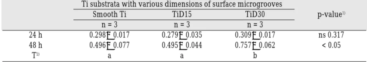
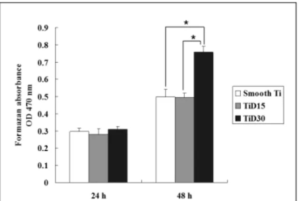
significantly related between those obtained at 48 h incubation. Multiple comparison of the fibrob- last proliferation data from the XTT assay at 48 h incubation showed the mean OD value of TiD30 to be significantly greater compared to that of smooth Ti and TiD15 (p<0.05) (Table II and Fig.
2). All other comparisons between groups were not statistically significant.
BrdU ASSAY
In ANOVA, the mean OD values of the BrdU absorbance at 24 h incubation were significantly different between and within all groups (p<0.05).
According to the data using the Ti discs with
various dimensions of surface microgrooves as cul- ture substrata, the results from the BrdU assay were significantly related between those obtained at 24 h incubation. Multiple comparison of the fibrob- last viability and proliferation data from the BrdU assay at 24 h incubation showed the mean OD value of TiD30 to be significantly greater compared to that of smooth Ti (p<0.05) (Table III and Fig. 3). All other comparisons between groups were not statistically significant.
RT-PCR
Increase in the expression of transcripts in TiD30 was noted with the genes encoding FN, Table II. Comparison of fibroblast proliferation on Ti substrata at 24 and 48 h incubation by structural dimensions of surface microgrooves using XTT assay
Ti substrata with various dimensions of surface microgrooves
Smooth Ti TiD15 TiD30 p-value1)
n = 3 n = 3 n = 3
24 h 0.298±0.017 0.279±0.035 0.309±0.017 ns 0.317
48 h 0.496±0.077 0.495±0.044 0.757±0.062 < 0.05
T2) a a b
1) Statistical significances were tested by one-way analysis of variance among groups.
2) The same letters indicate non-significant difference between groups based on Tukey’s multiple comparison tests.
TiD: Titanium Disc
Table III. Comparison of fibroblast proliferation on Ti substrata at 24 h incubation by struc- tural dimensions of surface microgrooves using BrdU assay
Ti substrata with various dimensions of surface microgrooves
Smooth Ti TiD15 TiD30 p-value1)
n = 3 n = 3 n = 3
24 h 0.152±0.013 0.199±0.033 0.220±0.017 < 0.05
T2) a a,b b
1) Statistical significances were tested by one-way analysis of variance among groups.
2) The same letters indicate non-significant difference between groups based on Tukey’s multiple comparison tests.
TiD: Titanium Disc
CDK6, and p21cip1at 48 h incubation compared to that in the groups of smooth Ti or TiD15. On the other hand, decrease in p27kip1gene expression levels was noted in TiD30 compared to that in the groups of smooth Ti or TiD15. RhoA, TGF-βR-II, FGFR1 genes showed similar results in the expres- sion levels except, with the CDK4 gene, increase in the expression was obvious in TiD15 and TiD30 compared to the expression in the smooth Ti group (Fig. 4).
DISCUSSION
One of the most frequently used colorimetric assays, XTT, used in this study is a method well established in investigating the influence of a specific substance/material and/or process on cell survival. The results of the XTT assay for cell viability and proliferation are influenced not only by the number but also by the metabolic activ- ity of cells. From the XTT assay in this study, sur- face microgrooves of 30 μm in width and 3.5 μm Fig. 2.Effect of structural dimensions of surface microgrooves on human gingival fibroblast prolifer- ation on Ti substrata. The mean optical density (OD) values and standard deviations of formazan absorbance from XTT assay are presented.
*: significant difference between groups (p<0.05), OD: optical density, TiD: titanium disc
Fig. 3.Effect of structural dimensions of surface microgrooves on human gingival fibroblast viability and proliferation on Ti substrata. The mean optical den- sity (OD) values and standard deviations of BrdU absorbance are presented.
*: significant difference between groups (p<0.05), OD: optical density, TiD: titanium disc
Fig. 4.Analysis on expression of genes in RT-PCR. See text for description and Table I for abbreviations.
TiD: titanium disc, RT-PCR: reverse transcriptase- polymerase chain reaction.
in depth on Ti substrata were verified to signifi- cantly enhance the proliferation of cultured human gingival fibroblasts. This result is in con- trast to the result of a previous study that either the presence of surface microgrooves or groove dimensions were verified to increase the prolif- erating activity of adhered fibroblasts.5However, the authors used microgrooves of 1-10 μm in width, which were considered significantly nar- rower than the diameter of a single human fibrob- last. In a recent study analyzing on microgroove- related cell migration and alignment, the authors strongly implied that the width of 30 μm rough- ly represents the main diameter of the cells.13 The importance of the presence of cell contact on the bottom of the grooves in relation to contact guidance has previously been suggested.6It was concluded in the study that, at confluence, microgrooves with relatively wider grooves com- pared to the narrower ones were able to sup- port greater numbers of cells. The TiD30 sub- strata in this study were considered to have enabled the cells to contact the bottom of the grooves and supported the greater number of cells compared to that on the smooth Ti or the TiD15 substrata at 48 h incubation. However, we found no significant difference in the proliferation between groups at 24 h. To confirm this, we compared the amount of DNA synthesis between groups using BrdU assay at 24 h incubation.
BrdU assay was developed as a non-radioactive alternative to the [3H]-thymidine incorporation assay. It is a colorimetric immunoassay for the quantification of cell proliferation, based on the measurement of BrdU incorporation during DNA synthesis. The developed color and the absorbance values from the assay directly correlate to the amount of DNA synthesis and the number of proliferating cells. From the BrdU assay in this study, the amount of DNA synthesis of the cells was significantly increased on the TiD30
substrata compared to those on the other groups of substrata. Taken together, we suggest that surface microgrooves of 30 μm in width and 3.5 μm in depth on Ti substrata enhance the prolif- eration and increase the amount of DNA synthesis of cultured human gingival fibroblasts.
A linear linkage exists from the extracellular matrix (ECM) such as fibronectin outside of a cell to the actin cytoskeleton via integrin receptors. It is essential for an efficient signaling connection for cells to respond to extracellular cues. Integrin receptors can transduce signals alone or collab- oratively with other membrane receptors such as growth factor stimulation of receptor tyrosine kinases (RTKs) in an adhered cell.14Therefore, we evaluated the expression of the genes encoding FN (ECM), RhoA (RhoA GTPase-control of actin cytoskeleton), and TGF-βR-II/FGFR1 (RTKs) at 48 h incubation, at which the human gingival fibrob- lasts had been verified by the XTT assay to show significant difference in proliferation. We had no reason to confirm that the TiD30 substrata increased the expression levels of the genes except with the FN gene, increase in the expres- sion of transcripts was obvious. To confirm that the genes involved proliferation, namely, G1/S cell cycle progression, would show difference in the expression levels between groups, we evaluated the expression of the genes encoding CDK4/6 (effecter-molecules in G1/S cell cycle progression) and their inhibitors, p27kip1/p21cip1. The result suggested clearly that the TiD30 substrata increased the expression levels of the genes involved in G1/S cell cycle progression. However, the p21cip1 gene showed a different pattern of expression compared with that of the p27kip1 gene. The p21kip1 gene actually showed an identical expression profile to that of the FN gene or the CDK6 gene. A previous study demonstrated that up-regulation of the p21kip1 gene was noted in 3-dimensional (3D) cultures compared to the gene-expression regu-
lation in 2D cultures.15Taken together, we suggest that surface microgrooves of 30 μm in width and 3.5 μm in depth on Ti substrata increase the expression levels of the genes involved in G1/S cell cycle progression, ECM synthesis, and pos- sibly, the genes involved in 3D cultures.
CONCLUSION
Human gingival fibroblasts were plated and incubated on three groups of titanium discs as the smooth Ti substrata, the Ti substrata with microgrooves of 15 μm in width and 3.5 μm in depth, and the Ti substrata with microgrooves of 30 μm in width and 3.5 μm in depth. From the results of the proliferation analysis using XTT and BrdU assay, and the gene expression analysis in RT-PCR, it can be concluded that the Ti substrata with microgrooves of 30 μm in width and 3.5 μm in depth enhance proliferation and alter gene expression of cultured human gingival fibroblasts.
REFERENCES
1. den Braber ET, de Ruijter JE, Ginsel LA, von Recum AF, Jansen JA. Quantitative analysis of fibroblast morphology on microgrooved surfaces with various groove and ridge dimensions. Biomater 1996;17:2037-2044.
2. Walboomers XF, Ginsel LA, Jansen JA. Early spreading events of fibroblasts on microgrooved substrates. J Biomed Mater Res 2000;51:529-534.
3. Chou L, Firth JD, Uitto VJ, Brunette DM. Substratum surface topography alters cell shape and regu- lates fibronectin mRNA level, mRNA stability, secretion and assembly in human fibroblasts. J Cell Sci 1995;108:1563-1573.
4. Dalby MJ, Riehl, MO, Yarwood SJ, Wilkinson CD Curtis AS. Nucleus alignment and cell signaling in fibroblasts: response to a micro-grooved topography.
Exp Cell Res 2003;284:274-282.
5. den Braber ET, de Ruijter JE, Smits HT, Ginsel LA, von Recum AF, Jansen JA. Quantitative analy- sis of cell proliferation and orientation on sub-
strata with uniform parallel surface micro-grooves.
Biomater 1996;17:1093-1099.
6. Walboomers XF, Monaghan W, Curtis AS, Jansen JA. Attachment of fibroblasts on smooth and mi- crogrooved polystyrene. J Biomed Mater Res 1999;46:212-220.
7. Walboomers XF, Croes HJ, Ginsel LA, Jansen JA.
Contact guidance of rat fibroblasts on various implant materials. J Biomed Mater Res 1999;47:204- 212.
8. Zhang C, Meng XF, Zhu ZH, Yang X, Deng AG.
Role of connective tissue growth factor in plas- minogen activator inhibitor-1 and fibronectin ex- pression induced by transforming growth factor β 1 in renal tubular cells. Chinese Med J 2004;117:990- 996.
9. Moran CJ, Friel AM, Smith TJ, Cairns M, Morrison JJ. Expression and modulation of Rho kinase in hu- man pregnant myometrium. Molecular Human Reproduction Mol Hum Reprod. 2002;8:196-200.
10. Boume´diene K, Takigawa M, Pujol JP. Cell density- dependent proliferative effects of transforming growth factor (TGF)-beta 1, beta 2, and beta 3 in hu- man chondrosarcoma cells HCS-2/8 are associated with changes in the expression of TGF-beta receptor type I. Cancer Invest 2001;19:475-486.
11. Takayama S, Yoshida J, Hirano H, Okada H, Murakami S. Effects of basic fibroblast growth factor on human gingival epithelial cells. J Periodontol 2002;73:1467-1473.
12. Wong H, Riabowol K. Differential CDK-inhibitor gene expression in aging human diploid fibroblasts.
Exp Gerontol 1996;3:311-325.
13. Kaiser JP, Reinmann A, Bruinink, A. The effect of topographic characteristics on cell migration velocity.
Biomater 2006;27:5230-5241.
14. Lee JW, Juliano R. Mitogenic signal transduction by integrin- and growth factor receptor-mediated pathways. Mol Cells 2004;17:188-202.
15. Li S, Lao J, Chen BP, Li YS, Zhao Y, Chu J, Chen KD, Tsou TC, Peck K, Chien S. Genomic analysis of smooth muscle cells in 3-dimensional collagen matrix. FASEB J 2003;17:97-99.
Reprint request to:
KEUN-WOOLEE, D.D.S., M.S.D., Ph.D.
DEPARTMENT OFPROSTHODONTICS,COLLEGE OFDENTISTRY, YONSEIUNIVERSITY
134, SHINCHON-DONG,SEODAEMUN-GU,SEOUL,120-095, KOREA [email protected]