38
J. Appl. Biol. Chem. 52(1), 38-40 (2009)
Short Communication
Green Alga Ulva pertusa Inhibits Nitric Oxide and Prostaglandin-E
2Formation in Murine Macrophage RAW 264.7 Cells
Ji-Young Kim1, Dong Sam Kim1, Eun-Jin Yang1, Weon-Jong Yoon1, Jong Seok Baik1, Wook Jae Lee1, Nam Ho Lee2, and Chang-Gu Hyun1*
1Research Group for Cosmetic Materials, Jeju Biodiversity Research Institute (JBRI) and Jeju Hi-Tech Industry Development Institute (HiDI), Jeju 697-943, Korea
2Department of Chemistry, Cheju National University, Jeju 690-756, Korea
Accepted February 11, 2009
Key words:inflammation, nitric oxide, prostaglandin-E2, Ulva pertusa
Macrophages play an important role in inflammatory disease through the release of factors such as nitric oxide (NO), prostaglandins, and cytokines involved in the immune response (Hibbs et al., 1987; Palmer et al., 1988; Hseu et al., 2005).
Activated macrophages transcriptionally express inducible nitric oxide synthase (iNOS), which catalyzes the oxidative deamination of L-arginine to produce NO, and is responsible for the prolonged and profound production of NO. Overproduction of NO by iNOS can provoke deleterious consequences such as septic shock and inflammatory diseases (Xie and Nathan, 1994;
Titheradge, 1999; Zamora et al., 2000; Zhou et al., 2008).
Prostaglandins also play a major role as mediators of the inflammatory response. The rate-limiting enzyme in the synthesis of prostaglandins is cyclooxygenase (COX). Two isoforms of COX have been identified: COX-1 and COX-2.
COX-2 is induced by several stimuli, and is responsible for the production of large amounts of pro-inflammatory prostaglandins at the inflammatory site. On the basis of these observations, it has been hypothesized that inhibiting NO and prostaglandin production in macrophages could serve as the basis for the potential development of anti-inflammatory drugs.
Ulva pertusa, dominant seaweed in the coastal zone of Jeju Island, Korea, is distributed in various coastal waters including
the brackish waters of the inner bays. It often contributes to the formation of ‘green tides’, which cause ecological and indirect economic damages. Recently, there have been an increasing number of reports on the formation of huge masses of algae due to eutrophication (Hiraoka et al., 2004; McAvoy and Klug, 2005; Lin et al., 2008). These excessive masses largely consist of various species of green algae, and U. pertusa has been a representative case in Jeju Island since 2007. As a result, in some areas, the excessive growth of the algae impeded the growth of aquatic crops and affected the scenic beauty of the beaches. Therefore, it is worthwhile to determine how to make use of the ‘green tides’, especially U. pertusa, in an economically productive manner. This study was designed to estimate the anti- inflammatory effect of the solvent fraction constituents of U.
pertusa by measuring the production of NO and prostaglandin E2 (PGE2) in murine macrophage RAW 264.7 cells. In April 2008, specimens of U. pertusa were collected from Jeju Island.
A voucher specimen (JBRI-08025) has been deposited at the herbarium of Jeju Biodiversity Research Institute. The materials for extraction were cleaned, dried at room temperature for 2 weeks, and ground into a fine powder. The dried alga (50 g) was extracted with 80% ethanol (EtOH; 2 L) at room temperature for 24 h and then vacuum-evaporated. The evaporated EtOH extract (16 g) was suspended in water (2 L) and partitioned three times with solvents -n-hexane (2 L), dichloromethane (CH2Cl2; 2 L), ethyl acetate (EtOAc; 2 L), and butanol (BuOH; 2 L). The yields of these solvent partitions were as follows: n-hexane, 1.6 g;
CH2Cl2, 2.6 g; EtOAc, 0.5 g; BuOH, 1.5 g; and H2O, 9.6 g. NO is an endogenous free radical species and, in small amounts, is an important regulator of the physical homeostasis, whereas large amounts of NO has been closely correlated with the pathophysiology of a variety of diseases and inflammation. After exposure to inducers, such as lipopolysaccharide (LPS) from Gram-negative bacteria, inducible NOS (iNOS) can be induced in various cells, including macrophages, smooth muscle cells, and hepatocytes, to trigger cytotoxicity, tissue damage, inflammation sepsis, and stroke (Marletta, 1993; Jiang et al., 2006; Tung et al., 2008). Thus, measuring NO production could be a possible method for assessing the anti-inflammatory effects of seaweed extracts. The nitrite assay was performed to determine whether the solvent extracts of U. pertusa modulated the NO production from murine macrophage RAW264.7 activated by LPS (1µg/mL). NO production was expressed as the ratio of the amount of NO produced from the solvent fraction with LPS to that from LPS alone (Fig. 1). No basal NO production was observed in the incubation with only the crude extract from U. pertusa without LPS (data not shown). On the other hand, NO production in the cells incubated with LPS and
*Corresponding author
Phone: +82-64-720-2811; Fax: +82-64-720-2801 E-mail: [email protected]
doi:10.3839/jabc.2009.007
Anti-inflammatory activity of Ulva pertusa 39
the solvent fraction of the U. pertusa extracts decreased. The addition of the CH2Cl2 fraction (50µg/mL) to the medium with LPS largely inhibited the production of NO with ratio of 62.45 (Fig. 1). The number of viable activated macrophages was not altered by the solvent fractions as determined by lactate dehydrogenase (LDH) assays, indicating that the inhibition of NO synthesis by the solvent fractions was not simply due to cytotoxic effects. The CH2Cl2 fraction of U. pertusa (25 to 100µg/mL) inhibited NO production in a dose-dependent manner.
The activation of macrophages plays a critical role in the inflammatory process by releasing a variety of inflammatory mediators. In addition to NO production, PGE2 is also an important inflammatory mediator involved in pathogenesis.
Thus, determining the inhibitory effects on the abnormal accumulation of PGE2 is another method by which the anti- inflammatory effects of seaweed extracts could be assessed. To examine whether the extract and the solvent fractions of the U.
pertusa extract inhibit PGE2, the cells were activated with LPS (1µg/mL) and then incubated with the extract and the solvent
Fig. 1. Effects of U. pertusa extract and solvent fractions on the production of nitric oxide and cytotoxicity in RAW 264.7 cells.
Production of nitric oxide was assayed from the culture medium of cells stimulated with LPS (1µg/mL) in the presence of ethanol extract and solvent fractions of U. pertusa (A). NO production was determined using ELISA method. After the cells were treated with dichloromethane (B) fractions (25 to 100µg/mL), nitrite production by RAW 264.7 mouse macrophage cells was inhibited in a dose- dependent manner. Cytotoxicity was determined using LDH method.
Values are means±SEM of triplicate experiments. *p<0.05; **p<0.01.
Fig. 2. Inhibitory effects of ethanol extract and solvent fractions of U. pertusa on PGE2 production and COX-2 expression in RAW 264.7 cells. RAW 264.7 cells (1.5×10⁵ cells/mL) were stimulated by LPS (1µg/mL) with ethanol extract and solvent fractions (50µg/mL) of U. pertusa for 24 h. Supernatants were collected after 24 h, and PGE2 concentration of supernatants was determined using ELISA method (A). The COX-2 levels were determined by immunoblotting.
After the cells were treated with dichloromethane fraction (25 to 100
µg/mL), COX-2 expression by RAW 264.7 mouse macrophage cells was inhibited in a dose-dependent manner (B). Values are means±
SEM of triplicate experiments. *p<0.05; **p<0.01.
Fig. 3. Cell viability of HaCaT Cells treated with ethanol extract and solvent fractions. Ethanol extract and solvent fractions exhibited relatively low cytotoxicity, with cell viability almost 100% at 100µg/
mL (A). MTT assay was performed after incubation of HaCaT cells treated with various concentrations of CH2Cl2 fraction for 24 h at 37oC in a 5% CO2 atmosphere (B). Absorbance was measured at 570 nm with a spectrophotometer (Power Wave, Bio-tek, Winooski, VT).
40 Ji-Young Kim et al.
fractions of U. pertusa for 24 h. The CH2Cl2 (50µg/mL) fraction of U. pertusa exhibited reduction in PGE2 up to 79.02% (Fig.
2A). The level of COX-2 was detected by immunoblotting. The CH2Cl2 fraction of U. pertusa (25 to 100µg/mL) inhibited the level of COX-2, in a dose-dependent manner (Fig. 2B). We next examined the cytotoxic effects of the ethanol extract and the solvent fractions of U. pertusa on human keratinocyte HaCaT cells (Fig. 3). Whereas the ethanol extract and the solvent fractions of U. pertusa at concentrations lower than 100µg/mL showed almost 100% cell viability except for the CH2Cl2
fraction, which at 100µg/mL showed only about 80% cell viability. In the human dermal fibroblast cells, such as HaCaT cells, the ethanol extract and the solvent fractions of U. pertusa oil did not show significant cytotoxic effect (data not shown).
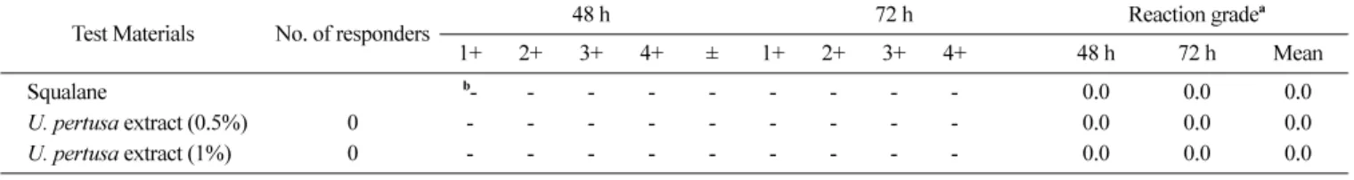
These data suggest that U. pertusa has a low cytotoxicity against the mammalian cells. Finally, to clinically evaluate the irritation effect of the U. pertusa extracts for applications on the human skin, a patch test invoving topical treatment of the U. pertusa extracts was performed. None of the 32 subjects experienced adverse reactions such as erythema, burning or pruritus based on the 48 and 72 h readings (Table 1). In conclusion, results of the present study demonstrated that U. pertusa extracts can reduce NO and PGE2 production and are safe for use as a possible anti- inflammatory agent for the management of the human skin health.
Acknowledgments
This research was supported by the Regional Technology Innovation Program (RTI 04-02-07) and the JEJU SEA-GREEN PROJECT, which are managed by the Ministry of Knowledge and Economy, Korea.
References
Hibbs Jr JB, Taintor RR, and Vavrin Z (1987) Macrophage cytotoxicity: Role for L-arginine deiminase and imino nitrogen
oxidation to nitrite. Science235, 473-476.
Hiraoka M, Ohno M, Kawaguchi S, and Yoshida G (2004) Crossing test among floating Ulvathalli forming ‘green tide’ in Japan. Hydrobiologia512, 239-245.
Hseu YC, Wu FY, Wu JJ, Chen JY, Chang WH, Lu FJ, Lai YC, and Yang HL (2005) Anti-inflammatory potential of Antrodia Camphorata through inhibition of iNOS, COX-2 and cytokines via the NF-kappaB pathway. Int Immunopharmacol 5, 1914- 1925.
Jiang JS, Shih CM, Wang SH, Chen TT, Lin CN, and Ko WC (2006) Mechanisms of suppression of nitric oxide production by 3-O-methylquercetin in RAW 264.7 cells. J Ethnopharmacol
103, 281-287.
Lin A, Shen S, Wang J, and Yan B (2008) Reproduction diversity of Enteromorpha prolifera. J Integr Plant Biol 50, 622-629.
Marletta MA (1993) Nitric oxide synthase structure and mechanism. J Biol Chem 268, 12231-12234.
McAvoy KM and Klug JL (2005) Positive and negative effects of riverine input on the estuarine green alga Ulva intestinalis (syn.
Enteromorpha intestinalis) (Linneaus). Hydrobiologia545, 1-9.
Palmer RMJ, Ashton DS, and Moncada S (1988) Vascular endothelial cells synthesize nitric oxide from L-arginine. Nature
333, 664-666.
Titheradge MA (1999) Nitric oxide in septic shock. Biochim Biophys Acta1411, 437-455.
Tung YT, Chua MT, Wang SY, and Chang ST (2008) Anti- inflammation activities of essential oil and its constituents from indigenous cinnamon (Cinnamomum osmophloeum) twigs.
Bioresour Technol99, 3908-3913.
Xie Q and Nathan C (1994) The high-output nitric oxide pathway:
role and regulation, J. Leukoc Biol 56, 576-582.
Zamora R, Vodovotz Y and Billiar TR (2000) Inducible nitric oxide synthase and inflammatory diseases. Mol Med 6, 347- Zhou HY, Shin EM, Guo LY, Youn UJ, Bae K, Kang SS, Zou LB,373.
and Kim YS (2008) Anti-inflammatory activity of 4- methoxyhonokiol is a function of the inhibition of iNOS and COX-2 expression in RAW 264.7 macrophages via NF- kappaB, JNK and p38 MAPK inactivation. Eur J Pharmacol
586, 340-349.
Table 1. Results of human skin primary irritation test (n=32)
Test Materials No. of responders 48 h 72 h Reaction gradea
1+ 2+ 3+ 4+ ± 1+ 2+ 3+ 4+ 48 h 72 h Mean
Squalane b- - - - - - - - - 0.0 0.0 0.0
U. pertusa extract (0.5%) 0 - - - - - - - - - 0.0 0.0 0.0
U. pertusa extract (1%) 0 - - - - - - - - - 0.0 0.0 0.0
aReaction grade=Σ[{Grade×no. of responders}/{4 (maximum grade)×32 (total subjects)}]×100×(1/2).
bNo reaction.
Thirty-two healthy female Korean subjects were selected based on inclusion and exclusion criteria, and written consent was obtained in each case. The average age was 39.94±6.81 years (range: 20 to 49). The subjects had no history of allergic contact dermatitis and had not used topical or systemic irritant preparations within the previous 1 month. The 80% ethanol extract of U. pertusa formulated with squalane (negative control) was prepared and applied at 0.5 and 1%. The patches (chambers) remained in place for 48 h. Once the patches were removed, the site was read 30 min and 24 h later;
the readings were scored according to the Cosmetic, Toiletry, and Fragrance Association (CTFA) guidelines.