10.3988/jcn.2011.7.1.10 J Clin Neurol 2011;7:10-18
A Common Pathogenic Mechanism Linking Type-2 Diabetes and Alzheimer’s Disease: Evidence from Animal Models
Sun Ah Park
Department of Neurology, Soonchunhyang University College of Medicine, Bucheon Hospital, Bucheon, Korea
Received August 26, 2010 Revised September 25, 2010 Accepted September 25, 2010 Correspondence Sun Ah Park, MD, PhD Department of Neurology, Soonchunhyang University College of Medicine, Bucheon Hospital, 1174 Jung-dong, Wonmi-gu, Bucheon 420-767, Korea Tel +82-32-621-5221 Fax +82-32-621-5016 E-mail [email protected]
The failure of large-scale drug trials targeting the amyloidogenic pathway in Alzheimer’s disease (AD) is increasing the need to identify a novel pathogenic mechanism. Studies finding a relation- ship between sporadic AD and type-2 diabetes mellitus (T2DM) are now receiving more atten- tion. The risk for developing both T2DM and sporadic AD increases exponentially with age, and having T2DM doubles the risk of developing AD. The postmortem brains of AD patients show altered activities of insulin receptors and downstream molecules, as well as reduced protein and mRNA levels of insulin. More-recent laboratory research using animal models has identified mechanisms that are shared by diabetes and AD. Exogenous application of streptozotocin, which disrupts systemic insulin secretion, results in insulin deficiency, increased tau phosphorylation, and cognitive impairments that can be reversed by exogenous insulin supplementation. Howev- er, AD pathology is more severe in T2DM animal models exhibiting hyperinsulinemia and insu- lin resistance, and this is not modulated by insulin. The symptoms of this AD pathology included increased tau phosphorylation at multiple sites, increased tau cleavage, and greater neuronal and synaptic damage, even with increased amyloid β protein production. It has therefore been sug- gested that hyperinsulinemia and insulin resistance represent major factors underlying AD in T2DM. A recent study involving cross-mating ob/ob and amyloid precursor protein transgenic mice provided evidence that T2DM and AD aggravate each other, and suggested that cerebral vessels constitute an important substrate that is commonly damaged by the two major disorders.
Given the evidence provided by animal models, further investigation of the mechanisms underly- ing T2DM in AD should help to identify potential treatment targets in AD.
J Clin Neurol 2011;7:10-18 Key Wordszz Alzheimer’s disease, animal model, diabetes, insulin resistance, mechanism.
Introduction
Alzheimer’s disease (AD), which is the most-prevalent neuro- degenerative disease, results in progressive cognitive dysfunc- tion and inability to perform complex daily activities. As a re- sult of the incapacity experienced by those with AD, a huge burden is inevitably placed on caregivers, which makes AD a major social problem.1 There is strong evidence from genetic studies of familial AD that amyloid β proteins, which are pro- duced by sequential cleavages of amyloid precursor proteins (APPs) by the β-site of the APP-cleaving enzyme (BACE), fol- lowed by γ-secretase, play central roles in AD.2 There have been repeated tests of therapeutic strategies directly targeting Aβ production, such as inhibiting or modulating BACE and γ- secretase, using aggregation blockers, clearance-enhancing,
and vaccinating with Aβ peptides or antibodies against vari- ous Aβ epitopes.3 However, in contrast to the results obtained by studies on transgenic animals with familial AD mutation, no evidence of the efficacy of these approaches has been produced in human AD patients.4 Therefore, it is highly likely that familial and sporadic AD are characterized by different ty- pes of pathogenesis.
Several studies have produced epidemiological, clinical, and pathological evidence of the relationship between AD and type-2 diabetes mellitus (T2DM). Longitudinal epidemiologi- cal studies have revealed that the risk of developing late-onset AD is 1.4-4.3 higher among those with T2DM,5-10 and that AD patients showed elevated plasma but lower cerebrospinal fluid (CSF) insulin levels, disrupting the relationship between pe- ripheral and central insulin.11 Furthermore, the postmortem
brains of individuals who had been diagnosed with AD showed decreased insulin, insulin-like growth factor 1 (IGF-1), and re- ceptor activities in proportion to the severity of disease.12 This relationship was greater in the elderly and in non-apolipopro- tein-E4 carriers.11 Therefore, the mechanism linking T2DM and AD is expected to provide an important clue toward eluci- dating the process underlying sporadic AD, especially in those AD patients not carrying the known major risk factor for spo- radic AD, apolipoprotein-E4. This paper reviews the evidence of the close relationship between T2DM and AD obtained by various animal studies, and summarize the proposed pathogen- ic mechanism shared by these two major disorders.
Studies in Diabetes Mellitus Models
(Table 1)Streptozotocin-injection model
Streptozotocin [STZ; 2-deoxy-2-(3-(methyl-3-nitrosoureido))- D-glucopyranose], a betacytotoxic drug, selectively destroys insulin-secreting pancreatic β cells and thereby causes type-1 diabetes mellitus (T1DM).13 Although the STZ-injection mu- rine model has been the most-widely used to investigate the re- lationship between diabetes mellitus (DM) and AD, studies re- lying on this model have focused on T1DM, which has pro- duced less evidence of a relationship with AD than has T2DM.
Nonetheless, increased tau phosphorylation with decreased binding to microtubules has been noted consistently in research using the STZ-injection murine model;14-16 this result has prob- ably been due to inhibited phosphatase activities rather than to enhanced kinase activities.14 This effect was substantially re- versed by supplying insulin. However, contradictory results have emerged with respect to Aβ pathology.
STZ itself cannot pass through the blood-brain barrier due to the absence of the STZ transporter GLUT2. Therefore, the sys- temic-injection model can reveal only the effect of systemic hypoinsulinemia on the brain. Direct injection of STZ into the brain has thus been attempted.
Inside the mammalian brain, GLUT2 is distributed heteroge- neously;17 therefore, intracerebroventricular (i.c.v.) injection of STZ can selectively decrease the level of insulin in the brain without disturbing systemic insulin and glucose levels. In ad- dition, the expression levels of insulin and IGF-1 receptors were reduced, showing that insulin signaling was impaired,18,19 which is similar to the condition observed in the postmortem brains of AD patients with so-called type-3 diabetes,20 characterized by simultaneously decreased insulin production and resistance to insulin reception in the brain. Although increased phosphoryla- tion of tau was noted consistently after i.c.v. injection of STZ, this phenomenon was present at more sites, inhibiting micro- tubule-binding activities. Overactivation of glycogen synthase
kinase-3β (GSK3β) and decreased O-linked N-acetylglucos- amine glycosylation (O-GlcNAcylation) resulting from im- paired insulin signaling have been found to be the primary causes of hyperphosphorylation of tau. In relation to Aβ pathol- ogy, the change in brain parenchyma was subtle, but increased Congo-red-positive aggregates were observed in brain capillar- ies, suggesting that Aβ-related impairment of the microcircula- tion is present in this animal model.
In summary, increased tau phosphorylation and microtubule instability resulting from decreased phosphatase activity has been noted consistently in studies using the STZ-injection mod- el, with these consequences being reversed by exogenous insu- lin supplementation. Significant Aβ pathology was not observed on the basis of this systemic insulin-deficient model, suggest- ing that insulin deficiency itself is not sufficient for the emergen- ce of AD. On the other hand, i.c.v. STZ-injected subjects who experience problems with both insulin production and insulin signaling similar to those seen in the brains of animals with AD demonstrated more-extensive tau and Aβ-related pathology in microvessels. Thus, insulin resistance is thought to be an impor- tant contributor to the emergence of AD pathology in DM.
The spontaneous DM model
The spontaneous DM model may be more suitable for validat- ing the relationship between DM and AD pathology since al- lowing a subject’s experience to progress from the preclinical to the fully developed stage more closely replicates the prog- ress of actual patients. Several different spontaneous DM mod- els have been developed through specialized in-breeding. BB/
Wor rats represent the T1DM model, in which autoimmune pro- blems destroy insulin-secreting β cells in the pancreas. Cogni- tive dysfunction, along with typical neurodegenerative chang- es such as gliosis and neuronal and synaptic loss, have been noted in studies using this approach.21 In relation to AD pathol- ogy, increased tau phosphorylation as well as increased APP, BACE, and amyloid β proteins were present in this rodent model. All of this pathology was reversible by the application of insulinomimetic C-peptide, suggesting that insulin deficien- cy is the major mechanism underlying AD-related pathology in this model.
However, there was much less pathology in this model than in Bio-Breeding Zucker diabetic rat/Wor rats, which is a T2DM spontaneous model that was evaluated at the same time.21 AD pathology has been evaluated extensively in two widely avail- able T2DM spontaneous models: Bio-Breeding Zucker dia- betic rat/Wor rats and db/db mice.14,21 It was found that the neurodegenerative changes, consisting of neuronal loss, glio- sis, and synaptic loss, were more prominent in these models. In addition, the diabetes-associated increase in tau phosphorylation was noted to be more widespread and more extensive than in
the T1DM model. Interestingly, increased tau cleavage, which is known to be particularly toxic to neurons and to form a nu- cleation for neurofibrillary tangles,22 occurred exclusively with the T2DM model.14
From the greater AD pathology in the T2DM model it can be
suggested that an important pathogenic factor intimately relat- ed to T2DM plays a critical role in AD pathology and that in- vestigation into this factor will provide important clues as to the identity of the mechanism responsible for sporadic AD.
T2DM is characterized by insulin resistance and consequent Table 1. Animal studies using the diabetes mellitus model in the literature
Animal model AD pathology Behavior outcome
STZ-injection model STZ systemic injection to C57BL/6J, JAX mice14
↑Tau phosphorylation at S199/202, T231 No tau cleavage
* But less-prominent tau pathology than T2DM model, db/db mice compared at the same time
ND
STZ systemic injection to Swiss Webster mice16
↑Tau phosphorylation at T231
↑Aβ, but similar level of APP and CTF Partial reversal of the changes by insulin
Impaired learning on Barnes circular-maze task STZ systemic injection
to C57BL/6NJcl mice15
↑ Tau phosphorylation at the S199, S396, S404 (prevented by supplement of insulin)
→↓Tau binding to microtubules,
p-tau is limited to the neuropil and axons, But, no difference in APP, CTF, and Aβ levels
ND
STZ i.c.v. injection to Wistar rats19
↑ Tau phosphorylation at S199, T212, S396 in the cerebrum, but not at S202, T205, S214, S217, S262, S422
↑Phosphorylation of neurofilaments
↓Microtubule-binding activity of tau
↑Neurofibrillary degeneration
ND
STZ i.c.v. injection to Wistar rats18
↑ Congo-red-positive aggregates in the brain capillaries, suggesting Aβ peptide-like aggregates
↓Memory function on Morris water-maze test Spontaneous DM model
BBZDR/Wor rats, T2DM model compared with BB/WOR, T1DM model21
Neuronal loss in both, but worse in BBZDR/Wor Gliosis in both, but more in BBZDR
↓Synaptophysin stain
↑Dystrophic neuritis, but worse in BBZDR
↑APP, BACE, β-amyloid, but more in BBZDR
↑Tau phosphorylation in both, but more in BBZDR
ND
db/db mice (T2DM) compared with STZ-injection model14
↑Tau cleavage
↑ More prominent tau phosphorylation at more sites, S199/202, T231, and Ser396, compared with STZ
ND
Genetically engineered model targeting insulin signaling Neuron-specific insulin
receptor KO mice24
Complete loss of activation of PI-3K Markedly decreased p-Akt and p-GSK3β
↑Tau phosphorylation at T231 but not at S202 No alteration in neuronal proliferation/survival
No memory impairments No change in brain glucose metabolism on PET
IRS-2-deletion mice25 ↓ Neuronal proliferation during development by 50%, but not increased apoptosis
↑Tau phosphorylation at S202 in cytoplasmic deposits
ND
Double Tg2576/Irs2–/– mice26 ↓Amyloid deposition
but ↑tau phosphorylation (at S409, S396/404, S235, S202) with reduced tau phosphatase PP2A
Improved behavioral deficits IDE-KO mice27 >50% decrease in Aβ degradation
↑Cerebral accumulation of endogenous Aβ
ND
*T2DM model was evaluated and compared at the same time.
Aβ: amyloid β protein, AD: Alzheimer’s disease, APP: amyloid precursor protein, BACE: β-site of the APP-cleaving enzyme, BBZDR: bio-breed- ing zucker diabetic rat, CTF: c-terminal fragment, i.c.v.: intracerebroventricular, DM: diabetes mellitus, IDE: insulin-degrading enzyme, IRS- 2: insulin receptor substrate 2, KO: knockout, ND: no test for cognition or behavior, PET: positron-emission tomography, p-GSK-3β: phos- phorylated glycogen synthase kinase-3β, PI-3K: phosphatidylinositol 3-kinase, PP2A: protein phosphatase 2A, p-tau: phosphorylated tau, STZ: streptozotocin, T1DM: type-1 DM, T2DM: type-2 DM, Tg: transgenic.
impaired glucose metabolism and utilization. The reduced abil- ity of insulin receptors to respond to insulin characterizes the insulin resistance suffered by most AD patients.23 For this rea- son, insulin signaling has been the prime target of investigations into the factors linking T2DM and AD.
Genetically engineered model targeting insulin signaling Studies performed using genetically modified animals and tar- geting insulin signaling have attempted to identify the steps in- volved in insulin signaling that have the greatest relevance for AD pathology.24-26
Neuron-specific insulin receptor knockout mice24
A mouse with selectively deficient insulin receptors in the brain shows complete inhibition of phosphatidylinositol 3-kinase (PI-3K) and dramatic inhibition of downstream Akt molecules.
Blocking Akt activation is expected to decrease the phosphor- ylation of GSK3β, thereby inducing the phosphorylation of tau.24 However, the patterns of tau phosphorylation differ with the location of the phosphorylation characterized by the decre- ase at Thr231 and the increase at Ser201, even though both sites have been reported to be GSK3β substrates. Moreover, there is as yet no evidence of neuronal cell damage, cognitive dysfunc- tion on the Morris water-maze test, or changes in glucose me- tabolism as detected on positron-emission tomography (PET) scans, which strongly suggests that the abolition of insulin re- ceptors does not significantly affect AD pathology.
Insulin-receptor-substrate-2-knockout mice
Insulin receptor substrate (IRS) is the molecule connecting the receptors for insulin and IGF-1 to various downstream effec- tors including PI-3K, Akt, and extracellular signal-regulated ki- nase cascades. Consistent with other experiments conducted using DM models, studies of IRS-2-/-mice have shown highly increased phosphorylation of tau, but no disturbance of neuro- nal survival and gliosis.25 The questionable effect of IRS-2 on AD pathology was also demonstrated in a study of Tg2576/
IRS-2-/-, which was conducted by cross-mating the IRS-2 dele- tion model with Tg2576, the APPswe-overexpressing AD model.26 Contrary to expectations, IRS-2 deletion significant- ly decreased the Aβ burden and improved cognitive function- ing. However, increased tau phosphorylation at 396/404, 235, and 231 without the formation of tangles was also noted. The clear dissociation of tau pathology from Aβ pathology and cog- nition clearly shows that increased tau phosphorylation per se is not the critical factor linking T2DM and AD. Together the findings obtained using different spontaneous T2DM models show that specific ablation of individual steps of insulin signal- ing does not result in evident AD pathology, which suggests that simple impairment of the insulin signaling does not play
an important role in AD pathology.
Insulin degradation enzyme knockout mice
Insulin degradation enzyme (IDE) is the major enzyme in- volved in insulin destruction through a direct interaction. It is also known to play a critical role in Aβ clearance.27 Its affinity for insulin is higher than for Aβ; therefore, more IDE tends to bind to insulin in the milieu of hyperinsulinemia, actually re- sulting in decreased Aβ degradation. A study using IDE-defi- cient mice clearly demonstrated the potent regulating effect of IDE on Aβ metabolism. Indeed, the level of cerebral Aβ in- creased by 10-65% in IDE-/- mice.27 Furthermore, IDE degrad- ed primarily to soluble Aβ, the most-neurotoxic form of Aβ.28 Thus, the relative shortage of usable IDE as a result of the hy- perinsulinemia associated with T2DM and the consequent in- crease in soluble Aβ may play central roles in the linkage be- tween T2DM and AD.
Studies with Alzheimer’s Disease Models
(Table 2)In addition to the aforementioned investigations involving on DM animal models, an increasing number of recent studies of the significance of the concurrent pathologies of DM and AD and their common mechanisms have relied on AD models that include diabetes. A review of recent well-designed studies us- ing AD models enables understanding of the current status and future directions of research in this domain.
The effect of STZ injection in the transgenic mouse model of AD
The effects of DM as a result of systemic-STZ-induced insulin deficiency were evaluated in two transgenic models: pR5 mice with P301L tau and triple transgenic (3xTG) AD.29,30 Aggrava- tion of tau pathology by concomitant DM was evident, with markedly increased phosphorylation and insoluble fraction of tau forming neurofibrillary tangles.29 The same effect of DM on APP and soluble Aβ was also noted, but not on the expres- sion of total tau in the 3xTG AD model.30 Interestingly the am- yloidogenic effect of DM was decreased by continuous sup- plementation of a glucagon-like peptide-1 agonist, one of the antidiabetic medications.
As described above, i.c.v. injection of STZ results in an insulin- resistant state in the brain.18,19 Therefore, this model is useful for evaluating the effect of insulin resistance. Reduced cognition as well as increased cerebral aggregated Aβ fragments, total tau proteins, and congophilic amyloid deposits were observed when Tg2576 mice injected with STZ at 3 months of age were evalu- ated at 9 months of age.31 Thus, both insulin deficiency and in- sulin resistance are thought to accelerate AD pathology.
AD model with a high-fat diet32-34
The ingestion of a high-fat diet over a long period leads to in- sulin resistance and obesity, which are core findings of T2DM.
Functional abnormalities in insulin signaling, such as decreased insulin receptor β subunit autophosphorylation and reduced PI- 3K-Akt signaling, are evident in this line of research. Feeding Tg2576 (APPswe-overexpressing) mice a high-fat diet for sev- eral months induced increased Aβ generation, Aβ plaque bur- den, and γ-secretase activity, with decreased IDE activities.32,33 Decreased phosphorylation of GSK-3α and GSK-3β, which in- crease tau phosphorylation, was found, and the extent of this de- crease was strongly correlated with the levels of γ-C-terminal fragment. In addition, impaired behavioral performance ac- companied this pathologic abnormality. The aggravating effect of T2DM on AD pathology via ingestion of a high-fat diet was reinforced by a study using 3xTG AD mice,34 which showed in- creases in Aβ40, Aβ42, and soluble tau.
Cross-mated double-transgenic models of AD and DM Recent data obtained from cross-mated double-transgenic an- imals with APP23-ob/ob and APP23-NSY mice show the im- pact of T2DM on AD pathology, providing evidence of dif-
ferent pathogenic mechanisms.35 APP23 is a well-established AD model that overexpresses the Swedish mutation form of APP with a Thy-1 promoter. An ob/ob mouse is leptin deficient, and an NSY mouse is a polygenic T2DM model.36 Double- transgenic mice exhibited an earlier onset of diabetes and more- severe cognitive impairment. Interestingly, prominent micro- vascular changes, characterized by increased inflammation with up-regulated receptors for advanced glycation end prod- ucts (RAGE) expression and amyloidosis, were prominent in these brains, and these signs correlated strongly with other neu- ropathological changes such as reactive gliosis and decreased cholinergic fibers. However, no increase in brain Aβ levels was observed. Consistent findings have been found in studies of APP23-NSY animals fed a high-fat diet; these include remark- able vascular inflammation with amyloidosis in the absence of an altered Aβ burden in the brain. RAGE are known mediate the pro-oxidant effects precipitated by Aβ acting on neuronal and cerebrovascular cells in the context of AD.37 Given the early and marked increase in RAGE expression combined with the Aβ found in microvessels, RAGE-Aβ interactions in small cerebral vessels probably play major roles in the pathogenesis linking T2DM and AD when both disorders are present.
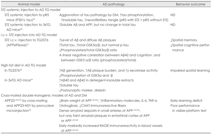
Table 2. Animal studies using an AD transgenic animal model in the literature
Animal model AD pathology Behavior outcome
STZ systemic injection to AD TG model STZ systemic injection to pR5 mice (P301L tau)29
Aggravation of tau pathology by DM; ↑tau phosphorylation, ↑Insoluble tau, ↑neurofibrillary tangle (pR5 with STZ > pR5 without STZ)
ND STZ systemic injection to 3xTG
AD mice30
↑Soluble Aβ and APP, but no change in total tau ND
i.c.v. STZ injection into AD TG model STZ i.c.v. injection to TG2576 (APP695swe)31
↑Level of Aβ and diffuse Aβ plaques
↑Total tau, ↑total GSK3α/β, but normal p-tau
↓Phosphorylated/total GSK3α/β ratio
A linear negative correlation between Aβ42 and cognition, and between GSK3 α/β ratio (phosphorylated/total)
↓Spatial memory,
↓Spatial cognitive perfor- mance
High-fat diet in AD TG model
In TG257633 ↑Aβ generation, ↑Aβ plaque burden, and ↑γ-secretase activity
↓Phosphorylation of GSK3α and -β
Impaired spatial learning In 3xTG AD mice34 ↑Aβ40 and Aβ42 in detergent-insoluble extracts
↑Soluble tau
↓Postsynaptic marker, drebrin Cross-mated double-transgenic models of AD and DM
APP23–ob/ob by cross-mating and APP23-NSY by pronuclear microinjection35
↓Brain weight of APP–ob/ob, ↑inflammatory molecules, IL-6, TNF-a
↑Astrogliosis, ↓CHAT-immunoreactive fibers Dense amyloid deposits in small arteries of APP–ob/ob, but only faint amyloid plaques in entorhinal cortex of APP or APP–ob/ob
Early markedly increased RAGE immunoreactivity in blood vessels of APP–ob/ob
Early learning deficit Poor performance in visible-platform test
Aβ: amyloid β protein, AD: Alzheimer’s disease, APP: amyloid precursor protein, CHAT: choline acetyltransferase, DM: diabetes mellitus, i.c.v.: intracerebroventricular, IL: interleukin, ND: no test for cognition or behavior, p-tau: phosphorylated tau, RAGE: receptor for advanced glycation end products, STZ: streptozotocin, Tg: transgenic, TNF-a: tumor necrosis factor alpha, 3xTG: triple transgenic.
Potential mechanisms linking T2DM and AD Data from animal studies illustrating the intimate relationship between T2DM and AD provide insights into the mechanisms shared by these conditions. This section summarizes the mech- anisms shared by T2DM and AD (Table 3), which were initial- ly suspected to be epiphenomenal but are now considered to constitute the molecular mechanisms that link and mutually in- tensify the disorders.
Insulin resistance
Insulin resistance is characterized by reduced responsiveness of insulin receptors and decreased downstream signaling for the purpose of insulin stimulation. To compensate for these dysfun- ctions, the islet β-cells of the pancreas secrete more insulin, the- reby creating a state of hyperinsulinemia. Insulin resistance with hyperinsulinemia constitutes the core feature of T2DM and is frequently also observed in AD patients.
Impaired insulin receptor signaling
There is evidence that Aβ oligomers can directly induce neuro- nal insulin resistance in the AD brain by inhibiting the insulin signaling targeting the insulin/Akt pathway38,39 and by remov- ing insulin receptors after binding at the dendrites of synaptic sites.40,41 Impaired insulin signaling cannot efficiently inhibit GSK3; therefore, the activated GSKα triggers APP γ-secretase activity, while the activated GSK3β increases tau phosphoryla- tion,42,43 which simultaneously aggravates the two major path- ological substrates of AD. In addition, insulin signaling is im- portant for the facilitation of memory via its regulation of sy- naptic structure and functioning and its promotion of neuronal survival.44 This effect is eliminated under conditions of imped- ed insulin functioning, resulting in neurodegeneration.
When prolonged systemic hyperinsulinemia is combined with insulin resistance, insulin-related activities in the brain be- come more impaired due to the decreased transport of insulin across the blood-brain barrier.45 Furthermore, hyperinsulinemia in the peripheral blood supply is thought to raise CSF Aβ42 lev- els.46 Therefore, a pathological feed-forward relationship be-
tween insulin resistance and AD pathology operates in the pres- ence of comorbid T2DM.
Impairment of Aβ clearance via IDEs
AD and T2DM share their major catalytic enzyme, IDE. As demonstrated in IDE-/- animals,27 deficiency in IDE increases Aβ and aggravates AD pathology in the brain, and the enhance- ment of IDE activity reverses the exacerbated Aβ pathology in IDE-/- and APP double-transgenic mice.47 In states of hyperin- sulinemia, increased insulin is more likely to efficiently bind to IDE than to Aβ; therefore, the level of native IDE that is avail- able for binding to Aβ in the service of degradation markedly decreases, resulting in exacerbation of AD pathology. Postmor- tem analyses of the brains of individuals with late-onset AD show decreased levels of IDE expression in the hippocampus,48 suggesting that the decreased availability of IDE is a common pathophysiology linking T2DM and AD.
Impaired glucose metabolism
T2DM inevitably results in chronic hyperglycemia, which im- pairs glucose metabolism via ineffective glucose transport, de- priving the neuronal cells of their major mechanism for metab- olism and thereby producing serious complications that con- tribute to AD pathology. In addition, decreased glucose meta- bolism can be detected in AD even before the clinical onset of dementia, and is typically noted in fluorodeoxyglucose (FDG)- PET data. For this reason, FDG-PET can be a valuable neuro- imaging marker for the early diagnosis of AD.49
Decreased O-GlcNAcylation
Diminished O-GlcNAcylation is considered to be a major mo- lecular mechanism through which impaired glucose metabolism confounds T2DM and AD. A rat model produced by i.c.v. injec- tion of STZ19 showed down-regulation of O-GlcNAcylation, impaired insulin signaling, and decreased glucose transporter activity. Decreased glucose metabolism in neurons lowers the level of UDP-GlcNAcylation, thereby decreasing tau O-Glc- NAcylation. Decreased O-GlcNAcylation is also found in the brains of those with AD50 and is inversely related to the phos- phorylation of tau. Therefore, diminished O-GlcNAcylation may result in the hyperphosphorylation of tau, leading to the tau-related pathology associated with AD.
Increased advanced glycation end products, inflammation, and oxidative stress
Chronic hyperglycemia induces the creation of advanced gly- cation end products (AGEs) via Maillard reactions, during whi- ch reducing sugars can react with the amino groups of proteins to produce cross-linked complexes and unstable compounds.51 Therefore, AGEs mediate various complications of diabetes by Table 3. Proposed common mechanisms linking AD and T2DM
Insulin resistance
Impaired insulin receptor signaling Impairment of Aβ clearance via IDEs Impaired glucose metabolism
Decreased O-GlcNAcylation
Advanced glycation end products, inflammation, and oxidative stress
Enhanced glucocorticoid effect Cerebrovascular insufficiency
Aβ: amyloid β protein, AD: Alzheimer’s disease, IDE: insulin-degrad- ing enzyme, O-GlcNAcylation: O-linked N-acetylglucosamine gly- cosylation, T2DM: type-2 DM.
interacting with the receptors for AGE present in the vascular cells and microglia, which enhances various inflammatory pro- cesses and oxidative stress. Furthermore, increased AGEs can contribute to amyloidosis52 and tau phosphorylation in AD.53 Indeed, the immunoreactivity of AGEs was markedly in- creased in Aβ plaques and neurofibrillary tangles. Therefore, increased AGEs may be another important factor shared by T2DM and AD. On the other hand, increased inflammatory me- diators such as tumor necrosis factor α, interleukin-1β, and in- terleukin-6 are induced not only by AGEs, but also by hyperin- sulinemia itself.54 Specifically, the concentration of F2-iso- prostane, a marker of lipid peroxidation, was increased in the CSF during insulin infusion in normal adults, and this was cor- related with changes in CSF levels of Aβ42.54 Increased levels of inflammatory cytokines along with oxidative stress derived from different mechanisms would be expected to lead to AD pathology through a synergistic process.55
Enhanced glucocorticoid effect
In chronic uncontrolled DM, the concentration of circulating cortisol increases,56,57 which adversely affects cognitive func- tioning.58 In db/db mice (the well-known T2DM model), enhan- ced corticoid activity has been shown to result in impaired syn- aptic plasticity and decreased neurogenesis, thereby yielding learning and memory deficits59 and providing evidence that in- creased cortisol contributes to the neurodegenerative complica- tions of T2DM. With respect to AD, elevated levels of cortisol have been correlated with the extent of hippocampal atrophy, decreased cognitive performance, and rapid decline.60,61 To- gether these findings indicate that enhanced glucocorticoid ac- tivity is a confounding factor in both T2DM and AD.
Cerebrovascular insufficiency
T2DM increases cerebrovascular disease via an atherosclerotic process,8,63 and data revealing the influence of cerebrovascular disease on the development and severity of AD have been re- ported.63 After excluding the influence of overt cerebrovascu- lar disease, the intimate relationship between T2DM and AD persists, underpinned by the aforementioned common mecha- nisms. Recent data obtained from ob/ob or polygenic NSY mice and from APP23 transgenic mice indicate that another pathology in the cerebral microvessels leading to cerebrovascu- lar insufficiency constitutes a major contributor to the cognitive decline linked to both T2DM and AD.35 Up-regulation of RAGE and inflammatory changes followed by increased amyloid de- position in the cerebral small vessels have been shown to be im- portant in producing cognitive impairments in these models, but not parenchymal Aβ burdens. Therefore, cerebrovascular insuf- ficiency caused by increased RAGE-Aβ interactions, leading to inflammation and amyloid angiopathy, can be considered as a
shared mechanism of T2DM and AD in addition to the overt cerebrovascular disorder resulting from increased vascular risk factors related to T2DM.
Conclusion
Animal models provide strong evidence of a relationship be- tween T2DM and AD, and they suggest several potential mech- anisms linking the two disorders. It is strongly suggested that AD pathology in T2DM derives from various mechanisms op- erating synergistically rather than from a single mechanism op- erating independently. Pathogenic alterations in insulin signal- ing, Aβ clearance by IDE, glucose metabolism, O-GlcNA- cylation, Aβ aggregation by AGEs, inflammation, oxidative stress, circulating cortisol, and cerebral vascular insufficiency are considered to contribute to both T2DM and AD. The inci- dence of comorbidity of T2DM and AD increases with age.
Therefore, this common pathophysiology probably constitutes a major underpinning of late-onset sporadic AD, and a novel therapeutic approach targeting this pathological process could contribute to the development of a more efficient and effective treatment for AD.
Conflicts of Interest
The author has no financial conflicts of interest.
Acknowledgements
This study is supported by a grant of the Korean Health Technology R
& D Project, Ministry for Health, Welfare & Family Affairs, Republic of Korea (A092004), and by the Korea Research Foundation (KRF) grant funded by the Korea government (MEST, 2009-0067850) The author has no conflict of interest.
REFERENCES
1. Suh GH, Knapp M, Kang CJ. The economic costs of dementia in Ko- rea, 2002. Int J Geriatr Psychiatry 2006;21:722-728.
2. Hardy J, Selkoe DJ. The amyloid hypothesis of Alzheimer’s disease:
progress and problems on the road to therapeutics. Science 2002;297:
353-356.
3. Rafii MS, Aisen PS. Recent developments in Alzheimer’s disease ther- apeutics. BMC Med 2009;7:7.
4. Mangialasche F, Solomon A, Winblad B, Mecocci P, Kivipelto M. Al- zheimer’s disease: clinical trials and drug development. Lancet Neurol 2010;9:702-716.
5. Leibson CL, Rocca WA, Hanson VA, Cha R, Kokmen E, O’Brien PC, et al. Risk of dementia among persons with diabetes mellitus: a popu- lation-based cohort study. Am J Epidemiol 1997;145:301-308.
6. Ott A, Stolk RP, van Harskamp F, Pols HA, Hofman A, Breteler MM.
Diabetes mellitus and the risk of dementia: The Rotterdam Study. Neu- rology 1999;53:1937-1942.
7. Grodstein F, Chen J, Wilson RS, Manson JE; Nurses’ Health Study.
Type 2 diabetes and cognitive function in community-dwelling elder- ly women. Diabetes Care 2001;24:1060-1065.
8. Peila R, Rodriguez BL, Launer LJ; Honolulu-Asia Aging Study. Type 2 diabetes, APOE gene, and the risk for dementia and related pathologies:
The Honolulu-Asia Aging Study. Diabetes 2002;51:1256-1262.
9. Arvanitakis Z, Wilson RS, Bienias JL, Evans DA, Bennett DA. Diabe-
tes mellitus and risk of Alzheimer disease and decline in cognitive func- tion. Arch Neurol 2004;61:661-666.
10. Xu WL, Qiu CX, Wahlin A, Winblad B, Fratiglioni L. Diabetes melli- tus and risk of dementia in the Kungsholmen project: a 6-year follow- up study. Neurology 2004;63:1181-1186.
11. Craft S, Peskind E, Schwartz MW, Schellenberg GD, Raskind M, Porte D Jr. Cerebrospinal fluid and plasma insulin levels in Alzheimer’s dis- ease: relationship to severity of dementia and apolipoprotein E geno- type. Neurology 1998;50:164-168.
12. Rivera EJ, Goldin A, Fulmer N, Tavares R, Wands JR, de la Monte SM.
Insulin and insulin-like growth factor expression and function deterio- rate with progression of Alzheimer’s disease: link to brain reductions in acetylcholine. J Alzheimers Dis 2005;8:247-268.
13. Szkudelski T. The mechanism of alloxan and streptozotocin action in B cells of the rat pancreas. Physiol Res 2001;50:537-546.
14. Kim B, Backus C, Oh S, Hayes JM, Feldman EL. Increased tau phos- phorylation and cleavage in mouse models of type 1 and type 2 diabe- tes. Endocrinology 2009;150:5294-5301.
15. Planel E, Tatebayashi Y, Miyasaka T, Liu L, Wang L, Herman M, et al.
Insulin dysfunction induces in vivo tau hyperphosphorylation through distinct mechanisms. J Neurosci 2007;27:13635-13648.
16. Jolivalt CG, Lee CA, Beiswenger KK, Smith JL, Orlov M, Torrance MA, et al. Defective insulin signaling pathway and increased glycogen synthase kinase-3 activity in the brain of diabetic mice: parallels with Alzheimer’s disease and correction by insulin. J Neurosci Res 2008;86:
3265-3274.
17. Brant AM, Jess TJ, Milligan G, Brown CM, Gould GW. Immunologi- cal analysis of glucose transporters expressed in different regions of the rat brain and central nervous system. Biochem Biophys Res Commun 1993;192:1297-1302.
18. Salkovic-Petrisic M, Tribl F, Schmidt M, Hoyer S, Riederer P. Alzheim- er-like changes in protein kinase B and glycogen synthase kinase-3 in rat frontal cortex and hippocampus after damage to the insulin signall- ing pathway. J Neurochem 2006;96:1005-1015.
19. Deng Y, Li B, Liu Y, Iqbal K, Grundke-Iqbal I, Gong CX. Dysregula- tion of insulin signaling, glucose transporters, O-GlcNAcylation, and phosphorylation of tau and neurofilaments in the brain: implication for Alzheimer’s disease. Am J Pathol 2009;175:2089-2098.
20. Steen E, Terry BM, Rivera EJ, Cannon JL, Neely TR, Tavares R, et al.
Impaired insulin and insulin-like growth factor expression and signal- ing mechanisms in Alzheimer’s disease--is this type 3 diabetes? J Al- zheimers Dis 2005;7:63-80.
21. Li ZG, Zhang W, Sima AA. Alzheimer-like changes in rat models of spontaneous diabetes. Diabetes 2007;56:1817-1824.
22. de Calignon A, Fox LM, Pitstick R, Carlson GA, Bacskai BJ, Spires- Jones TL, et al. Caspase activation precedes and leads to tangles. Nature 2010;464:1201-1204.
23. Janson J, Laedtke T, Parisi JE, O’Brien P, Petersen RC, Butler PC. In- creased risk of type 2 diabetes in Alzheimer disease. Diabetes 2004;53:
474-481.
24. Schubert M, Gautam D, Surjo D, Ueki K, Baudler S, Schubert D, et al.
Role for neuronal insulin resistance in neurodegenerative diseases. Proc Natl Acad Sci U S A 2004;101:3100-3105.
25. Schubert M, Brazil DP, Burks DJ, Kushner JA, Ye J, Flint CL, et al. In- sulin receptor substrate-2 deficiency impairs brain growth and promotes tau phosphorylation. J Neurosci 2003;23:7084-7092.
26. Killick R, Scales G, Leroy K, Causevic M, Hooper C, Irvine EE, et al.
Deletion of Irs2 reduces amyloid deposition and rescues behavioural deficits in APP transgenic mice. Biochem Biophys Res Commun 2009;
386:257-262.
27. Farris W, Mansourian S, Chang Y, Lindsley L, Eckman EA, Frosch MP, et al. Insulin-degrading enzyme regulates the levels of insulin, amyloid beta-protein, and the beta-amyloid precursor protein intracellular do- main in vivo. Proc Natl Acad Sci U S A 2003;100:4162-4167.
28. Sudoh S, Frosch MP, Wolf BA. Differential effects of proteases invol-
ved in intracellular degradation of amyloid beta-protein between deter- gent-soluble and -insoluble pools in CHO-695 cells. Biochemistry 2002;
41:1091-1099.
29. Ke YD, Delerue F, Gladbach A, Götz J, Ittner LM. Experimental diabe- tes mellitus exacerbates tau pathology in a transgenic mouse model of Alzheimer’s disease. PLoS One 2009;4:e7917.
30. Li Y, Duffy KB, Ottinger MA, Ray B, Bailey JA, Holloway HW, et al.
GLP-1 receptor stimulation reduces amyloid-beta peptide accumula- tion and cytotoxicity in cellular and animal models of Alzheimer’s dis- ease. J Alzheimers Dis 2010;19:1205-1219.
31. Plaschke K, Kopitz J, Siegelin M, Schliebs R, Salkovic-Petrisic M, Rie- derer P, et al. Insulin-resistant brain state after intracerebroventricular streptozotocin injection exacerbates Alzheimer-like changes in Tg2576 AbetaPP-overexpressing mice. J Alzheimers Dis 2010;19:691-704.
32. Ho L, Qin W, Pompl PN, Xiang Z, Wang J, Zhao Z, et al. Diet-induced insulin resistance promotes amyloidosis in a transgenic mouse model of Alzheimer’s disease. FASEB J 2004;18:902-904.
33. Kohjima M, Sun Y, Chan L. Increased food intake leads to obesity and insulin resistance in the tg2576 Alzheimer’s disease mouse model. En- docrinology 2010;151:1532-1540.
34. Julien C, Tremblay C, Phivilay A, Berthiaume L, Emond V, Julien P, et al. High-fat diet aggravates amyloid-beta and tau pathologies in the 3xTg-AD mouse model. Neurobiol Aging 2010;31:1516-1531.
35. Takeda S, Sato N, Uchio-Yamada K, Sawada K, Kunieda T, Takeuchi D, et al. Diabetes-accelerated memory dysfunction via cerebrovascular inflammation and Abeta deposition in an Alzheimer mouse model with diabetes. Proc Natl Acad Sci U S A 2010;107:7036-7041.
36. Ueda H, Ikegami H, Kawaguchi Y, Fujisawa T, Nojima K, Babaya N, et al. Age-dependent changes in phenotypes and candidate gene analy- sis in a polygenic animal model of Type II diabetes mellitus; NSY mouse. Diabetologia 2000;43:932-938.
37. Yan SD, Chen X, Fu J, Chen M, Zhu H, Roher A, et al. RAGE and am- yloid-beta peptide neurotoxicity in Alzheimer’s disease. Nature 1996;
382:685-691.
38. Townsend M, Mehta T, Selkoe DJ. Soluble Abeta inhibits specific sig- nal transduction cascades common to the insulin receptor pathway. J Biol Chem 2007;282:33305-33312.
39. Lee HK, Kumar P, Fu Q, Rosen KM, Querfurth HW. The insulin/Akt signaling pathway is targeted by intracellular beta-amyloid. Mol Biol Cell 2009;20:1533-1544.
40. Zhao WQ, De Felice FG, Fernandez S, Chen H, Lambert MP, Quon MJ, et al. Amyloid beta oligomers induce impairment of neuronal insu- lin receptors. FASEB J 2008;22:246-260.
41. De Felice FG, Vieira MN, Bomfim TR, Decker H, Velasco PT, Lambert MP, et al. Protection of synapses against Alzheimer’s-linked toxins: in- sulin signaling prevents the pathogenic binding of Abeta oligomers.
Proc Natl Acad Sci U S A 2009;106:1971-1976.
42. Phiel CJ, Wilson CA, Lee VM, Klein PS. GSK-3alpha regulates pro- duction of Alzheimer’s disease amyloid-beta peptides. Nature 2003;
423:435-439.
43. Hooper C, Killick R, Lovestone S. The GSK3 hypothesis of Alzheim- er’s disease. J Neurochem 2008;104:1433-1439.
44. Zhao WQ, Alkon DL. Role of insulin and insulin receptor in learning and memory. Mol Cell Endocrinol 2001;177:125-134.
45. Craft S, Peskind E, Schwartz MW, Schellenberg GD, Raskind M, Porte D Jr. Cerebrospinal fluid and plasma insulin levels in Alzheimer’s dis- ease: relationship to severity of dementia and apolipoprotein E geno- type. Neurology 1998;50:164-168.
46. Watson GS, Peskind ER, Asthana S, Purganan K, Wait C, Chapman D, et al. Insulin increases CSF Abeta42 levels in normal older adults. Neu- rology 2003;60:1899-1903.
47. Leissring MA, Farris W, Chang AY, Walsh DM, Wu X, Sun X, et al. En- hanced proteolysis of beta-amyloid in APP transgenic mice prevents plaque formation, secondary pathology, and premature death. Neuron 2003;40:1087-1093.
48. Cook DG, Leverenz JB, McMillan PJ, Kulstad JJ, Ericksen S, Roth RA, et al. Reduced hippocampal insulin-degrading enzyme in late-onset Alzheimer’s disease is associated with the apolipoprotein E-epsilon4 allele. Am J Pathol 2003;162:313-319.
49. Mistur R, Mosconi L, Santi SD, Guzman M, Li Y, Tsui W, et al. Current Challenges for the Early Detection of Alzheimer’s Disease: Brain Imag- ing and CSF Studies. J Clin Neurol 2009;5:153-166.
50. Liu F, Shi J, Tanimukai H, Gu J, Gu J, Grundke-Iqbal I, et al. Reduced O-GlcNAcylation links lower brain glucose metabolism and tau pathol- ogy in Alzheimer’s disease. Brain 2009;132:1820-1832.
51. Brownlee M. Advanced protein glycosylation in diabetes and aging.
Annu Rev Med 1995;46:223-234.
52. Vitek MP, Bhattacharya K, Glendening JM, Stopa E, Vlassara H, Buca- la R, et al. Advanced glycation end products contribute to amyloidosis in Alzheimer disease. Proc Natl Acad Sci U S A 1994;91:4766-4770.
53. Necula M, Kuret J. Pseudophosphorylation and glycation of tau protein enhance but do not trigger fibrillization in vitro. J Biol Chem 2004;
279:49694-49703.
54. Fishel MA, Watson GS, Montine TJ, Wang Q, Green PS, Kulstad JJ, et al. Hyperinsulinemia provokes synchronous increases in central inflam- mation and beta-amyloid in normal adults. Arch Neurol 2005;62:
1539-1544.
55. Wyss-Coray T. Inflammation in Alzheimer disease: driving force, by- stander or beneficial response? Nat Med 2006;12:1005-1015.
56. Desrocher M, Rovet J. Neurocognitive correlates of type 1 diabetes
mellitus in childhood. Child Neuropsychol 2004;10:36-52.
57. Lee ZS, Chan JC, Yeung VT, Chow CC, Lau MS, Ko GT, et al. Plasma insulin, growth hormone, cortisol, and central obesity among young Chinese type 2 diabetic patients. Diabetes Care 1999;22:1450-1457.
58. Lupien S, Lecours AR, Lussier I, Schwartz G, Nair NP, Meaney MJ.
Basal cortisol levels and cognitive deficits in human aging. J Neurosci 1994;14:2893-2903.
59. Stranahan AM, Arumugam TV, Cutler RG, Lee K, Egan JM, Mattson MP. Diabetes impairs hippocampal function through glucocorticoid-me- diated effects on new and mature neurons. Nat Neurosci 2008;11:
309-317.
60. Csernansky JG, Dong H, Fagan AM, Wang L, Xiong C, Holtzman DM, et al. Plasma cortisol and progression of dementia in subjects with Al- zheimer-type dementia. Am J Psychiatry 2006;163:2164-2169.
61. Huang CW, Lui CC, Chang WN, Lu CH, Wang YL, Chang CC. Ele- vated basal cortisol level predicts lower hippocampal volume and cog- nitive decline in Alzheimer’s disease. J Clin Neurosci 2009;16:1283- 1286.
62. Schmidt R, Launer LJ, Nilsson LG, Pajak A, Sans S, Berger K, et al.
Magnetic resonance imaging of the brain in diabetes: the Cardiovascu- lar Determinants of Dementia (CASCADE) Study. Diabetes 2004;53:
687-692.
63. Esiri MM, Nagy Z, Smith MZ, Barnetson L, Smith AD. Cerebrovascu- lar disease and threshold for dementia in the early stages of Alzheimer’s disease. Lancet 1999;354:919-920.