This case report demonstrates the beneficial effects of cupping therapy (CT) in a 35-year-old man who is diagnosed with a fracture of the radial shaft due to a motorcycle accident. One year after the treatment started, pseudoarthrosis developed in the radius and an autogenous iliac bone graft was performed.
However, extension dysfunction in the wrist became evident. After another 6 months of physical therapy and rehabilitation, no improvements were observed. Therefore, CT and adjunctive electrostimulation were performed, after 30 days of treatment, marked recovery of muscle function and full wrist extension were observed, as determined by electromyography and a grade 5/5 on the Medical Research Council power of wrist extension scale. The results in this case study suggest that CT in conjunction with adjunctive electrostimulation, may accelerate functional recovery from postoperative radial palsy, and provide a useful alternative treatment in this situation.
©2018 Korean Acupuncture & Moxibustion Medicine Society. This is an open access article under the CC BY- NC-ND license (http://creativecommons.org/licenses/by-nc-nd/4.0/).
Article history:
Submitted: December 29, 2016 Revised: February 1, 2017 Accepted: February 6, 2017 Keywords:
cupping therapy, radial palsy, physical therapy,
rehabilitation
J Acupunct Res 2018;35(1):1-3
Case Report
Cupping Therapy Combined with Rehabilitation for the Treatment of Radial Palsy: a Case Report
Ali Ramazan Benli
1,*, Demir Yazici Senay
2, Mustafa Koroglu
3, Tansel Mutlu
4, Selman Erturhan
5, Muhammet Nur Ogun
6, Didem Sunay
11 Department of Family Medicine, Faculty of Medicine, Karabuk University, Karabuk, Turkey 2 Department of Physical Examination and Rehabilitation, Aydin Ataturk State Hospital, Aydin, Turkey 3 Department of Hematology, Faculty of Medicine, Karabuk University, Karabuk, Turkey
4 Department of Orthopedics, KBU Karabuk Educational and Research Hospital, Karabuk, Turkey 5 Family Medicine, Yildizeli State Hospital, Yildizeli, Sivas, Turkey
6 Department of Neurology, Faculty of Medicine, Abant Izzet Baysal University, Bolu, Turkey
ARTICLE INFO ABSTRACT
Journal of Acupuncture Research
Journal homepage: http://www.e-jar.org
*
Corresponding author.Karabuk University, Faculty of Medicine, Department of Family Medicine, Karabuk, Turkey E-mail : [email protected]
https://doi.org/10.13045/jar.2016.02957 pISSN 2586-288X eISSN 2586-2898
©2018 Korean Acupuncture & Moxibustion Medicine Society. This is an open access article under the CC BY-NC-ND license (http://creativecommons.org/licenses/by-nc- nd/4.0/).
Introduction
Cupping therapy (CT) is a traditional and supplementary practice that has been used for hundreds of years. Wet and dry cupping are the most commonly used methods for implementing CT. The basis of dry CT is to increase blood flow in subcutaneous tissue by generating a negative pressure on the skin [1]. CT is used in various diseases with different etiologies, such as migraine, chronic back pain, metabolic syndrome, hypertension, carpal tunnel syndrome, and fibromyalgia [2, 3].
Fractures of the radius and the humerus are common in high- energy traumas, like traffic accidents [4]. Although radial nerve injury may occur with radial fractures and surgeries, it is more commonly observed with contusion and compression, and it generally resolves with physical therapy and rehabilitation (PTR).
If recovery is not achieved, surgical exploration is performed
[5]. In such cases, supplementary methods may be used as an alternative or adjunctive therapy to PTR. In this case report, we present the effect of CT in a patient with postoperative radial palsy, who had not achieved recovery after 6 months of PTR.
Case Report
A 35-year-old man was brought to the emergency department with a fracture of the radial shaft incurred in a motorcycle accident. A fixation plate was applied to the radius by the orthopedic department. At the end of the first year of treatment, pseudoarthrosis developed in the radius, and an autogenous iliac bone graft was applied. After the iliac bone graft to the radius, the function of wrist extension function was lost (grade 1/5 on the Medical Research Council power of wrist extension scale).
Anti-inflammatory and vitamin B12 treatments were started in
J Acupunct Res 2018;35(1):1-3 2
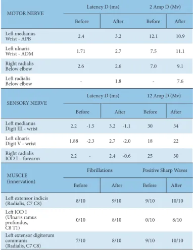
the acute phase for edema. At the end of the acute phase, the patient underwent physical therapy for the left radial posterior interosseous nerve (RPIN) palsy. A PTR program of 30 sessions (three sessions per week) was initiated, which included transcutaneous electrical nerve stimulation (TENS) (20 minutes, frequency 100 Hertz, current rate 100 µs), galvanic stimulation (5 × 1 ms pulses at 300 Hz), range of motion exercises, and isometric strength and relaxation exercises. Although 90 sessions of physical therapy were completed, RPIN amplitude stimulation could not be perceived, this represented severe axonal injury (Table 1). The absence of a beneficial response on radial palsy following this treatment indicated that nerve compression, damage, or laceration during surgery might be the cause of this condition.
The patient was referred to the Department of Orthopedics for tendon transfer due to the failure of the current treatments, he was then admitted to our clinic for alternative treatment instead of undergoing surgery. After assessment of the nerve palsy, CT was considered as a suitable therapy for the patient.
Dry CT was applied over the RPIN daily for 15-20 minutes (Fig. 1). After 20 days of dry CT, there was minimal wrist mobility on extension, therefore adjunctive electro stimulation therapy was performed. When dry CT was combined with TENS using the electric stimulation protocol (20 minutes, 5 × 1 ms pulses at 300 Hz) for 30 days, normal muscle contraction was observed (grade 5/5 on the Medical Research Council power of wrist extension scale), and the nerve and muscle function results were recorded by
electromyogram (EMG) and recovery was observed especially in interosseous dorsalis of the left radial nerve (Table 1). No adverse events were observed with the combined therapy. These results suggest that the effects of increasing blood flow and creating negative pressure using CT, together with electrostimulation, speed up recovery of the radial nerve.
Discussion
Forearm fractures occur for multiple reasons; direct trauma being the most common cause. In high-energy traumas like traffic accidents, open fractures and soft tissue damage occur together.
According to the fracture type, various treatment methods can be applied to radial shaft fractures. Surgical treatments of open humeral shaft fractures are preferable due to better control of fracture fragments, reduced time to union, and increased functional recovery; however, for closed humeral shaft fractures with radial palsy, recovery can be achieved using noninvasive therapeutic methods [6]. Indications for external fixation of the forearm are restricted to open fractures, in which soft tissue loss is high and bone loss exists. Internal fixation with open reduction and a single plate placement with 4 screws is preferable to achieve additional functional recovery. If fragmentation accounts for more than 50%, grafting is recommended [7]. Nerve damage occurs rarely after radial shaft fractures. The radial nerve can be damaged primarily (due to trauma), or through secondary (iatrogenic) damage following surgical investigation [8] as was observed in the present case.
Following nerve damage, reinnervation potential is usually observed by EMG within 6-8 weeks. Detection of reinnervation potential within 6-8 weeks supports a diagnosis of axonotmesis.
However, if reinnervation potential is not detected, then the nerve damage is classified as neurotmesis and requires surgical exploration [8]. The decision on whether nerve damage requires surgery is controversial [9]. In this case, the first EMG was performed at 7 months after surgery, and severe axonal injury in the left RPIN was detected.
After nerve damage occurs, muscle atrophy begins and progresses daily. For this reason, starting rehabilitation early is very important. Brown et al have shown that for every 6 days of denervation, a 1% loss of function occurs [10]. Pain and edema also cause function loss in the acute phase [11]. In this case, anti- inflammatory medication and physical therapy were utilized in the acute phase to relieve pain and remove edema. In addition to the treatment methods for prevention of muscle atrophy, therapeutic interventions were performed for the repair of nerve damage.
Fig. 1. Applying cupping therapy along the radial nerve trace.
Left medianus
Wrist - APB 2.4 3.2 12.1 10.9
Left ulnaris
Wrist - ADM 1.71 2.7 7.5 11.1
Right radialis
Below elbow 2.6 2.6 7.0 9.1
Left radialis
Below elbow - 1.8 - 7.6
Left medianus
Digit III - wrist 2.2 -1.5 3.2 -1.1 30 34
Left ulnaris
Digit V - wrist 1.88 -2.3 2.7 -2.0 18 22
Right radialis
IOD I – forearm 2.2 - 2.4 -0.6 25 30
Left extensor indicis
(Radialis, C7 C8) 8/10 9/10 9/10 10/10
Left IOD I (Ulnaris ramus profundus, C8 T1)
0/10 8/10 0/10 8/10
Left extensor digitorum communis
(Radialis, C7 C8) 7/10 8/10 9/10 10/10
ADM, abductor digiti minimi; APB, abductor pollicis brevis; IOD I, interosseous dorsalis 1; ms, millisec- onds; mV, microvolt; D, dispersion.
Table 1. Electromyogram Results of the Patient’s Wrist Function Before and After Cupping Therapy.
MOTOR NERVE
SENSORY NERVE
MUSCLE (innervation)
Before After Before After Before After Before After
Latency D (ms) 2 Amp D (Mv)
Latency D (ms) 12 Amp D (Mv)
Fibrillations Positive Sharp Waves Before After Before After
Ali Ramazan Benli et al / Cupping Therapy in Radial Palsy 3