ORIGINAL ARTICLE
Journal of the Korean Surgical Society
JKSS
pISSN 2233-7903ㆍeISSN 2093-0488 J Korean Surg Soc 2011;81:195-204
http://dx.doi.org/10.4174/jkss.2011.81.3.195
Received April 11, 2011, Revised June 8, 2011, Accepted June 17, 2011 Correspondence to: Jang Yong Jeon
Department of Surgery, Hallym University Sacred Heart Hospital, Hallym University College of Medicine, 896 Pyeongchon-dong, Dongan-gu, Anyang 431-070, Korea
Tel: +82-31-380-3772, Fax: +82-31-385-0157, E-mail: [email protected]
cc Journal of the Korean Surgical Society is an Open Access Journal. All articles are distributed under the terms of the Creative Commons Attribution Non-Commercial License (http://creativecommons.org/licenses/by-nc/3.0/) which permits unrestricted non-commercial use, distribution, and reproduction in any medium, provided the original work is properly cited.
Management of endoscopic retrograde
cholangiopancreatography-related perforations
Byung Seup Kim, In-Gyu Kim, Byoung Yoon Ryu
1, Jong Hyeok Kim
2, Kyo Sang Yoo
2, Gwang Ho Baik
1, Jin Bong Kim
1, Jang Yong Jeon
Department of Surgery, Hallym University Sacred Heart Hospital, Hallym University College of Medicine, Anyang, 1Department of Gastroenterology & Hepatology, Hallym University Chuncheon Sacred Heart Hospital, Hallym University College of Medicine, Chuncheon, 2Department of Gastroenterology & Hepatology, Hallym University Sacred Heart Hospital, Hallym University College of Medicine, Anyang, Korea
Purpose: The purpose of this study is to analyze the treatment strategies of patients with endoscopic retrograde chol- angiopancreatography (ERCP)-related perforations. This is a retrospective study. Methods: We experienced 13 perforations associated with ERCP. We reviewed the medical recordsand classified ERCP-related perforations according to mechanism of injury in terms of perforating device. Injury by endoscopic tip or insertion tube was classified as type I, injury by cannulation catheter or sphincterotomy knife as type II, and injury by guidewire as type III. Results: Of four type I injuries, one case was managed by conservative management after primary closure with a hemoclip during ERCP. The other three patients under- went surgical treatments such as primary closure orpancreatico-duodenectomy. Of five type II injuries, two patients under- went conservative management and the other three cases were managed by surgical treatment such as duodenojejunostomy, duodenal diverticulization and pancreatico-duodenectomy. Of four type III injuries, three patients were managed con- servatively and the remaining patient was managed by T-tube choledochostomy. Conclusion: Type I injuries require immedi- ate surgical management after EPCP or immediate endoscopic closure during ERCP whenever possible. Type II injuries re- quire surgical or conservative treatment according to intra- and retro-peritoneal dirty fluid collection findings following ra- diologic evaluation. Type III injuries almost always improve after conservative treatment with endoscopic nasobilliary drainage.
Key Words: Endoscopic retrograde cholangiopancreatography, Perforation, Surgery
INTRODUCTION
Endoscopic retrograde cholangiopancreatography (ERCP) is commonly performed to treat hepato-pancreato-biliary disease. The rate of ERCP-related bowel perforation is 0.3
to 1.0% [1-3]. The mortality rate in perforated patients is as high as 25% [4]. Many previous reports have described the management of perforation injuries associated with ERCP.
Some have characterized treatment strategies according to location and mechanism of bowel perforation [5,6]. How-
Our proposal Stapfer et al. [5] Howard et al. [6]
Type I Injury by endoscope before approach to papilla
Type I Lateral or medial wall perforation Type III Duodenal perforation remote from the papilla
Type II Injury by cannulation catheter for puncture of papilla or knife for sphincterotomy
Type II Peri-Vaterian injury Type II Periampullary retroperitoneal perforation
Type III Injury by guidewire of bile duct or pancreatic duct after puncture of papilla
Type III Distal bile duct injury related to wire/basket instrumentation
Type I Guidewire perforation
None - Type IV Retroperitoneal air alone None -
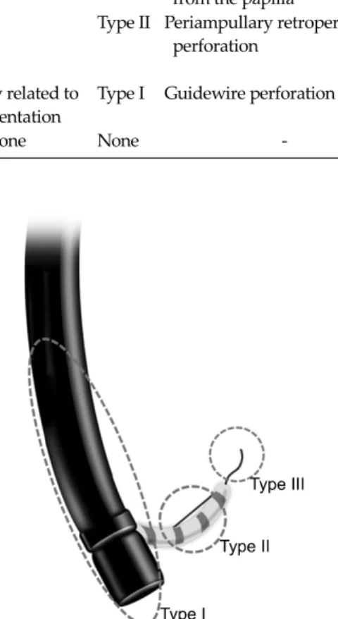
Table 1. Classification of endoscopic retrograde cholangiopancreatography-related perforations
Fig. 1. Classification of endoscopic retrograde cholangiopan- creatography-related perforations according to injury mechanism.
ever, these classifications may be difficult to apply to real clinical situations because of their ambiguity, and there- fore the most appropriate management strategy for ERCP- related perforations remains unclear. The purpose of this study is to analyze the treatment strategies and outcomes of patients with ERCP-related perforations based on a new classification.
METHODS
Between April 1994 and December 2009, 7,638 cases of ERCP were performed. Among these patients, twelve pa- tients (0.16%) experienced perforations that were asso- ciated with ERCP. One patient with suspected injury dur- ing ERCP was transferred to our hospital for management.
The patient was included in our study. We retrospectively reviewed the medical records of 13 patients who were managed for perforations associated with ERCP.
We classified ERCP-related perforations according to mechanism of injury in terms of the perforating device. If bowel perforation was identified while the endoscope was inserted into the second portion of duodenum or while it was withdrawn from duodenum, the perforation was caused by the endoscopic blind tip or insertion tube. We classified this type of injury as type I.
If the injury was caused by a cannulation catheter or a knife for sphincterotomy, the injury was classified as type II.
Injuries caused by guidewires after cannulation of the papilla during exploration of the bile duct or pancreatic duct was classified as type III (Table 1, Fig. 1).
We analyzed data regarding the clinical manifestations,
diagnostic methods, radiologic findings, methods of man- agement, and clinical outcomes of all patients.
RESULTS
Demographic characteristics
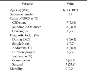
The sample included six male and seven female patients with a median age of 65.1 (±10.7) years. The objectives of ERCP were common bile duct stones (53.8%), jaundice with suspicious malignancy (38.5%) and cholangitis with- out stone (7.7%). Diagnoses of perforation were made dur- ing ERCP in six patients (46.2%). The other seven patients were diagnosed after ERCP by plain X-ray, abdominal computed tomography (CT) or sonography. Six of 13 pa- tients (46.2%) were managed conservatively, while the
Variable Value
Age (yr) (±SD) 65.1 (±10.7)
Sex (male:female) 6:7
Cause of ERCP, n (%)
CBD stone 7 (53.8)
Jaundice, R/O Cancer 5 (38.5)
Cholangitis 1 (7.7)
Diagnostic tool, n (%)
During ERCP 6 (46.2)
Simple X-ray 1 (7.7)
Abdominal CT 5 (38.5)
Ultrasonography 1 (7.7)
Treatment, n (%) Conservative Surgical
6 (46.2) 7 (53.8) Mortality 0 (0.0)
ERCP, endoscopic retrograde cholangiopancreatography; CBD, common bile duct; R/O, rule out; CT, computed tomography.
Table 2. Patients’ demography
Type I (n = 4)
Type II (n = 5)
Type III (n = 4) Time to diagnosis after ERCP
Immediate (<2 hr) (77%) 4 3 3
Delayed (>24 hr) (23%) 0 2 1
Method of treatment
Non-operation 1 2 3
Operation 3 3 1
ERCP, endoscopic retrograde cholangiopancreatography.
Table 3. Time to diagnosis and method of treatment
Management method Conservatively Surgically
Patient No. 1 2 3 4
Sex/age M/75 F/72 M/73 M/68
Cause of ERCP CBD stone Klatskin tumor R/O Distal CBD stone R/O AOV tumor
Possible predisposing factor Duodenal deformitiy &
stricture
None S/P subtotal gastrectomy, Billoth II
Diverticulum
Time to diagnosis <1 hr <1 hr <1 hr <1 hr
Symptom Abd. tenderness Abd. pain Abd. pain, fever Abd. pain, fever
Radiologic finding Minimal contrast extravasion
Free air Free air Emphysema
Diagnostic tool During ERCP During ERCP During ERCP During ERCP
Leukocytosis Yes No Yes Yes
Treatment Endoscopic clipping Primary closure Primary closure T-tube choledochostomy
Whipple`s operation
Post-ERCP stay (day) 9 32 15 36
Time interval from ERCP to operation
3 hr 2 hr 3 day
Operative finding Lateral perforation at
2nd part of duodenum Mild inflammation
Afferent loop perforation Mild inflammation
Lateral perforation at 2nd part of duodenum, medial duodenaldivericular perrforation, periduodenal abscess, severe inflammation
Post-operative complication None None P-J leakage, wound
dehiscence
Outcome Well Well Well Well
ERCP, endoscopic retrograde cholangiopancreatography; CBD, common bile duct; R/O, rule out; AOV, ampulla of Vater; S/P, status post;
Abd., abdominal; P-J, pancreatico-jejunostomy.
Table 4. Treatment of type I injuries
other seven patients (53.8%) were managed by surgical treatment. There was no mortality (Table 2).
All four patients with type I injury were diagnosed with bowel perforation during ERCP procedure. One case im- proved with conservative management, while the other three cases were managed surgically. Of five type II in- juries, three cases were detected immediately and the oth- er two cases were detected late. Two cases improved with conservative management, while the other three cases were managed surgically. Of four type III injuries, most were detected immediately. Three cases improved with
Management method Conservatively Surgically
Patient No. 5 6a) 7 8 9
Sex/age F/58 M/79 F/80 M/49 F/49
Cause of ERCP Cholangitis R/O Pancreatic cancer with CBD invasion
Gall bladder cancer R/O IPMN of pancreatic head
R/O CBD cancer
Possible predisposing factor
Diverticulum Obscure ampulla Diverticulum None CBD stricture
Time to diagnosis 2 hr 2 hr <1 hr 1 day 3 day
Symptom Abd. pain None Abd. pain Abd. pain Abd. pain
Radiologic finding Retroperitoneal air no fluid collection
Perirenal free air, no fluid collection
Free air Morison pouch
fluid collection
Retroperitoneal air and dirty fluid collection
Diagnostic tool CT CT During ERCP Ultrasonography CT
Leukocytosis No No Yes Yes Yes
Treatment Levin tube No drainage Duodenojejunostomy,
T-tube
choledochostomy, cholecystectomy
Duodenostomy, T-tube
choledochostomy, cholecystectomy, multiple drainage
PPPD
Post-ERCP stay 16 day 4 day 18 day 40 day 37 day
Time interval from ERCP to operation
3 hr 2 day 10 day
Operative finding Anteriomedial wall
perforation of 2nd part of duodenum, Mild inflammation
Retroperitoneal bile staining large amount ascites (1,500 mL), saponification, No definite perforation site
Severe duodenal edema, swelling, retroperitoneal inflammation &
mesenteric thickening, No definite perforation site Post-operative
complication
None Sepsis, 2nd
operation on POD 11 for pancreatic fistula; duodenal diverticulization
None
Outcome Well Well Well Well Well
ERCP, endoscopic retrograde cholangiopancreatography; R/O, rule out; IPMN, intraductal papillary mucinous neoplasm; CBD, common bile duct; Abd., abdominal; CT, computed tomography; PPPD, pylorus preserving pancreaticoduodenectomy; POD, post-operative days.
a)Patient no. 6 This patient was transfer to other hospital at the request of the patient after 4 days of conservative management.
Table 5. Treatment of type II injuries
conservative management, while the last was managed surgically (Table 3).
Methods of treatment
Type I injuries (Table 4)
Of four patients, only one was managed conservatively with endoscopic treatment. Endoscopic clipping was per- formed just after bowel perforation during ERCP. This pa-
tient improved without antibiotics or any drainage pro- cedures.
Three patients underwent surgical treatment. Two pa- tients underwent immediate surgery within threehours after ERCP. The operations were performed to achieve pri- mary closure of the perforation site. One patient (patient No. 2), who was thought to have Klatskin tumor of Bisthmus type IIIbin pre-operative radiologic finding, re- ceived percutaneous transhepatic biliary drainage after
primary closure of the perforation site. None of the pa- tients who underwent immediate surgery experienced any complications or had problematic outcomes. One pa- tient underwent a delayed operation because he had stable vital signs and the possibility of ampulla of Vater cancer requiring extensive operation. We considered elective Whipple's operation for this patient. In the operative find- ings, the tissue around the lesion was fragile and severely inflamed. Post-operative complications such as pancrea- tico-jejunostomy site leakage and wound dehiscence were observed. Biopsy reported duodenal wall defect with peri- duodenal abscess, epithelial hyperplasia in common bile duct and no tumor. Fortunately, the patient recovered well after wound closure under general anesthesia and con- servative treatment for anastomosis site leakage.
Type II injuries (Table 5)
Of five patients, two were managed conservatively. The cannulation of the ampulla of Vater failed in these pa- tients. After ERCP, follow-up CT and simple X-ray showed retroperitoneal air and perirenal free air, but no fluid collection. Neither patient had leukocytosis. One patient received Levin tube drainage and the other was managed without any drainage procedures. Neither patient experi- enced any complications during hospital stay. One case was transferred to another hospital at the request of the patient.
Of five patients, three were managed by surgical treat- ment. One case underwent an immediate operation. Two cases were detected later and underwent delayed opera- tions. In the immediately operated case, the perforation occurred because the cannula punctured the anterio-me- dial wall of the duodenum instead of the ampulla of Vater.
The patient complained of abdominal pain immediately after ERCP and free air was detected on simple X-ray. After a little dissection of pancreatico-duodenal junction, we found an approximately 5 mm-sized perforation site.
Duodeno-jejunostomy through perforation site, T-tube choledochostomy and cholecystectomy were performed, and the patient’s condition improved after the operation.
The reported biopsy was adenocarcinoma in fundus of gallbladder, invasion into perimuscular connective tissue and no tumor in resection margin of cystic duct. Another
case was detected within a day after ERCP. Fluid collection in Morison pouch was detected inultrasonography and greatly increased in follow-up sonography two days after ERCP. The fluid looked like complicated ascites. Surgery was performed andwe detected retroperitoneal bile stain- ing and saponification, but were unable to locate the bowel perforation. We believe that pancreatic and bile duct in- jury may have occurred as the cannula passed through the ampulla of Vater. Although we carried out multiple drain- age procedures, the patient’s condition worsened due to the development of a pancreatic fistula. We re-operated on postoperative day 11 and performed a duodenal diverti- culization. The patient improved after the second opera- tion. The last patient’s injury was detected three days after ERCP. A common bile duct stricture was seen in the CT scan before ERCP. ERCP was performed with some diff- iculty. The endoscopist did not recognize the perforation during the procedure. Three days from ERCP, a CT was performed due to severe abdominal pain. The CT showed retroperitoneal air and fluid collection. Because the vital signs and symptoms of the patient were tolerable and common bile duct cancer was suspected, we decided to perform delayed extensive operation after conservative management. This operation was performed 10 days after ERCP. The perforation site was not identified, severe retro- peritoneal inflammation remained and the duodenal wall was severely edematous. We carried out a pylorus-pre- serving pancreatico-duodenectomy. Biopsy reported stric- tureand chronic active inflammation with epithelial cell hyperplasia in common bile ductand chronic active in- flammation with extensive abscess formation and serositis in duodenum. There was no tumor. The patient was dis- charged at post-operative day 37 without any com- plications.
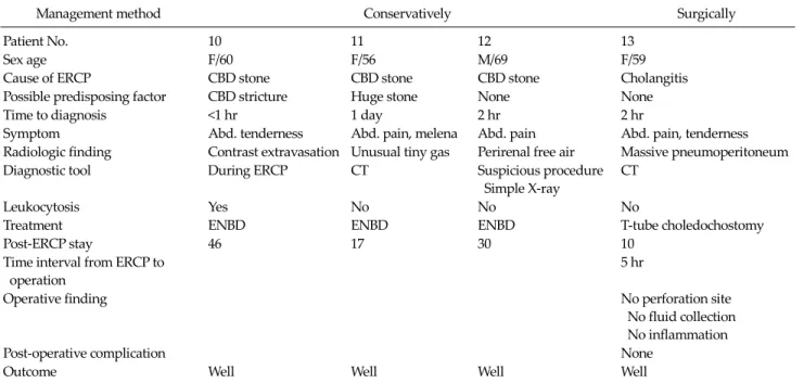
Type III injuries (Table 6)
Of four patients, three were successfully managed con- servatively with endoscopic nasobilliary drainage (ENBD).
Perforations by guidewire were identified by contrast ex- tra-vasation during ERCP, unusual gas on CT and peri- renal free air on simple X-ray. All three cases experienced no difficulties during the ERCP procedure.
One underwent surgical treatment. The patient experi-
Management method Conservatively Surgically
Patient No. 10 11 12 13
Sex age F/60 F/56 M/69 F/59
Cause of ERCP CBD stone CBD stone CBD stone Cholangitis
Possible predisposing factor CBD stricture Huge stone None None
Time to diagnosis <1 hr 1 day 2 hr 2 hr
Symptom Abd. tenderness Abd. pain, melena Abd. pain Abd. pain, tenderness
Radiologic finding Contrast extravasation Unusual tiny gas Perirenal free air Massive pneumoperitoneum
Diagnostic tool During ERCP CT Suspicious procedure
Simple X-ray
CT
Leukocytosis Yes No No No
Treatment ENBD ENBD ENBD T-tube choledochostomy
Post-ERCP stay 46 17 30 10
Time interval from ERCP to operation
5 hr
Operative finding No perforation site
No fluid collection No inflammation
Post-operative complication None
Outcome Well Well Well Well
ERCP, endoscopic retrograde cholangiopancreatography; CBD, common bile duct; Abd., abdominal; CT, computed tomography; ENBD, endoscopic nasobilliary drainage.
Table 6. Treatment of type III injuries
enced severe abdominal pain after ERCP. CT was per- formed immediately after ERCP and revealed massive pneumoperitoneum. The endoscopist had not experi- enced any difficulty during ERCP, and therefore did not recognize the perforation. We performed an immediate di- agnostic laparotomy, but were unable to determine the ex- act location of the perforation. There was no fluid collec- tion and no inflammation. We suspected bile duct injury, and performed a T-tube choledochostomy. After surgery, the patient was discharged without problems or any complications.
DISCUSSION
ERCP-related bowel perforations are very rare and unpredictable. There have been some reports about possi- ble predisposing factors. Enns et al. [2] reported that fac- tors associated with increased risk of ERCP-related bowel perforation included suspected sphincter of Oddi dys- function, older age, a dilated bile duct, sphincterotomy, and longer duration of the procedure. Kayhan et al. [7] re- ported that the presence of duodenal anatomic abnormal-
ities and peripapillary diverticulum were associated with complication. In the present study, among three patients with duodenal diverticulae, one patient (No. 4) experi- enced a duodenal diverticular perforation by endoscopic tip. Another patient (No. 1) had duodenal stricture due to a previous duodenal ulcer and experienced a duodenal perforation during endoscopic approach. Another patient (No. 3) underwent subtotal Billroth II gastrectomy due to gastric cancer and had an anatomic variation of the affer- ent jejunal loop. This afferent jejunal loop was torn by en- doscopy during insertion. Another patient (No. 6) had an obscure ampulla. The cannula entered the ampulla only after several attempts. We believe that the injury around the ampulla was caused by the cannulation catheter dur- ing this procedure. Another patient (No. 9) experienced a cannula puncture of the ampulla of Vater but the guide- wire did not enter the bile duct through the cannula. The endoscopist was unable to advance the endoscope to con- tinue the procedure. We suspected that the retroperitoneal perforation around the ampulla was caused by the cannu- lation catheter.
Two previous studies introduced classifications of ERCP-related perforations based on anatomical location
and mechanism of injury. Stapfer et al. [5] defined type I (lateral or medial wall perforation of duodenum), type II (peri-Vaterian injury), type III (distal bile duct injury) and type IV (retroperitoneal air alone) injuries. Howard et al.
[6] classified type I (guidewire perforation), type II (peri- ampullary retroperitoneal perforation) and type III (duo- denal perforation remote from the papilla) injuries. We found Stapfer et al.’s classification to be ambiguous re- garding the boundary between the anterior or posterior duodenum and the peri-Vaterian area. And Howard et al.’s classification was limited because the definition of
‘remote’ in type III injuries was not clear. Stafer's type I and Howard's type III also mentioned perforation within the duodenum. In our experience, one patient (No. 3) had ana- tomic variation due to a previous operation. It was diffi- culty to classify this patient according to Stapfer et al.'s or Howard et al.'s classification. This patient had a perfo- ration not in duodenum but in jejunum of afferent loop.
Previous reports focused on perforated location but we fo- cusedon perforation size. The perforation size varied ac- cording to the device causing perforation. We simplified the classification of ERCP-related bowel perforations by basing our classification only on the mechanism of injury (Table 1, Fig. 1).
ERCP course could be divided to 3 steps. The 1st step is approaching the second portion of the duodenum. The 2nd step is cannulation of ampulla of Vater by catheter.
Sphincterotomy could be done using sphincterotomy knife. The 3rd step is investigation of bile duct and pancre- atic duct. The main device used differs from step to step.
Type I injuries are induced by endoscopic tip or insertion tube. The diameter of endoscopic tip for ERCP is approx- imately equal to that of a finger, and the camera view is from the side unlike the usual gastroduodenoscopic tip view [8]. ERCP endoscopes are so thick and stiff that bowel injuries may be aggravated in proportion to the size of perforation. During advancement of the endoscope, the side of the bowel could be torn by insertion tube. Large perforations are not expected to heal without surgery due to severe intra-peritoneal contamination and sepsis.
Therefore, we propose that exploratory laparotomy is a better choice for treatment of Type I injuries. In the present study, patients with type I injuries were all treated surgi-
cally except for one (No. 1). When that patient suffered an endoscopic injury on the duodenal wall, immediate endo- scopic clipping was performed to limit intra-peritoneal contamination. The patient improved with conservative management. Siebert reported the successful use of an en- doscopic clipping device to treat a duodenal perforation that occurred during an endoscopic ultrasound examina- tion [9]. Mutignani et al. [10] described a duodenal perfo- ration that occurred during ERCP that was sealed with fi- brin glue and managed conservatively. If immediate clo- sure by endoscopic methods is possible, conservative management without surgery may be the best treatment method. Of course, some cases require surgery after endo- scopic clipping due to hemodynamic instability [8]. It is necessary to closely observe patients’ vital signs after en- doscopic closure of bowel perforations. Ryozawa et al. [11]
reported that the development of double-balloon endo- scopes had resulted in improved success rates for ERCP in patients with Roux-en-Y reconstruction. In our patient sample, one patient experienced duodenal perforation with Billroth II anasotmosis. We believe that it is advisable to use double-balloon endoscopy in cases with Billroth II anatomic variations.
Type II injuries are induced by sphincterotomy knives or cannulation catheters. Generally, the diameter of ERCP cannulation catheters is 5 to 7 Fr. This diameter is so small that perforations by cannulae may seal naturally. Howev- er, if significant bile or pancreatic juice leakage occurs, the healing of injured tissue due to irritant fluid would be dif- ficult and emergency surgery should be considered. We believe that fluid collection in the intra- or retro-peritoneal cavity is a significant operative indication of type II injures induced by sphincterotomy knives or cannulation cathe- ters. Stapfer et al. [5] reported that fluid collection in the retroperitoneal or peritoneal cavity is an indicator for sur- gery after ERCP-related duodenal perforation. Morgan et al. [8] reported that persistent collection of infected fluids collection can prevent the healing of the perforation site.
Husain et al. [12] reported that 33% (7/21) of patients showed extra-luminal retroperitoneal air following endo- scopic sphincterotomy and that this observation was not clinically significant. Stapfer et al. [5] insisted that retro- peritoneal air alone probably requires no additional treat-
ment or further work-up, if the findings of abdominal ex- aminations are normal and there is no evidence or suspi- cion of contrast extravasation. In two of our patients (No.
5 and No. 6), CT findings showed retroperitoneal air after ERCP. But the patients' symptoms were mild. Vital signs and laboratory values were also normal. Although retro- peritoneal air was observed in CT, there were no fluid col- lections in retroperitoneal or intra-abdominal cavity. We tried conservative managementand these treatments were done successfully. In type II injuries when the patient is stable and has no fluid collection and only retroperitoneal air, conservative management may be possible. In the case of patient No.7, she showed not retroperitoneal air but in- tra-peritoneal air. Because of suspected panperitonitis on physical examination, we decided on emergency oper- ation without follow-up CT scan. We found bile leakage in the pancreatico-duodenal junction. If the patient had CT evaluation, fluid collection would have shown because bile leakage was observed at that time. We dissected the pancreatico-duodenal junction while towing the duode- num. After a minor dissection, we could find an approx- imately 5 mm sized perforation site in the anterio-medial portion of the duodenum. The perforation was caused by cannulation catheter according to the endoscopist and the diameter of catheter was 1.8 mm. We thought that the per- foration size could have been enlarged to 5 mm due to the procedures of lateral traction and dissection. We per- formed duodenojejunostomy for duodenal perforation.
Some surgeons recommend primary closure and drainage in the case of early detection [13]. However, we prefer duodenojejunostomy. This procedure is thought to have the benefit of decompression of duodenal pressure through side-to-side anastomosis. The procedure is not so difficult and does not take very long; about 15 minutes.
Duodenojejunostomy could be another method in duode- nal injury that requires operation.
Type III injuries are induced by guidewires after cannu- lation of the ampulla. The diameter of the guidewire is smaller than that of a cannulation catheter, and therefore perforations may be small and the location of perforation might be in the common bile duct or pancreatic duct pass- ing through the ampulla of Vater. If ENBD was main- tained, the pressure and flow in injured ducts might be
lower than in injured ducts without ENBD. The possibility of being sealed-off is high and inflammation may be mild.
Therefore, the success rate of conservative management may be higher than in other types of bowel perforation.
Howard et al. [6] reported that patients who suffered guidewire perforations resolved with conservative treat- ment. In our study, all cases, except one (No. 13), were treated by conservative management with ENBD. This ex- ceptional case had immediate operation due to severe ab- dominal pain and intra-abdominal free air. We presumed that the perforation was caused by the guidewire, because insertion of endoscopy to the duodenum was smooth and the cannulation of the ampulla was uneventful. In oper- ation, we observed little inflammation around the oper- ative field and were unable to find the location of the bow- el perforation. The patient improved after T-tube choledo- chostomy. We thought that the effect of this operation would be similar to ENBD to decrease flow and pressure of common bile duct. Chung et al. [14] reported that im- provement of symptoms within 24 hours was correlated with spontaneous recovery. Neither the presence of retro- peritoneal air nor contrast leaks was predictive of the need for surgery. In our cases, we did not observe retroperi- toneal air, but observed intra-abdominal free air. It was difficult to decide whether the immediate exploratory lap- arotomy of our patient was truly necessary. We think that conservative management with ENBD might have been sufficient after 24 hours observation in patient number 13.
When intra-abdominal free air occurs due to injury by guidewire, it is thought that conservative management with ENBD is possible.
One patient (No. 4) in type I injuries and two patients (No. 8 and No. 9) in type II injuries underwent delayed operations. We performed a delayed operation due to the preparation required for extensive surgery on suspicious malignancies. We were immediately unable to operate on the patients with suspicious malignancy because we con- sidered the preparations for anesthesia and operation team of one-stage operations of malignancy to be insuf- ficient. But the results of delayed operation were unsatis- factory. These patients had severe inflammation in spite of conservative management and difficulties were encoun- tered during surgery due to fragile tissues and adhesion.
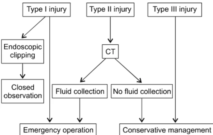
Fig. 2. Algorithm for the management of endoscopic retrograde cholangiopancreatography-related perforations. CT, computed tomography.
And post operative complications like leakage and fistula occurred in delayed operation. In contrast, the patients who received an immediate operation (No. 2, 3, 7) experi- enced only mild inflammation and had relatively fresh tis- sue, so surgeries were uneventful and post-operative re- coveries were satisfactory. Avgerinos et al. [15] insisted that the interval between perforation and operation was of great significance. Mortality rates increase dramatically with delayed surgical management (>24 hours). During conservative management, patients’ vital signs remained stable and pains were alleviated. We thought that the rea- son for the resolved pain and stable vital signs during con- servative management would be not intra-peritoneal per- foration but, retroperitoneal perforation. Because the ret- roperitoneal area is a trapped space, the abscess is likely to be localized. When the operation was done in the 3 or 10 days of conservative management, it was difficult to deal with remaining inflammatory tissues. We could not per- form the previously planned extensive surgery in patient- number 8 because of severe tissue inflammation. Conside- ring the operative findings in patients with delayed oper- ation (No. 4, 8, 9), if these patients hadn’t undergone sur- gery, they would not have recovered within a short period.
So, we propose that when we are aware of ERCP related perforation, emergency operation would be better than delayed operation because of the poor prognosis for can- cer leakage as well as high morbidity for delayed opera- tion. It would be best if ERCP is performed under prepara- tion for extensive surgery just in case of suspicious malig- nancy. Sometimes it is necessary to perform ERCP before completion of preparation for extensive surgery. Using frozen biopsy in cases of suspicious malignancy will help to minimize the extent of operation of transduodenal phincteroplasty or primary closure.
Sarli et al. [16] reported a wide range of operative proce- dures for the treatment of ERCP-associated perforations, including simple retroperitoneal drainage, duodenal re- pair around a T-tube inserted into the perforation, com- mon bile duct exploration and T-tube placement, duode- nal diversion by antrectomy and gastrojejunostomy or gastrojejunostomy with pyloric exclusion, and pancrea- ticoduodenectomy. We employed different methods in- cluding primary closure, T-tube choledochostomy, duode-
nal diverticulization, and the classic Whipple operation based on radiologic and operative findings. We believe that the operative modality is best decided on a case- by-case basis.
In conclusion, type I injuries require immediate surgical management after ERCP or immediate endoscopic closure during ERCP. When type II injuries occur, CT evaluation is needed for evaluation of fluid collection. If there is dirty fluid collection in the intra- and retro-peritoneal area, sur- gical management should be considered. If there is no flu- id collection, conservative treatment is possible. Type III injuries almost always require conservative treatment with ENBD drainage.
When surgery is recommended, immediate surgery is preferable to delayed surgery due to high morbidity (Fig.
2).
CONFLICTS OF INTEREST
No potential conflict of interest relevant to this article was reported.
REFERENCES
1. Christensen M, Matzen P, Schulze S, Rosenberg J.
Complications of ERCP: a prospective study. Gastrointest Endosc 2004;60:721-31.
2. Enns R, Eloubeidi MA, Mergener K, Jowell PS, Branch MS,
Pappas TM, et al. ERCP-related perforations: risk factors and management. Endoscopy 2002;34:293-8.
3. Pungpapong S, Kongkam P, Rerknimitr R, Kullavanijaya P.
Experience on endoscopic retrograde cholangiopancrea- tography at tertiary referral center in Thailand: risks and complications. J Med Assoc Thai 2005;88:238-46.
4. Booth FV, Doerr RJ, Khalafi RS, Luchette FA, Flint LM Jr.
Surgical management of complications of endoscopic sphincterotomy with precut papillotomy. Am J Surg 1990;
159:132-5.
5. Stapfer M, Selby RR, Stain SC, Katkhouda N, Parekh D, Jabbour N, et al. Management of duodenal perforation af- ter endoscopic retrograde cholangiopancreatography and sphincterotomy. Ann Surg 2000;232:191-8.
6. Howard TJ, Tan T, Lehman GA, Sherman S, Madura JA, Fogel E, et al. Classification and management of perfo- rations complicating endoscopic sphincterotomy. Surgery 1999;126:658-63.
7. Kayhan B, Akdoğan M, Sahin B. ERCP subsequent to ret- roperitoneal perforation caused by endoscopic sphinc- terotomy. Gastrointest Endosc 2004;60:833-5.
8. Morgan KA, Fontenot BB, Ruddy JM, Mickey S, Adams DB. Endoscopic retrograde cholangiopancreatography gut perforations: when to wait! When to operate! Am Surg 2009;75:477-83.
9. Seibert DG. Use of an endoscopic clipping device to repair a duodenal perforation. Endoscopy 2003;35:189.
10. Mutignani M, Iacopini F, Dokas S, Larghi A, Familiari P, Tringali A, et al. Successful endoscopic closure of a lateral duodenal perforation at ERCP with fibrin glue. Gastroint- est Endosc 2006;63:725-7.
11. Ryozawa S, Iwamoto S, Iwano H, Ishigaki N, Taba K, Sakaida I. ERCP using double-balloon endoscopes in pa- tients with Roux-en-Y anastomosis. J Hepatobiliary Pan- creat Surg 2009;16:613-7.
12. Husain S, Garmager K, McPhee MS, Jacob KM, Fisher JK, Helzberg JH. The significance of retroperitoneal air follow- ing endoscopic sphincterotomy [abstract]. Gastrointest Endosc1995;41:400.
13. Cho MS, Park DE, Chae KM. Management for duodenal perforation caused by endoscopic retrograde cholangio- pancreatography (ERCP). J Korean Surg Soc 2007;72:210-5.
14. Chung RS, Sivak MV, Ferguson DR. Surgical decisions in the management of duodenal perforation complicating en- doscopic sphincterotomy. Am J Surg 1993;165:700-3.
15. Avgerinos DV, Llaguna OH, Lo AY, Voli J, Leitman IM.
Management of endoscopic retrograde cholangiopancrea- tography: related duodenal perforations. Surg Endosc 2009;23:833-8.
16. Sarli L, Porrini C, Costi R, Regina G, Violi V, Ferro M, et al.
Operative treatment of periampullary retroperitoneal per- foration complicating endoscopic sphincterotomy. Surgery 2007;142:26-32.