Laparoscopic common bile duct exploration in patients with previous upper abdominal operations
Keong Won Yun1,2, Young Joon Ahn1, Hae Won Lee1, In Mok Jung1, Jung Kee Chung1, Seung Chul Heo1, Ki-Tae Hwang1, and Hye Seong Ahn1
Department of Surgery, 1Seoul Metropolitan Government Seoul National University Boramae Medical Center,
2Seoul National University College of Medicine, Seoul, Korea
Backgrounds/Aims: We aimed to to evaluate the feasibility of laparoscopic common bile duct exploration (LCBDE) in patients with previous upper abdominal surgery. Methods: Retrospective analysis was performed on data from the attempted laparoscopic common bile duct exploration in 44 patients. Among them, 5 patients with previous lower ab- dominal operation were excluded. 39 patients were divided into two groups according to presence of previous upper abdominal operation; Group A: patients without history of abdominal operation. (n=27), Group B: patients with history of upper abdominal operation. Both groups (n=12) were compared to each other, with respect to clinical characteristics, operation time, postoperative hospital stay, open conversion rate, postoperative complication, duct clearance and mortality. Results: All of the 39 patients received laparoscopic common bile duct exploration and choledochotomy with T-tube drainage (n=38 [97.4%]) or with primary closure (n=1). These two groups were not statistically different in gen- der, mean age and presence of co-morbidity, mean operation time (164.5±63.1 min in group A and 134.8±45.2 min in group B, p=0.18) and postoperative hospital stay (12.6±5.7 days in group A and 9.8±2.9 days in group B, p=0.158).
Duct clearance and complication rates were comparable (p>0.05). 4 cases were converted to open in group A and 1 case in group B respectively. In group A (4 of 27 (14.8%) and 1 of 12 (8.3%) in group B, p=0.312) Trocar or Veress needle related complication did not occur in either group. Conclusions: LCBDE appears to be a safe and effective treatment even in the patients with previous upper abdominal operation if performed by experienced laparoscopic sur- geon, and it can be the best alternative to failed endoscopic retrograde cholangiopancreatography for difficult cholelithiasis. (Korean J Hepatobiliary Pancreat Surg 2012;16:154-159)
Key Words: Laparoscopic common bile duct exploration; Previous surgery
Received: October 12, 2012; Revised: October 15, 2012; Accepted: October 16, 2012 Corresponding author: Young Joon Ahn
Department of Surgery, Seoul Metropolitan Government Seoul National University Boramae Medical Center, 20, Boramae 5-gil, Dongjak-gu, Seoul 156-707, Korea
Tel: +82-2-870-2274, Fax: +82-2-870-3863, E-mail: [email protected]
Copyright Ⓒ 2012 by The Korean Association of Hepato-Biliary-Pancreatic Surgery Korean Journal of Hepato-Biliary-Pancreatic Surgery ∙ pISSN: 1738-6349
INTRODUCTION
Traditionally, the management of common bile duct (CBD) stones required open laparotomy and bile duct ex- ploration which was first performed by Courvoisier and Thomton separately in 1889.1 Since the introduction of endoscopic retrograde cholangiopancreatography (ERCP) and endoscopic sphincterotomy (EST) in the 1970s, endo- scopic management of CBD stones has gained popularity.2,3 After the introduction of laparoscopic cholecystectomy (LC) from France in 1987, the mainstream treatment of CBD stones with gallbladder stones is ERCP combined with LC.2,3 But, this is generally performed in two stages, which makes the patients uncomfortable not only in clin-
ical but also in an economical respect . It also can result in the need for repeated procedures if it fails, and some- times surgery is needed.
Recently, laparoscopic skills have developed quite rapidly. In the case of CBD stones, there are many reports involving laparoscopic procedures. Since the introduction of laparoscopic CBD exploration (LCBDE) in 1991, a sin- gle-stage approach without attempting ERCP has been suggested. These days, the majority of patients suffering from concomitant gallstones and CBD stones have been managed by a single-stage approach. It has been proven not only feasible but also effective in many reports. This procedure appears to be shorter hospital stay and a similar stone clearance rate, relative cost-effectiveness while pre-
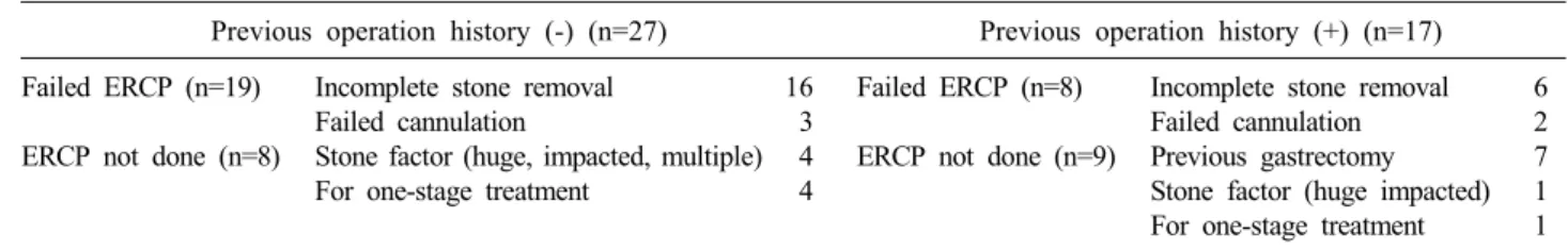
Table 1. Indications of laparoscopic common bile duct exploration
Previous operation history (-) (n=27) Previous operation history (+) (n=17) Failed ERCP (n=19)
ERCP not done (n=8)
Incomplete stone removal Failed cannulation
Stone factor (huge, impacted, multiple) For one-stage treatment
16 3 4 4
Failed ERCP (n=8) ERCP not done (n=9)
Incomplete stone removal Failed cannulation Previous gastrectomy Stone factor (huge impacted) For one-stage treatment
6 2 7 1 1 serving the function of the sphinter of Oddi and less
ERCP-related complications.4,5 But, in cases with history of prior abdominal operation, the laparoscopic approach had potential difficulties - manipulation of instruments, potential risks of general anesthesia in patients who had medical problems, and technical demands.6-11
Previous abdominal surgery has been considered as a relative contraindication for laparoscopy.12-14 With devel- opment of surgical skills and instruments recently, the lap- aroscopic procedures were performed as a common treat- ment in CBD stones even in complicated cases. The use- fulness of LCBDE has been reported with success rates in the range of 93.3-100% in general patients.12 The im- pact of previous abdominal operations on LC and urologic procedures has been studied, and the outcomes were not found to be affected by prior surgery.12-16 However, only a few reports about the impact of previous abdominal op- erations on LCBDE have been published. We aimed to evaluate the feasibility of LCBDE even in patients with previous abdominal surgery by a single surgeon who ex- perienced LC from over 200 cases in patients with upper abdominal operations.
METHODS
Patients and grouping
From December 2003 to March 2012, 44 patients with a diagnosis of CBD stone underwent LCBDE at the Department of Surgery, Boramae Medical Center, Seoul, Korea. All patients were diagnosed with abdominal com- puted tomography (CT), received ERCP or magnetic reso- nance cholangiopancreatography (MRCP), and informed about their health conditions. Among them, 27 patients were without history of previous abdominal surgery, while 17 patients had received various kinds of abdominal oper- ation before. LCBDE were applied to (1) the patients with failed ERCP (n=27): incomplete stone removal (n=22),
failed cannulation (n=5), and (2) the patients who did not have ERCP (n=17): stone factor (multiple, huge, im- pacted) (n=5), anatomic variation came from gastric re- section (n=7), for one- stage treatment (n=5) (Table 1).
Among them, we excluded the 5 patients with previous lower abdominal operations who did not meet the criteria in this study.
We divided the remaining 39 patients into two groups;
27 patients without any history of previous abdominal op- eration were in Group A and 12 patients who underwent previous upper abdominal surgery were in Group B. Both groups were compared to each other with respect to their clinical characteristics (sex, age, body weight and co-mor- bidity), operation time, postoperative hospital stay, post- operative complication and conversion rate to open surgery.
Operation techniques
Under general anesthesia, the patient was laid in a 15 degree back up and 15 degree right tilt position. We used a four-trocar technique; 11-mm infra-umbilical port for laparoscope, one 11-mm port in subxiphoid for working port and future choledochoscope, one 5-mm port 5cm in- ferior to the right subcostal margin on the right mid- clavicular line, one 5 mm post on the right flank for assistance. If the previous incision scar was more than 3 cm far from the umbilicus, we used an infra-umbilical port, otherwise we made a camera port 3 cm right lateral to the umbilicus. After exposure of the rectus fascia, up- ward lifting of the full thickness of the rectus fascia with a Cocher clamp was followed by a careful insertion of a Veress needle. Access into the peritoneal cavity by a Veress needle could be confirmed by free dropping of normal saline and initial low intraabdominal pressure.
After insufflations of CO2 gas to 10 mmHg, we incised the anterior rectus fascia to expose the peritoneal layer.
Regurgitation of insufflated CO2 gas with simple puncture
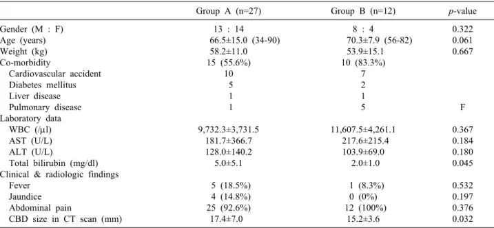
Table 3. Characteristics of patients in both groups
Group A (n=27) Group B (n=12) p-value
Gender (M : F) Age (years) Weight (kg) Co-morbidity
Cardiovascular accident Diabetes mellitus Liver disease Pulmonary disease Laboratory data WBC (/μl) AST (U/L) ALT (U/L)
Total bilirubin (mg/dl) Clinical & radiologic findings Fever
Jaundice Abdominal pain
CBD size in CT scan (mm)
13 : 14
66.5±15.0 (34-90) 58.2±11.0 15 (55.6%)
10 5 1 1 9,732.3±3,731.5
181.7±366.7 128.0±140.2
5.0±5.1 5 (18.5%) 4 (14.8%) 25 (92.6%) 17.4±7.0
8 : 4
70.3±7.9 (56-82) 53.9±15.1 10 (83.3%)
7 2 1 5 11,607.5±4,261.1
217.6±215.4 103.9±69.0
2.0±1.0 1 (8.3%)
0 (0%) 12 (100%)
15.2±3.6
0.322 0.061 0.667
F 0.367 0.184 0.180 0.045 0.532 0.197 0.376 0.032 Group A, absence of previous abdominal operation; Group B, presence of upper abdominal operation; AST, aspartate trans- aminase; ALT, alanine transaminase; CBD, common bile duct; CT, computed tomography
Table 2. Types of previous abdominal operations in group B
Incision Operation Number
Upper midline
Laparoscopy (3 holes) McBurney Low midline
Gastrectomy (total, subtotal) Primary repair of gastic ulcer Feeding jejunostomy
Laparoscopic cholecystectomy Appendectomy*
Hysterectomy*
7 2 1 2 3 2 Group B, presence of upper abdominal operation
*Not included in this study
of peritoneum could assure the access to the intra-abdomi- nal cavity. Finger dissection was a safe and useful method for clearance of adhesion without bowel injury.
After the identification of CBD, choledochotomy was performed with a vertical incision approximately 1 to 1.5 cm in length with electrocautery. Both edges of the opened common bile duct were stitched-up with Vicryl suture and retracted in the opposite direction for easy en- trance of choledochoscope. Through the 5-mm flexible choledochoscope, we identified the CBD stones, and re- moved them by flushing method with sterile saline, a stone basket, and sometimes crashed them by electro- hydraulic lithotripsy.
The choledochotomy site was closed by continuous su- ture, using vicryl 3-0 with T-tube insertion and chol-
ecystectomy was performed. We ended the operation with a Jackson-Pratt drain insertion into the operation site. All the operations were performed by a single surgeon.
Statistical analysis
Statistical significance was analyzed using the Chi-square test, Student’s t-tests. A p-value<0.05 was considered significant.
RESULTS
During the study period, a total number of 44 patients underwent LCBDE initially for CBD stones in our hospital. This population comprised 24 (54.5%) males and 20 (45.5%) females with a mean age of 67.5±14.0 years (range, 34 to 90 years). There were 17 cases with previous operation history. Of 17 cases, 10 had upper abdominal operation for gastric disease (n=9) and temporary jejunos- tomy (n=1), 2 underwent laparoscopic cholecystectomy and another 5 cases had gynecologic procedures via low midline incision (n=2) and open appendectomy with McBurney incision (n=3) which were excluded in this study (Table 2).
Preoperative status is presented in Table 3. In terms of male to female ratio, mean age, body weight, there were no significant differences. 15 cases in group A and 10
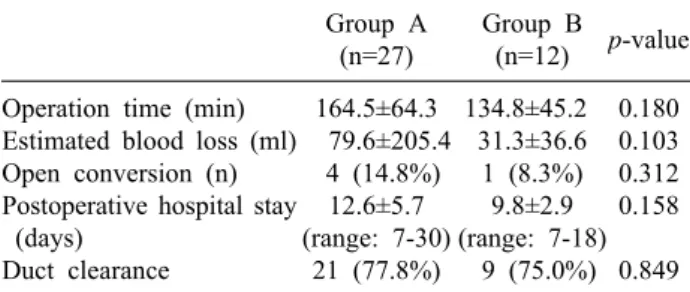
Table 4. Comparison of operative results in both groups Group A
(n=27)
Group B (n=12) p-value Operation time (min)
Estimated blood loss (ml) Open conversion (n) Postoperative hospital stay (days)
Duct clearance
164.5±64.3 79.6±205.4
4 (14.8%) 12.6±5.7 (range: 7-30)
21 (77.8%)
134.8±45.2 31.3±36.6 1 (8.3%) 9.8±2.9 (range: 7-18)
9 (75.0%) 0.180 0.103 0.312 0.158 0.849 Group A, absence of previous abdominal operation; Group B, presence of upper abdominal operation
Table 5. Postoperative complications
Group A Group B Postoperative complication
Biliary stricture Wound problem Bile leakage
3 (11.1%) 1 1*
1
1 (8.3%) 0 0 1 Group A, absence of previous abdominal operation; Group B, presence of upper abdominal operation
*A case of open conversion cases in group B had an additional underlying disease
such as hypertension, cerebral disease, diabetes mellitus, chronic liver diseases, pulmonary disease like tuberculosis or pneumonia without a significant difference between the two groups (p=0.151).
The mean operation time was 164.5±63.1 minutes in group A and 134.8±45.2 minutes in group B, but there was no significant difference (p=0.180). For estimated blood loss (EBL), there was no significant difference (79.6±205.4 ml in group A and 31.3±36.6 ml group B, p=0.103) though it showed more blood loss in group A than in group B. No significant difference in the post- operative hospital stay was observed between the two groups (p=0.158). There were no complications due to port placement in either group. In 3 patients, minor bile leakage was shown during postoperative cholangiography at postoperative 7 days. But no additional procedure was needed for the management of the minor bile leakage.
There was no mortality and no postoperative bleeding or bowel leakage A total of 5 cases were converted to open surgery (4 in group and 1 in group B). The reasons for open conversion in group A were severe intra-abdominal adhesion in 2 cases, an excessively small size of the ab- dominal cavity which impeded manipulation of the laparo- scopic instrument, and lastly for possible unexpected in- jury to CBD during dissection. In group B, just one case was converted to open surgery due to severe inflammation (Table 4 and 5).
DISCUSSION
As LC has became the gold standard for treatment of benign gallbladder disease with the development of skills and devices for laparoscopic procedures, the trend of treat-
ment for CBD stones has changed and various options are now prevalent. These options are ERCP with or without EST, LCBDE or open CBD exploration and radiologic intervention. With the advances in endoscopic and laparo- scopic technology, ERCP followed by LC has become the mainstream treatment for patients with concomitant GB and CBD stones in most centers.
However, ERCP has potential serious complications such as pancreatitis, cholangitis, duodenal perforation and life-threatening hemorrhage.17 The stone clearance rate of ERCP is reported as 75% notwithstanding the repeated ses- sions and additional procedures for the difficult CBD stones.
Moreover, ERCP fails in 3% to 10% of all patients. The superiority of the LCBDE with LC exists in the context of a single session procedure for concomitant GB and CBD stone and preservation of the function of sphincter of Oddi which prevents ERCP related complications and post-proce- dural pain. Therefore, LCBDE can be the best alternative to failed ERCP for difficult cholelithiasis,18,19 although an experienced laparoscopic surgeon is mandatory. LCBDE is well indicated in ‘difficult cholelithiasis’ which was defined by Tai et al.17 as a failure of endoscopic stone retrieval for the following reasons; (1) access and cannulation diffi- culty, (2) difficult nature of CBD stones in the aspect of number or size, (3) the presence of ERCP-related complica- tions, and (4) contraindication of EST.
In the early period of laparoscopic surgery, the laparo- scopic approach was contraindicated for patients with history of prior abdominal operation. However, not only with the development of surgical skills and instruments but with the accumulated experience of laparoscopic surgeons, compli- cated laparoscopic procedures can be overcome with or with- out previous operations.20-22 Several concerns that make sur- geons hesitate about the laparoscopic approach in previously operated abdomen include (1) possible risk of injury to organs
adjacent to the previous incision wound during insertion of the Veress needle or trocar (2) curtain-like severe adhesion that prevent access to the target organ from the prior operation as well as present inflammatory processes in spite of barely establishing pneumoperitoneum (3) risk of bowel injury dur- ing adhesiolysis and (4) higher probability of conversion to open surgery.
Site selection for the initial trocar is of importance with respect to both patient’s safety and exposure of the biliary tract. Li et al.20 reported that the blind puncture of a Veress needle is safe if the previous scar is more than 3 cm from the umbilicus, whereas the Hasson (open) technique is rec- ommended if the scar is less than 3 cm from the umbilicus.
In addition, we think the blind access with the trocar is not recommended even more than 3 cm from the previous scar, because the small bowel can be adherent to the innocent abdominal wall more than 3 cm from the previous incision in our experience. We applied the Veress needle method in almost all cases even less than 3 cm from the previous scar. Grasping the full layer of fascia of the rectus muscle with a Kocher clamp can facilitate abdominal wall lifting for safe insertion of the Veress needle. Confirmation of safe access into the peritoneal cavity can be achieved by free dropping of normal saline through the Veress needle followed by identifying initial low intraabdominal pressure after connecting CO2 gas. Mechanical insufflation of CO2
gas can detach multiple loose adhesions of bowel loops from the abdominal wall, which prevents possible injury to other organs not adherent to the previous scar. As usual, a simple puncture with the Veress needle to the adherent small bowel does not appear to cause serious problems, and injury from the Veress needle seems to be easily healed unless the bowel loop is severely distended. Hanney et al.23 reported two cases of aortic injury during use of the Hasson cannula and supported the Veress needle technique. Dunne et al.24 compared the Veress needle and Hasson technique for establishing pneumoperitoneum, which showed no sig- nificant difference between the technique chosen and in- cidence of complications. As aforementioned, we utilized an open technique with finger dissection after establishing pneumoperitoneum with a Veress needle (modified Hasson technique), which enabled avoidance of serious complica- tions possibly encountered at the stage of entrance to the abdominal cavity.
As noninvasive methods, Viscera slide ultrasound and
magnetic resonance imaging (MRI) have been suggested, and they can be considered for detecting intra-abdominal adhe- sions to the abdominal wall and adhesion-free areas so as to prevent injuries during the creation of pneumoperitoneum.22,25 They may serve as a diagnostic tool for future planning of laparoscopic surgery assuming that the clinical effi- ciency and economic feasibility are verified.
Once the abdominal cavity has been accessed, adhesiol- ysis should be performed enough to insert a second trocar to aid in acquisition of a view around the initial port, fol- lowed by retraction, and further dissection. Rather than adhesiolysis of all intra-abdominal adhesions, it is recom- mended to remove the adhesions interfering with the ap- proach to the operative field or laparoscopic procedures.
Excessive use of electrocautery should be refrained to pre- vent unwitting burn injury during dissection. Additionally, it should be considered that imprudent dissection with mere presumption of adhesion pattern beyond the visible area may give rise to unexpected injury to organs.
Once an upper gastrointestinal operation has been per- formed, especially biliary tract operations, dense adhe- sions around the hepatoduodenal ligament and upward ad- hesion of the 2nd portion of the duodenum are usually observed during re-exploration in laparoscopic fashion.
Identification of the gallbladder is most crucial for se- quential dissection along the right side of the common bile duct for bile duct exploration. If cholecystectomy has been performed in a previous operation, hepatic hilum should be approached by freeing the lateral parietes and beginning dissection on the right side along the lateral in- ferior border of the liver.20 Resultant down positioning of the transverse colon and duodenum enables finding of the common bile duct, hence the following procedure can be continued. In our opinion, complete dissection of the left side of the common bile duct is not recommended for pre- vention of injury to hepatic artery or portal vein injury.
Clearance of the anterior aspect of the common bile duct seems to be enough for the exploration with choledocho- scopy and other procedures for stone removal. We usually stitch up the opened edge of the bile duct for lateral re- traction in the opposite direction, and this procedure fixes the common bile duct and allows easy access of chol- edochoscope into the bile duct lumen.
In some cases, we encountered rather tedious situations owing to abundant muddy stones spilled out through the
opened bile duct. It took much operation time to clear up the sludge and stones from the abdominal cavity and the patient would suffer from postoperative fever. Accordingly, a patient with a highly fragile stone does not appear to be indicated for LCBDE.
The problem of T-tube insertion is still debatable and primary closure of the bile duct after exploration with or without biliary stent is in vogue at the moment.19,26-30 We inserted T-tubes in all cases as a route for identification and removal of the possible residual stones.
In summary, postoperative data of LCBDE in the pa- tients with previous upper abdominal operation on oper- ation time, conversion rate, postoperative complication rate was comparable to those in the patients without pre- vious history. With these results, LCBDE can be described as safe, minimally invasive procedures in the patient with previous abdominal operation as far as an experienced lap- aroscopic surgeon is available, and it can be the best alter- native to failed ERCP for difficult cholelithiasis.
REFERENCES
1. Beal JM. Historical perspective of gallstone disease. Surg Gynecol Obstet 1984;158:181-189.
2. Soper NJ. Laparoscopic general surgery--past, present, and future.
Surgery 1993;113:1-3.
3. Crawford DL, Phillips EH. Laparoscopic common bile duct exploration. World J Surg 1999;23:343-349.
4. Vandervoort J, Soetikno RM, Tham TC, et al. Risk factors for complications after performance of ERCP. Gastrointest Endosc 2002;56:652-656.
5. Poulose BK, Arbogast PG, Holzman MD. National analysis of in-hospital resource utilization in choledocholithiasis management using propensity scores. Surg Endosc 2006;20:186-190.
6. Franklin ME Jr, Pharand D, Rosenthal D. Laparoscopic common bile duct exploration. Surg Laparosc Endosc 1994;4:119-124.
7. Lezoche E, Paganini AM. Single-stage laparoscopic treatment of gallstones and common bile duct stones in 120 unselected, consec- utive patients. Surg Endosc 1995;9:1070-1075.
8. Millat B, Fingerhut A, Deleuze A, et al. Prospective evaluation in 121 consecutive unselected patients undergoing laparoscopic treatment of choledocholithiasis. Br J Surg 1995;82:1266-1269.
9. Rhodes M, Nathanson L, O'Rourke N, et al. Laparoscopic explora- tion of the common bile duct: lessons learned from 129 consecutive cases. Br J Surg 1995;82:666-668.
10. Urbach DR, Khajanchee YS, Jobe BA, et al. Cost-effective manage- ment of common bile duct stones: a decision analysis of the use of endoscopic retrograde cholangiopancreatography (ERCP), intra- operative cholangiography, and laparoscopic bile duct exploration.
Surg Endosc 2001;15:4-13.
11. Rhodes M, Sussman L, Cohen L, et al. Randomised trial of laparo- scopic exploration of common bile duct versus postoperative endo- scopic retrograde cholangiography for common bile duct stones.
Lancet 1998;351:159-161.
12. Karayiannakis AJ, Polychronidis A, Perente S, et al. Laparoscopic cholecystectomy in patients with previous upper or lower abdomi- nal surgery. Surg Endosc 2004;18:97-101.
13. Parsons JK, Jarrett TJ, Chow GK, et al. The effect of previous abdominal surgery on urological laparoscopy. J Urol 2002;168:2387-2390.
14. Schirmer BD, Dix J, Schmieg RE Jr, et al. The impact of previous abdominal surgery on outcome following laparoscopic cholecystectomy.
Surg Endosc 1995;9:1085-1089.
15. Yu SC, Chen SC, Wang SM, et al. Is previous abdominal surgery a contraindication to laparoscopic cholecystectomy? J Laparoendosc Surg 1994;4:31-35.
16. Law WL, Lee YM, Chu KW. Previous abdominal operations do not affect the outcomes of laparoscopic colorectal surgery. Surg Endosc 2005;19:326-330.
17. Tai CK, Tang CN, Ha JP, et al. Laparoscopic exploration of com- mon bile duct in difficult choledocholithiasis. Surg Endosc 2004;18:910-914.
18. Shin HS, Chun KS, Song IS. Laparoscopic common bile duct ex- ploration in patients with failed endoscopic stone extraction.
Korean J Hepatobiliary Pancreat Surg 2009;13:164-170.
19. Park YC, Jeong JS, Jeong JG, et al. The clinical outcome of laparo- scopic common bile duct exploration for the primary treatment of choledocholithiasis. Korean J Hepatobiliary Pancreat Surg 2011;15:13-18.
20. Li LB, Cai XJ, Mou YP, et al. Reoperation of biliary tract by laparoscopy: experiences with 39 cases. World J Gastroenterol 2008;14:3081-3084.
21. Tang CN, Tsui KK, Yang GP, et al. Laparoscopic exploration of com- mon bile duct in post-gastrectomy patients. Hepatogastroenterology 2008;55:846-849.
22. Zinther NB, Zeuten A, Marinovskij E, et al. Detection of abdominal wall adhesions using visceral slide. Surg Endosc 2010;24:
3161-3166.
23. Hanney RM, Carmalt HL, Merrett N, et al. Vascular injuries during laparoscopy associated with the Hasson technique. J Am Coll Surg 1999;188:337-338.
24. Dunne N, Booth MI, Dehn TC. Establishing pneumoperitoneum:
Verres or Hasson? The debate continues. Ann R Coll Surg Engl 2011;93:22-24.
25. Chung HJ, Park IY. Ultrasound can prevent visceral injuries during the creation of pneumoperitoneum in patients with previous abdomi- nal surgery. J Korean Soc Endosc Laparosc Surg 2006;9:45-48.
26. Wu JS, Soper NJ. Comparison of laparoscopic choledochotomy closure techniques. Surg Endosc 2002;16:1309-1313.
27. El-Geidie AA. Is the use of T-tube necessary after laparoscopic choledochotomy? J Gastrointest Surg 2010;14:844-848.
28. Zhang WJ, Xu GF, Wu GZ, et al. Laparoscopic exploration of common bile duct with primary closure versus T-tube drainage:
a randomized clinical trial. J Surg Res 2009;157:e1-5.
29. Chen CC, Wu SD, Tian Y, et al. Sphincter of Oddi-preserving and T-tube-free laparoscopic management of extrahepatic bile duct calculi. World J Surg 2011;35:2283-2289.
30. Jameel M, Darmas B, Baker AL. Trend towards primary closure following laparoscopic exploration of the common bile duct. Ann R Coll Surg Engl 2008;90:29-35.