Received: November 16, 2018 Revised: December 7, 2018 Accepted: December 7, 2018 CliniCAl
neurophysiology
Correspondence to Ki-Jong Park
Department of Neurology, Gyeongsang National University Changwon Hospital, 11 Samjeongja-ro, Seongsan-gu, Chang- won 51472, Korea
Tel: +82-55-214-3810 Fax: +82-55-214-3255 E-mail: pkjong@gnu.ac.kr
http://www.e-acn.org copyright © 2019 the Korean Society of clinical neurophysiology
Reference ranges for autonomic
function tests in healthy Korean adults
Kee Hong Park1, Byoung Joon Kim2, Sa-Yoon Kang3, Sun-Young Oh4, Eun Hee Sohn5, Kyeong-jin Song5, Jin-Hong Shin6, Kyoung Hwa Kang6, Eun Bin Cho7, Heejeong Jeong7, Hyung Lee8, Hyun Ah Kim8, Rock Bum Kim9, and Ki-Jong Park7,10
1Department of Neurology, Gyeongsang National University Hospital, Jinju, Korea
2Department of Neurology, Sungkyunkwan University School of Medicine, Seoul, Korea
3Department of Neurology, Jeju National University College of Medicine, Jeju, Korea
4Department of Neurology, Chonbuk National University School of Medicine, Jeonju, Korea
5Department of Neurology, Chungnam National University Hospital, Daejeon, Korea
6Department of Neurology, Pusan National University Yangsan Hospital, Pusan National University School of Medicine, Yangsan, Korea
7Department of Neurology, Gyeongsang National University Changwon Hospital, Changwon, Korea
8Department of Neurology, Keimyung University School of Medicine, Daegu, Korea
9Regional Cardiocerebrovascular Center, Gyeongsang National University Hospital, Jinju, Korea
10Department of Neurology, Gyeongsang National University School of Medicine, Jinju, Korea
Background: The standardized autonomic function test has become widely available. How- ever, there are no reference data for this test for the Korean population. This study explored reference data for sudomotor and cardiovagal function tests for the Korean population.
Methods: The sweat volume by quantitative sudomotor axon reflex test, heart-rate response to deep breathing (HRdb), expiration:inspiration (E:I) ratio, and Valsalva ratio (VR) were mea- sured in 297 healthy Korean volunteers aged from 20 to 69 years. Multivariate regression analysis was performed to evaluate the effects of age, sex, and body mass index on these variables. The 2.5th, 5th, 10th, 90th, 95th, and 97.5th percentile values were obtained for each investigation.
Results: The sweat volume was higher in males than in females. The HRdb and E:I ratio were negatively correlated with age, and were higher in males than in females. The VR was nega- tively correlated with age, but it was not correlated with sex.
Conclusions: This study has provided data on the reference ranges for sudomotor and car- diovagal function tests in healthy Korean adults.
Key words: Autonomic nervous system; Reference range, Sweating; Parasympathetic ner- vous system
ORCID
Kee Hong Park
https://orcid.org/0000-0001-5724-7432 Byoung Joon Kim
https://orcid.org/0000-0001-8424-881X Sa-Yoon Kang
https://orcid.org/0000-0001-6755-089X Sun-Young Oh
https://orcid.org/0000-0003-3174-1680 Eun Hee Sohn
https://orcid.org/0000-0001-5610-7606 Kyeong-jin Song
https://orcid.org/0000-0003-2330-8377 Jin-Hong Shin
https://orcid.org/0000-0002-5174-286X Kyoung Hwa Kang
https://orcid.org/0000-0002-6070-0821 Eun Bin Cho
https://orcid.org/0000-0003-2021-8587 Heejeong Jeong
https://orcid.org/0000-0001-6531-1860 Hyung Lee
https://orcid.org/0000-0003-0568-6104 Hyun Ah Kim
https://orcid.org/0000-0002-2140-4763 Rock Bum Kim
https://orcid.org/0000-0001-5868-0465 Ki-Jong Park
https://orcid.org/0000-0003-4391-6265
IntroductIon
The autonomic nervous system is involved in several bodily functions, including regulating the blood pressure, pulse, body temperature, respiration, digestion, urination, and sexual function, and it is affected by various diseases. An autonomic function test is used to objectively assess these functions, and noninvasive and quantifiable tests have recently been developed and used due to advancements in technology.1 Most of the previously published data on reference ranges are focused on Western populations,2,3 and there are almost no data available for Asians.4 However, autonomic function varies with race,5 and so reference rang- es for autonomic function tests for Koreans are needed. A single domestic institute recently published reference rang- es for sudomotor and cardiovagal functions according to age, sex, and body mass index (BMI).6 Rather than recruiting participants only from the single center, which would have been a limitation of the study, participants were additionally recruited from other centers. By so doing, the proposed ref- erence ranges for autonomic function tests would be more representative of the Korean population.
MaterIals and Methods
Participants
Tests were performed on 297 healthy volunteers (135 males and 162 females) in the neurology departments of six hospi- tals from March 2012 to May 2016. The included participants did not suffer from any neurological or systemic disorder and were not taking medication regularly. Participants with disorders that can affect the autonomic nervous system, such as diabetes, alcoholism, malnutrition, and anticancer therapy, were excluded. The participants were divided into groups based on sex and age. Clinical information such as age, sex, height, weight, and BMI were collected as the main outcome variables. Informed consent was obtained from all of the subjects.
Autonomic function tests
The tests were performed according to the “Guidelines for Autonomic Function Test” previously published in the Jour- nal of Pain and Autonomic Disorders. The protocols of the
tests are described below.7
The quantitative sudomotor axon reflex test (QSART) The QSART quantitatively assesses the postganglionic sym- pathetic cholinergic function and measures sweat volume after stimulating sweat glands using a 10% acetylcholine solution. In this study the QSART was applied to the medial forearm (at three-quarters of the distance from the ulnar epicondyle to the pisiform bone), proximal lateral leg (5 cm distal to the fibular head), distal medial leg (5 cm proximal to the medial malleolus), and proximal foot (above the exten- sor digitorum brevis muscle). The measurements were per- formed using the commercially available Q-Sweat
®
quanti-tative sweat measurement system (WR Medical Electronics Company, Maplewood, MN, USA), which was developed based on the principles of the QSART.
Heart-rate response to deep breathing (HRdb)
The HRdb test assesses the function of the parasympathetic nervous system based on the principle that the heart rate increases during inhalation and decreases during exhalation.
The heart rate is measured during 5 seconds of inhalation and 5 seconds of exhalation, with these measurements re- peated eight times. After a 2-minutes break, one more test is performed using a similar protocol. The five largest differ- ences in heart rate during inhalation and exhalation from among the first eight measurements are selected and aver- aged. The expiration:inspiration (E:I) ratio is found by dividing the longest RR interval during expiration by the shortest RR interval during inhalation.
Valsalva ratio (VR)
The VR is determined by measuring the blood pressure and heart rate while blowing into a mouthpiece at a pressure of at least 40 mmHg for 15 seconds. This is repeated two more times after a 3-minute break. The VR is calculated by dividing the highest heart rate late during phase 2 with the lowest heart rate during phase 4.
Statistical analyses
Statistical analyses were performed using IBM SPSS (version 21.0, IBM Corp., Armonk, NY, USA) and MedCalc for Windows (version 17.5.3, MedCalc Software, Ostend, Belgium). Stu- dent’s t-test or the Mann-Whitney U test was used to com-
pare continuous variables, while Fisher’s exact test was used to compare categorical variables. Univariate and multivariate regression analyses with stepwise backward selection were then applied to the main outcome variables (age, sex, and BMI). In cases where sex significantly affected the results, males and females were analyzed as separate groups. In cas- es where age and BMI significantly affected the results, the median, 2.5th, 5th, 10th, 90th, 95th, and 97.5th percentile values were obtained, and a scatter plot and regression fit were produced. Upon performing the regression analysis, the following model assumptions were tested: linearity be- tween the dependent and independent variables, normality of the error distribution, independence, and homoscedas- ticity of the errors. In cases where a model assumption was violated, data were transformed into logarithmic values to ensure that the assumption was satisfied. Internal validation was performed using the bootstrap method to confirm the appropriateness of the final model.
results
The participants were aged 41.6 ± 14.1 years (mean ± standard deviation), and the males were younger than the females (39.6 ± 13.5 vs. 43.3 ± 14.4 years, p = 0.036). The BMI was significantly higher in males than in females (23.1 ± 3.0 vs. 22.3 ± 3.0 kg/m2, p < 0.001; Table 1).
Sudomotor function
The sweat volume was higher in males than in females at all four measurement sites. The sweat volume for the fore- arm increased with age, but no association with age was observed for the other regions. The BMI showed a positive correlation with sweat volume in the proximal and distal legs. The regression equations for the four regions were as follows, where b0 is the intercept, b1 is Ln (age), b2 is sex (female), and b3 is Ln (BMI): forearm, b0 = -2.91, b1 = 0.62, and b2 = -0.57; proximal leg, b0 = -2.14, b2 = -0.72, and b3 = 0.76; distal leg, b0 = -2.72, b2 = -0.88, and b3 = 0.90; and foot, b0 = -0.95 and b2 = -0.64. The 2.5th, 5th, 10th, 90th, 95th, and 97.5th percentile values for the different measurement regions are presented in Table 2.
Table 1. Demographic characteristics of the participants
Total (n = 297) Males (n = 135) Females (n = 162) p-value
Age (years) 41.6 ± 14.1 39.6 ± 13.5 43.3 ± 14.4 0.036
Age group (years) 0.093
20-29 80 45 35
30-39 58 29 29
40-49 57 24 33
50-59 58 21 37
60-69 44 16 28
Height (cm) 165.5 ± 8.6 172.6 ± 5.8 159.6 ± 5.7 <0.001
Weight (kg) 63.6 ± 11.6 72.1 ± 9.7 56.6 ± 7.6 <0.001
Body mass index (kg/m2) 23.1 ± 3.0 24.2 ± 2.6 22.3 ± 3.0 <0.001
Values are presented as mean ± standard deviation or number.
Table 2. Sweat volume in the four tested regions according to sex
Percentile Forearm Proximal leg Distal leg Foot Male:female
2.5th 0.05:0.07 0.25:0.12 0.15:0.10 0.08:0.05 5th 0.08:0.09 0.36:0.17 0.17:0.11 0.08:0.05 10th 0.10:0.11 0.48:0.26 0.29:0.16 0.13:0.017 90th 1.85:1.14 2.90:1.41 2.54:1.25 1.07:0.56 95th 2.46:1.32 3.08:1.63 3.18:1.68 1.25:0.80 97.5th 2.77:1.57 3.24:2.69 3.94:2.37 1.34:0.91
Cardiovagal function
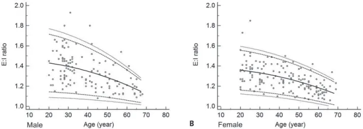
The HRdb and E:I ratio decreased as age increased, and were lower in females than in males. The VR showed a negative correlation with age only. None of the three variables were correlated with BMI. The regression equations for the three variables were as follows, where b0 is the intercept, b1 is Ln (age), and b2 is sex (female): HRdb, b0 = 62.63, b1 = -11.93, and b2 = -2.18 (Fig. 1); E:I ratio, b0 = 0.86, b1 = -0.16, and b2
= -0.04 (Fig. 2); and VR, b0 = 1.32 and b1 = -0.20 (Fig. 3). The 2.5th, 5th, 10th, 90th, 95th, and 97.5th percentile values for the different age groups and sexes in each test are present- ed in Tables 3-5.
dIscussIon
The results of the present study, which involved 297 par- ticipants, were similar to those of a previous domestic sin- gle-center study that involved 181 participants.6 Differences according to sex were clearly observed in all of the variables except for the VR. Decreased autonomic function with in- creasing age was shown in the cardiovagal function test but not in the measurements of sweat volume.
The Mayo Clinic has suggested reference ranges for the QSART.2 However, these reference ranges could not be used because most hospitals other than the Mayo Clinic use the
A B
Fig. 1. Heart-rate response to deep breathing (HRdb) as a function of age for males (A) and females (B). The HRdb was negatively correlated with age and was lower in females than in males. Lines show 2.5th, 5th, 50th, 95th, and 97.5th percentiles. bpm, beats per minute.
A B
Fig. 2. Expiration:inspiration (E:I) ratio as a function of age for males (A) and females (B). The E:I ratio was negatively correlated with age and was lower in females than in males. Lines show 2.5th, 5th, 50th, 95th, and 97.5th percentiles.
Q-Sweat system, which was developed using the QSART principle. The Mayo Clinic subsequently reported a relatively close correspondence between the Q-Sweat system and the QSART, but even that study did not suggest reference rang- es for the Q-Sweat system.3 In a study of the quantitative au- tonomic function test, Novak suggested reference ranges for the Q-Sweat system for different age groups, but no specific statistical methods or percentiles were described.8 Therefore, the present study is significant because it suggests reference ranges for the sweat volume when using the Q-Sweat sys- tem that were obtained by applying statistical methods.
This study found that sex was a significant factor influ- encing the sweat volume, which is consistent with previous studies.2,4,9,10 However, the effect of age on sweat volume
Table 3. Heart-rate response to deep breathing according to age group and sex
Percentile 20-29 years 30-39 years 40-49 years 50-59 years 60-69 years
Male:female
2.5th 11.99:12.90 10.20:7.27 9.83:9.37 7.46:6.16 6.84:4.19
5th 13.25:13.98 10.82:9.86 10.54:9.50 7.82:6.64 6.87:4.49
10th 14.29:14.79 12.18:12.88 11.08:9.79 7.98:8.98 7.02:5.48
90th 32.73:30.03 28.54:25.52 29.00:25.49 19.58:20.38 19.24:13.88
95th 34.05:32.36 31.20:25.96 31.92:25.87 21.96:21.58 21.44:14.81
97.5th 34.48:33.29 35.37:27.25 34.09:26.38 24.03:22.06 21.72:15.64
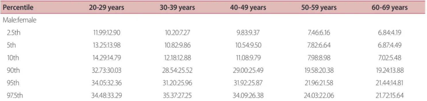
Table 4. Expiration:inspiration ratio according to age group and sex
Percentile 20-29 years 30-39 years 40-49 years 50-59 years 60-69 years
Male:female
2.5th 1.17:1.17 1.18:1.11 1.11:1.10 1.10:1.08 1.10:1.07
5th 1.20:1.17 1.20:1.14 1.14:1.12 1.12:1.10 1.10:1.07
10th 1.22:1.19 1.22:1.18 1.15:1.13 1.12:1.13 1.11:1.08
90th 1.64:1.52 1.52:1.44 1.50:1.43 1.41:1.34 1.33:1.23
95th 1.66:1.61 1.61:1.47 1.61:1.44 1.44:1.37 1.37:1.25
97.5th 1.66:1.75 1.72:1.49 1.70:1.45 1.49:1.39 1.39:1.29
Table 5. Valsalva ratio according to age group
Percentile 20-29 years 30-39 years 40-49 years 50-59 years 60-69 years
2.5th 1.37 1.34 1.42 1.32 1.25
5th 1.42 1.40 1.47 1.34 1.27
10th 1.54 1.45 1.52 1.40 1.31
90th 2.62 2.27 2.26 2.13 1.79
95th 2.73 2.47 2.34 2.15 1.89
97.5th 2.77 2.76 2.42 2.19 2.03
Fig. 3. Valsalva ratio (VR) as a function of age. The VR was negatively correlated with age. Lines show 2.5th, 5th, 50th, 95th, and 97.5th percen- tiles.
is unclear. Three studies conducted in the Mayo Clinic have produced variable results regarding the effect of aging.2,9,10 Furthermore, a study involving Chinese participants found a decreasing trend with age only in the foot region.4 This may be attributable to the effects of race on sudomotor function.11,12 In the present study, sweat volume increased with age in the forearm, whereas at other sites there was no influence of age. The effects of aging and race on the results obtained using the Q-Sweat system therefore need to be further evaluated.
HRdb was negatively correlated with age, which confirms the findings of previous studies.2,10,13,14 However, unlike in other studies, HRdb was lower in females than in males in the present study. The cutoff values of the 5th percentile in the USA and African studies were similar to those for the male group in our study.2,14 Aged females tended to show a lower HRdb, which may be caused by poor compliance.
The effect of age on VR is controversial,2,10,13,15 and our study found a negative correlation between age and VR.
The BMI was not considered when interpreting the au- tonomic function tests because it did not show a clear association, except partly when using the Q-Sweat system.
However, the participants of the present study showed a relatively uniform range of BMI values, with almost no obese participants. It has been reported that cardiovagal function deteriorates in people who are overweight or obese,16 and so caution is required when interpreting our results related to the BMI.
The lack of a disease control group in this study made it difficult to determine the optimal cutoff value. Based on pre- vious studies and our experience, we recommend reference values of the 5th percentiles for the Q-Sweat system, HRdb, E:I ratio, and VR.
The present study was subject to some limitations. First, the included subjects came mainly from a single hospital (GNUH). However, a standardized protocol was applied in all of the included centers, and so our findings can represent the reference ranges for the Korean population. Second, the validity or diagnostic accuracy of reference values in pa- tients with autonomic dysfunction was not assessed. Future studies should therefore include patient groups in order to confirm the findings of the present study.
The number of hospitals performing autonomic function tests is gradually increasing due to the increasing concern
about autonomic disorders. However, interpreting test re- sults has thus far been difficult due to discrepancies in using reference ranges obtained from studies involving only West- ern populations. We believe that the findings of the present study will be helpful not only for patient treatment but also for research into autonomic disorders.
Acknowledgements
This article is modified from an original article that was pub- lished in the Journal of Pain and Autonomic Disorders (the former official journal of the Korean Society of Pain and Au- tonomic Disorders) in June 2017 (titled “Reference range of autonomic function test in Korean healthy adults”).
Conflicts of Interest
The authors declare no competing financial interests.
reFerences
1. Low PA, Tomalia VA, Park KJ. Autonomic function tests: some clin- ical applications. J Clin Neurol 2013;9:1-8.
2. Low PA, Denq JC, Opfer-Gehrking TL, Dyck PJ, O’Brien PC, Slezak JM. Effect of age and gender on sudomotor and cardiovagal function and blood pressure response to tilt in normal subjects.
Muscle Nerve 1997;20:1561-1568.
3. Sletten DM, Weigand SD, Low PA. Relationship of Q-sweat to quantitative sudomotor axon reflex test (QSART) volumes. Mus- cle Nerve 2010;41:240-246.
4. Chen SF, Chang YT, Lu CH, Huang CR, Tsai NW, Chang CC, et al.
Sweat output measurement of the post-ganglion sudomotor response by Q-Sweat Test: a normative database of Chinese indi- viduals. BMC Neurosci 2012;13:62.
5. Goldstein IB, Shapiro D. The cardiovascular response to postural change as a function of race. Biol Psychol 1995;39:173-186.
6. Jeong H, Park KJ, Kang H, Choi NC, Kwon OY, Lim B. Effects of age, sex, and body mass index on sudomotor and cardiovagal func- tions in a healthy Korean population. Neurol Asia 2016;21:255- 260.
7. Park KJ, Lee H, Kim HA, Kang SY, Kim BJ, Nam TS, et al. Guidelines for autonomic function test. J Pain Aut Disord 2013;2:55-65.
8. Novak P. Quantitative autonomic testing. J Vis Exp 2011;(53):e2502.
9. Low PA, Caskey PE, Tuck RR, Fealey RD, Dyck PJ. Quantitative sudomotor axon reflex test in normal and neuropathic subjects.
Ann Neurol 1983;14:573-580.
10. Low PA, Opfer-Gehrking TL, Proper CJ, Zimmerman I. The effect of aging on cardiac autonomic and postganglionic sudomotor function. Muscle Nerve 1990;13:152-157.
11. McCance RA, Purohit G. Ethnic differences in the response of the sweat glands to pilocarpine. Nature 1969;221:378-379.
12. Vinik AI, Smith AG, Singleton JR, Callaghan B, Freedman BI, Tu- omilehto J, et al. Normative values for electrochemical skin con- ductances and impact of ethnicity on quantitative assessment of sudomotor function. Diabetes Technol Ther 2016;18:391-398.
13. Ewing DJ, Martyn CN, Young RJ, Clarke BF. The value of cardiovas-
cular autonomic function tests: 10 years experience in diabetes.
Diabetes Care 1985;8:491-498.
14. Torsvik M, Häggblom A, Eide GE, Schmutzhard E, Vetvik K, Win- kler AS. Cardiovascular autonomic function tests in an African population. BMC Endocr Disord 2008;8:19.
15. Vita G, Princi P, Calabro R, Toscano A, Manna L, Messina C. Cardio- vascular reflex tests. Assessment of age-adjusted normal range. J Neurol Sci 1986;75:263-274.
16. Thayer JF, Yamamoto SS, Brosschot JF. The relationship of auto- nomic imbalance, heart rate variability and cardiovascular dis- ease risk factors. Int J Cardiol 2010;141:122-131.