Treatment of Acute Acromioclavicular Joint Dislocation: Kirschner’s Wire Trans-acromial Fixation versus AO Locking Hook Plate Fixation
Young-Jun Kim, Yong-Min Chun
Department of Orthopaedic Surgery, Arthroscopy and Joint Research Institute, Severance Hospital, Yonsei University College of Medicine, Seoul, Korea
Background: The purpose of this study is to compare clinical and radiological outcomes between trans-acromial fixation with Kirschner’s wire (K-wire) and AO locking hook plate fixation for acute acromioclavicular (AC) joint dislocation.
Methods: This study included 61 patients who underwent either closed reduction and trans-acromial fixation with K-wire (group A, 23 patients) or open reduction and internal fixation with AO locking hook plate (group B, 38 patients). Pain on a visual analogue scale (VAS) score, the University of California Los Angeles (UCLA) shoulder score, the American Shoulder and Elbow Surgeons (ASES) score, and active range of motion (ROM) were used in the functional evaluation. For radiological evaluation, coracoclavicular distance (CCD) was measured on both clavicular anteroposterior view and compared between groups.
Results: At one-year follow-up, no significant differences in VAS pain score, UCLA shoulder score, ASES score, and active ROM were observed between groups, despite five cases (22.7%, 5/23) of complication in group A. The side-to-side difference between normal and affected CCD was 2.4 ± 2.2 mm in group A and 0.2 ± 0.7 mm in group B. This difference showed a statistical significance between groups (p<0.001).
Conclusions: For the treatment of acute AC joint dislocation, the K-wire trans-acromial fixation group showed a significantly greater CCD than the AO locking hook plate group. In addition, during the follow-up period, much higher incidence of complication related to implant was observed in the trans-acromial fixation group. Although clinical outcomes between groups were not significantly different, these results should be interpreted carefully.
(Clin Shoulder Elbow 2016;19(3):149-154) Key Words: Acromioclavicular joint; Dislocations
Clinics in Shoulder and Elbow Clinics in Shoulder and Elbow Vol. 19, No. 3, September, 2016
http://dx.doi.org/10.5397/cise.2016.19.3.149
Received November 22, 2015. Revised January 12, 2016. Accepted January 28, 2016.
Correspondence to: Yong-Min Chun
Department of Orthopaedic Surgery, Arthroscopy and Joint Research Institute, Severance Hospital, Yonsei University College of Medicine, 50-1 Yonsei-ro, Seodaemun-gu, Seoul 03722, Korea
Tel: +82-2-2228-5679, Fax: +82-2-363-6248, E-mail: [email protected] IRB approval (No. 4-2015-0408).
Financial support: None. Conflict of interests: None.
Introduction
Acromioclavicular (AC) joint injuries are common, accounting for approximately 9% of shoulder girdle injury.1) Males showed much more involvement than females, representing five to ten times, during the first three decades of life, and often in contact sports activities.2,3) While the AC joint and surrounding structures appear to be simple, the precise biomechanics and associated function between acromion and clavicle are not fully under- stood. This may be a reason for the substantial debate and lack of consensus regarding optimal treatment, despite introduction
of numerous surgical techniques for surgical management of this injury.4,5)
Among these surgical methods, trans-acromial fixation using Kirschner’s wire (K-wire) and AO locking hook plate have been widely used to stabilize the AC joint in recent decades. Although they are not anatomical repair or reconstruction of the coraco- clavicular (CC) ligament, they are relatively simple and easy to perform. Many studies have reported satisfactory outcomes us- ing these methods.6-14)
However, despite simplicity of the trans-acromial fixation us- ing K-wires, several complications have been reported, including
breakage, unexpected migration, and loss of reduction,6,15,16) which may be related to recent decrease in use. On the con- trary, the AO locking hook plate has recently been widely used and many studies have reported good clinical results.10-12) How- ever, there is a paucity of literature comparing these two non- anatomical stabilization methods.
The purpose of this study is to compare clinical and radiologi- cal outcomes between trans-acromial fixation with K-wires and locking hook plate fixation for acute AC joint dislocation. We hypothesize that clinical and radiological outcomes for K-wire trans-acromial fixation would be comparable to those of the AO locking hook plate fixation.
Methods
Seventy-nine patients who underwent either the trans- acromial fixation using K-wires or AO locking hook plate fixation (3.5 mm LCP clavicle hook plate; Synthes, Paoli, PA, USA) for acute (within two weeks after injury) AC joint dislocation from March 2009 to June 2014 in Severance Hospital were reviewed retrospectively. The patients assignments for each group were non-randomized; closed reduction and trans-acromial fixation (group A) was used during the early period of this study (between March 2009 and May 2011), group A were used; open reduc- tion and locking hook plate fixation (group B) was used during the remaining period. Regardless of period, Rockwood type IV AC joint dislocation was addressed by group B. In cases where a female patient was concerned about the postoperative scar, group A was performed.
The inclusion criteria were (1) acute Rockwood type III, IV, or V AC joint dislocation; (2) available follow-up data for a minimum of one-year after surgery. Exclusion criteria were (1) subacute (more than two weeks since injury) or chronic AC joint dislocation; (2) previous history of surgery on the affected shoul- der; (3) concomitant fracture around the ipsilateral shoulder.
Sixty-one patients (23 in group A and 38 in group B) met the inclusion and exclusion criteria. Severance Hospital Institutional Review Board approved this study and the requirement for in- formed consent was waived.
Functional and Radiological Evaluation
Pain on a visual analogue scale (VAS) score, the university of California Los Angeles (UCLA) Shoulder score, the American Shoulder and Elbow Surgeons (ASES) score, and active range of motion (ROM) were used for the functional evaluation. The active ROM included three movements; forward flexion in the scapular plane, external rotation with the arm at the side, and internal rotation. Internal rotation was estimated by determin- ing how far the patients could reach their thumb up the spinal segments. For ease of statistical analysis, the spinal segment was converted into numbers: segments at T1 through T12 were
designated as 1 through 12, segments at L1 through L5 were designated as 13 through 17, and the sacrum was designated as 18. Shoulder scores and active ROMs were measured by an independent examiner who was blinded to group assignment.
For the radiological evaluation, both clavicle anteroposterior (AP) views were taken regularly after surgery (two weeks, six weeks, 12 weeks, six months, and one year postoperatively), where the coracoclavicular distance (CCD) was measured by two independent examiners. The individual value was measured and then, the individual mean value was calculated. The CCD was defined as the perpendicular distance from the top of the coracoid process to the lower border of the clavicle.
Operative Procedures
All patients underwent surgery in 20o beach chair position on the ordinary operation table. For group A, closed reduction was performed under fluoroscopic guidance. The K-wire was insert- ed percutaneously at the lateral edge of the acromion, parallel to the acromion as possible. Passing the acromion, the K-wire was introduced into the clavicle, engaging its superior cortex. Two or three additional K-wires were inserted in the same manner.
Then, the ends of the K-wires were cut, bent into ‘J’ shape, and placed underneath the skin (Fig. 1). For group B, an approxi- mately 7- to 8-cm-sized skin incision was made on the distal clavicle and acromion, one fourth of width, from the posterior border of the clavicle. The dislocated AC joint was identified af- ter dissection, and a hook plate was placed under the acromion as well as upon the distal clavicle. The status of reduction, depth of the hook, and contour of the plate on the distal clavicle were checked under fluoroscopic guidance. Adjustments of the plate contour with appropriate depth of the hook was made until the optimal reduction and contour of the plate were achieved.
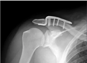
Then, locking screw fixation was performed (Fig. 2). Even though an additional CC ligament repair was not performed, the delto- trapezius fascial repair for reinforcement was done securely over the plate.
Postoperative Rehabilitation and Implant Removal Regardless of fixation methods, the affected arm was kept in a sling for six weeks after surgery. On the first day after surgery, pendulum exercise, self-assisted circumduction exercise, and gradual passive ROM as tolerable were begun. After six weeks postoperatively, active ROM exercise was begun as tolerated.
After three months postoperatively, the implant (K-wires or hook plate) was removed. Brisement under general anesthesia and subsequent arthroscopic capsular release were performed con- comitantly for patients who experienced shoulder stiffness at the time of the implant removal.
Statistical Analysis
The IBM SPSS Statistics ver. 20.0 program (IBM Co., Armonk,
NY, USA) was used for the statistical analyses. The Student’s t- test was used for between group comparisons of continuous or continuous ranked data including the VAS pain score, ROM, and shoulder UCLA and ASES scores. The paired t-test was used for comparison of preoperative and postoperative values within each group and Fisher’s exact test was used for comparison of categorical data including the presence of postoperative stiffness between groups. Statistical significance was set at p<0.05.
Results
Patient Demographics
Group A included 19 men and 4 women, and group B in- cluded 36 men and 2 women. The mean age at the time of surgery was 34.9 years (range, 21–56 years) in group A and 37.0 years (range, 19–63 years). In group A, 14 patients were injured on the right and the remaining 9 patients were injured on the left. In group B, 22 patients were injured on the right and 16 patients were injured on the left. In group A, six (26.1%, 6/23) patients were Rockwood type III and 17 (73.9%, 17/23) patients
were Rockwood type V; in group B, nine (23.7%, 9/38) patients were type III, two (5.3%, 2/38) patients were type IV, and 27 (71.1%, 27/38) patients were type V (Table 1).
Clinical and Radiological Assessments
At one-year follow-up, the mean VAS pain score was 1.2 ± 1.1 in group A and 0.9 ± 1.0 in group B with no significant dif- ference between groups. The mean UCLA shoulder score was 31.8 ± 3.2 in group A and 32.3 ± 2.4 in group B with no statis- tically significant difference. The mean ASES score was 91.4 ± 6.7 in group A and 93.3 ± 6.4 in group B with no significant dif- ference. The active ROM measured in both groups at one-year follow-up showed no significant differences in forward flexion, external rotation with arm at side, and internal rotation (Table 2).
Fig. 1. Trans-acromial fixation with Kirschner’s wires, right shoulder. Fig. 2. Locking hook plate fixation, right shoulder.
Table 1. Patients’ Demographics
Variable Group A (n=23) Group B (n=38) p-value
Sex (male/female) 19/4 36/2 0.187
Age (yr) 34.9 ± 10.5 37.0 ± 10.9 0.416
Injured side (right/left) 14/9 22/16 0.819
Rockwood type III 6 9 0.532
Rockwood type IV 0 2
Rockwood type V 17 27
Values are presented as number only or mean ± standard deviation.
Group A: closed reduction and percutaneous trans-acromial fixation with Kirschner’s wires, Group B: open reduction and internal fixation with AO locking hook plate.
Table 2. VAS Score, UCLA Shoulder Score, ASES Score, and Active Ranges of Motion for Both Groups at Final Follow-up
Variable Group A Group B p-value
VAS score 1.2 ± 1.1 0.9 ± 1.0 0.568
UCLA shoulder score 31.8 ± 3.2 32.3 ± 2.4 0.451
ASES score 91.4 ± 6.7 93.3 ± 6.4 0.362
Range of motion (°)
Forward flexion 152.8 ± 9.1 150.1 ± 9.9 0.647 Extenral rotation with arm at side 61.1 ± 10.3 58.9 ± 11.4 0.312 Internal rotation 9.3 ± 2.1 9.6 ± 2.5 0.514 Values are presented as mean ± standard deviation. The internal rotation was estimated by determining how far the patients could reach their thumb up the spinal segments. For ease of statistical analysis, the spinal segment was converted into numbers; segments at T1 through T12 were designated as 1 through 12, segments at L1 through L5 were designated as 13 through 17, and the sacrum was designated as 18.
VAS: visual analogue scale, UCLA: University of California at Los Angeles, ASES: American Shoulder and Elbow Surgeons, Group A: closed reduction and percutaneous trans-acromial fixation with K-wires, Group B: open reduc- tion and internal fixation with AO locking hook plate.
The mean preoperative CCD of the normal side was 7.4
± 2.5 mm in group A (intraclass correlation coefficient of in- terobserver reliability, ICC=0.873) and 7.6 ± 2.3 mm in group B (ICC=0.792) ; the mean affected CCD was 17.9 ± 5.5 mm in group A (ICC=0.899) and 17.3 ± 5.1 mm in group B (ICC=0.835). At the final follow-up, the mean affected CCD was 9.8 ± 3.1 mm in group A and 7.8 ± 2.3 mm in group B. A significant difference was observed between groups (p=0.006).
The side-to-side difference between normal and affected CCD at final follow-up was 2.4 ± 2.2 mm in group A and 0.2 ± 0.7 mm in group B, showing a statistical significance between groups (p<0.001).
Complications
One patient in group A had newly developed mild arthritis with heterotopic ossification around the AC joint, while there was no arthritis in group B. In six patients (15.8%) in group B, bony erosion under acromion was observed on plain X-ray. In group A, there were five complications (21.7%, 5/23): one case of K-wire breakage, one case of superficial infection followed by skin irritation by a bent end of K-wire migration, and three cases of reduction loss after K-wire removal. These five complications occurred in all Rockwood type V. In the wire-breakage case, the remaining K-wires maintained the acceptable reduction of the AC joint until removal of the K-wire, even though the CCD increased compared to immediate postoperative CCD. In cases of superficial infection, the infection was identified at four weeks after surgery. All K-wires were removed immediately and reduc- tion loss was followed. After resolving the infection, CC ligament reconstruction was recommended, but the patient did not want to undergo further surgery. In three patients of reduction loss, immediate postoperative plain X-ray just after removal showed well maintained CCD. However, at three months follow-up after removal, six months follow-up from the initial fixation, reduc- tion loss was observed. In group B, there was no complication such as reduction loss or infection, etc. during the follow-up period. For the first postoperative three months before implant removal, shoulder stiffness occurred in three patients (13.0%, 3/23) in group A and seven patients (18.4%, 7/38) in group B, who underwent both brisement under general anesthesia and subsequent arthroscopic capsular release at the time of implant removal. No significant difference in incidence of postoperative stiffness was observed between groups.
Discussion
This study was designed for comparison of clinical and radio- logical outcomes between trans-acromial fixation using K-wires and AO locking hook plate fixation for acute AC joint disloca- tion. The K-wire trans-acromial fixation showed comparable clinical outcomes to AO locking hook plate fixation, which was
consistent with part of our hypothesis. However, the remain- ing part of our hypothesis was not confirmed: the CCD in radiological assessment was significantly different; significantly greater CCD difference between normal and affected side at final follow-up was observed in the trans-acromial fixation group compared with the hook plate fixation group.
Among the methods for acute AC joint dislocation, trans-ac- romial fixation with pin or wire is a widely used method. Several investigators reported satisfactory outcomes after closed or open trans-acromial fixation with a pin or wire.17,18) Nevertheless, the pin or wire can migrate or be broken, and several complica- tions can follow such as skin irritation or reduction loss of the AC joint. Rhee et al.,13) who compared the tans-acromial fixation and AO hook plate, reported 8 cases (14%) of pin migration or breakage. In our study, five patients in the trans-acromial fixation group experienced a complication and, coincidentally, were all Rockwood type V AC joint injury. We think that this result may be attributable to unrepaired and unhealed soft tissue around the AC joint in Rockwood type V injury in closed reduction de- spite a three-month fixation period. In particular, among 17 pa- tients with Rockwood type V injury in the trans-acromial fixation group, these five-complication cases approach approximately 30%. In the difference of CCD between normal and affected side at final follow-up, the trans-acromial fixation group showed significantly inferior outcome, even though this was not directly related to clinical outcomes.
By contrast, there was no complication related to implant in the hook plate fixation group, although subacromial bony ero- sion was observed on the X-ray in some patients at the time of implant removal. While it appears that the trans-acromial fixa- tion with wires has fallen out of favor, popularity of locking hook plate fixation appears to have increased.10-14) Many studies have shown that subacromial bony erosion by hook plate and other complications such as impingement, rotator cuff lesion, and acromial fracture can occur after hook plate fixation. However, most cases of bony erosion, however, are asymptomatic and clinically insignificant.13-15,19,20) In practice, if the depth of the hook is too deep, it can cause impingement and rotator cuff injury; by contrast, if the depth of the hook is too shallow, it can result in subacromial erosion. Sim et al.15) reported that early implant removal can decrease this bony erosion. Despite our at- tempt to apply plates with an appropriate depth of hook and re- moved the implant after three months postoperatively, subacro- mial bony erosion occurred in 15.8% (6/38) patients in group B.
Rhee et al.13) bent the hook of the plate parallel to the acromion to prevent subacromial impingement or the hook encroaching the acromion; they also removed the implant at three to four months after fixation. Only two cases (10%) of subacromial bony erosion with any functional deficiency may result from these ef- forts. Kim and Jeon14) reported 36% subacromial bony erosion at the time of hook plate removal and in their study, the hook plate
was removed at about six months postoperatively.
Kim et al.21) recently reported an interesting study regarding the AC joint motion after hook plate fixation. In that study, the hook plate fixation of the AC joint can cause decreased motion of the distal clavicle with respect to the medial acromion. In addition, we know that hook plate fixation for the AC joint dis- location is indirect reduction of the AC joint by the lever arm of the hook. Considering these roles of the hook plate in AC joint fixation, a longer period of fixation can lead to higher incidence of subacromial bony erosion. Thus, as many investigators have indicated, removal of the implant after three to four months postoperatively would be appropriate.13,14)
In this study, among 61 patients included, there were only six (9.8%) female patients, and as indicated in previous literature, incidence was much lower in females, compared to males.2) In determining the surgical method for AC joint fixation in the cur- rent study, a relatively large scar after open reduction was an is- sue for female patients; of three cases of reduction loss after pin removal, one case was a female patient. Closed reduction and percutaneous pinning may have a cosmetic advantage in female patients; however care is required in application of this method in Rockwood type V injury.
We kept the affected arm in a sling for the first six weeks to relieve the load by arm weight on the AC joint regardless of the operation method; however we were concerned about shoul- der stiffness due to the relatively long period of wearing the sling. Despite immediate exercises to prevent stiffness, stiffness was observed in 16.4% (10/61) of patients at the time of implant removal who underwent both brisement under general anesthe- sia and subsequent arthroscopic capsular release at the time of implant removal.
This study has several limitations; first, this study has an inher- ent weakness as a retrospective comparative study. In addition, the patient assignment was not randomized; in general, closed reduction and trans-acromial fixation with K-wires was used initially and group B was used later; Second, even though our study showed no significant difference in clinical outcomes be- tween the two groups, we cannot exclude the possibility that this result may be attributed to the type II error. Thus, considering the aforementioned complications, care is required in interpret- ing our results; Third, the follow-up period was short and inci- dence of arthritis in the AC joint would be different in long-term follow-up; Fourth, we did not evaluate the AP translation of the AC joint via axial view. Considering that both methods could not reconstitute the AP stability of the AC joint, there would have been some differences in AP stability between the affected side and normal contralateral.
Conclusion
For the treatment of acute AC joint dislocation, the K-wire
trans-acromial fixation group showed a significantly greater CCD than the AO locking hook plate group at one-year follow-up after surgery. In addition, during the follow-up period, incidence of complication related to implant was much higher in the trans- acromial fixation group. Although clinical outcomes were not significantly different between the two groups, the clinical out- comes of this study should be interpreted carefully.
References
1. Mazzocca AD, Arciero RA, Bicos J. Evaluation and treatment of acromioclavicular joint injuries. Am J Sports Med. 2007;35(2):
316-29.
2. Fraser-Moodie JA, Shortt NL, Robinson CM. Injuries to the acromioclavicular joint. J Bone Joint Surg Br. 2008;90(6):697- 707.
3. Webb J, Bannister G. Acromioclavicular disruption in first class rugby players. Br J Sports Med. 1992;26(4):247-8.
4. Beitzel K, Cote MP, Apostolakos J, et al. Current concepts in the treatment of acromioclavicular joint dislocations. Arthros- copy. 2013;29(2):387-97.
5. Ceccarelli E, Bondì R, Alviti F, Garofalo R, Miulli F, Padua R.
Treatment of acute grade III acromioclavicular dislocation: a lack of evidence. J Orthop Traumatol. 2008;9(2):105-8.
6. Leidel BA, Braunstein V, Kirchhoff C, Pilotto S, Mutschler W, Biberthaler P. Consistency of long-term outcome of acute Rockwood grade III acromioclavicular joint separations after K- wire transfixation. J Trauma. 2009;66(6):1666-71.
7. O’Carroll PF, Sheehan JM. Open reduction and percutaneous Kirschner wire fixation in complete disruption of the acromio- clavicular joint. Injury. 1982;13(4):299-301.
8. Calvo E, López-Franco M, Arribas IM. Clinical and radiologic outcomes of surgical and conservative treatment of type III acromioclavicular joint injury. J Shoulder Elbow Surg. 2006;
15(3):300-5.
9. Verdano MA, Pellegrini A, Zanelli M, Paterlini M, Ceccarelli F. Modified Phemister procedure for the surgical treatment of Rockwood types III, IV, V acute acromioclavicular joint disloca- tion. Musculoskelet Surg. 2012;96(3):213-22.
10. Liu HH, Chou YJ, Chen CH, Chia WT, Wong CY. Surgical treat- ment of acute acromioclavicular joint injuries using a modified Weaver-Dunn procedure and clavicular hook plate. Orthope- dics. 2010;33(8). doi: 10.3928/01477447-20100625-10.
11. Kienast B, Thietje R, Queitsch C, Gille J, Schulz AP, Meiners J.
Mid-term results after operative treatment of rockwood grade III-V acromioclavicular joint dislocations with an AC-hook- plate. Eur J Med Res. 2011;16(2):52-6.
12. Eschler A, Gradl G, Gierer P, Mittlmeier T, Beck M. Hook plate fixation for acromioclavicular joint separations restores coraco- clavicular distance more accurately than PDS augmentation, however presents with a high rate of acromial osteolysis. Arch
Orthop Trauma Surg. 2012;132(1):33-9.
13. Rhee YG, Park JG, Cho NS, Song WJ. Clinical and radiologic outcomes of acute acromioclavicular joint dislocation: com- parison of kirschner’s wire transfixation and locking hook plate fixation. Clin Should Elbow. 2014;17(4):159-65.
14. Kim KC, Jeon YS. Treatment of acute acromioclavicular joint injuries using AO hook locking plate. Clin Should Elbow.
2014;17(3):114-9.
15. Sim E, Schwarz N, Höcker K, Berzlanovich A. Repair of com- plete acromioclavicular separations using the acromioclavicu- lar-hook plate. Clin Orthop Relat Res. 1995;(314):134-42.
16. Lizaur A, Sanz-Reig J, Gonzalez-Parreño S. Long-term results of the surgical treatment of type III acromioclavicular disloca- tions: an update of a previous report. J Bone Joint Surg Br.
2011;93(8):1088-92.
17. Lizaur A, Marco L, Cebrian R. Acute dislocation of the acro- mioclavicular joint. Traumatic anatomy and the importance of
deltoid and trapezius. J Bone Joint Surg Br. 1994;76(4):602-6.
18. Eskola A, Vainionpää S, Korkala O, Rokkanen P. Acute com- plete acromioclavicular dislocation. A prospective randomized trial of fixation with smooth or threaded Kirschner wires or cortical screw. Ann Chir Gynaecol. 1987;76(6):323-6.
19. Lin HY, Wong PK, Ho WP, Chuang TY, Liao YS, Wong CC. Cla- vicular hook plate may induce subacromial shoulder impinge- ment and rotator cuff lesion--dynamic sonographic evaluation.
J Orthop Surg Res. 2014;9:6.
20. Nadarajah R, Mahaluxmivala J, Amin A, Goodier DW. Clavicu- lar hook-plate: complications of retaining the implant. Injury.
2005;36(5):681-3.
21. Kim YS, Yoo YS, Jang SW, Nair AV, Jin H, Song HS. In vivo analysis of acromioclavicular joint motion after hook plate fixa- tion using three-dimensional computed tomography. J Shoul- der Elbow Surg. 2015;24(7):1106-11.