ISSN 0378-6471 (Print)⋅ISSN 2092-9374 (Online)
https://doi.org/10.3341/jkos.2019.60.12.1295
Case Report
양악수술 후 발생한 반사적 눈물분비의 소실 증례 보고 및 문헌 고찰
Loss of Reflex Tearing after Maxillary Orthognathic Surgery Case Report and Literature Review
방소라1⋅이태은1,2,3⋅유인천1,2,3⋅조남천1,2,3⋅안 민1,2,3
So Ra Bang, MD1, Tae Eun Lee, MD, PhD1,2,3, In Cheon You, MD, PhD1,2,3, Nam Chun Cho, MD, PhD1,2,3, Min Ahn, MD, PhD1,2,3
전북대학교 의학전문대학원 안과학교실1, 전북대학교 임상의학연구소2, 전북대학교병원 의생명연구원3 Department of Ophthalmology, Chonbuk National University Medical School1, Jeonju, Korea
Research Institute of Clinical Medicine, Chonbuk National University2, Jeonju, Korea Biomedical Research Institute, Chonbuk National University Hospital3, Jeonju, Korea
Purpose: To report a case of unilateral loss of reflex tearing after double-jaw surgery with anatomy and mechanism.
Case summary: A 32-year-old woman complained of a 3-year history of loss of tearing in her left eye. She had undergone dou- ble-jaw surgery 3 years prior to correct her malocclusion. In orbital computed tomography (CT) images, there were no specific findings, with the exception of a fractured left pterygoid plate, possibly caused by the double-jaw surgery. The tear break-up times of both eyes were similar, with non-specific findings on slit lamp examination. We diagnosed loss of reflex tearing due to pterygopalatine ganglion injury based on her history, physical examination and orbital CT findings.
Conclusions: Nerve damage due to artificial fracture may occur during double-jaw surgery and may result in loss of reflex tear- ing, thus, pre-operative evaluation of basic lacrimal secretion and a reflex tearing test are important. Once the loss of reflex tear- ing due to nerve injury occurrs, orbital CT scans are needed for diagnosis. Ophthalmologist examination is necessary to confirm the mechanism of loss of reflex tearing and changes in the anatomical structure.
J Korean Ophthalmol Soc 2019;60(12):1295-1300
Keywords: Absent reflex, Orthognathic surgery, Postoperative complication, Pterygopalatine fossa
■Received: 2019. 4. 25. ■ Revised: 2019. 6. 17.
■Accepted: 2019. 12. 6.
■Address reprint requests to Min Ahn, MD, PhD
Department of Ophthalmology, Chonbuk National University Hospital, #20 Geonji-ro, Deokjin-gu, Jeonju 54907, Korea Tel: 82-63-250-1965, Fax: 82-63-250-1960
E-mail: [email protected]
* This study was presented as a poster at the 119th Annual Meeting of the Korean Ophthalmological Society 2018.
* This paper was supported by Fund of Biomedical Research Institute, Chonbuk National University Hospital.
*Conflicts of Interest: The authors have no conflicts to disclose.
ⓒ2019 The Korean Ophthalmological Society
This is an Open Access article distributed under the terms of the Creative Commons Attribution Non-Commercial License (http://creativecommons.org/licenses/by-nc/3.0/) which permits unrestricted non-commercial use, distribution, and reproduction in any medium, provided the original work is properly cited.
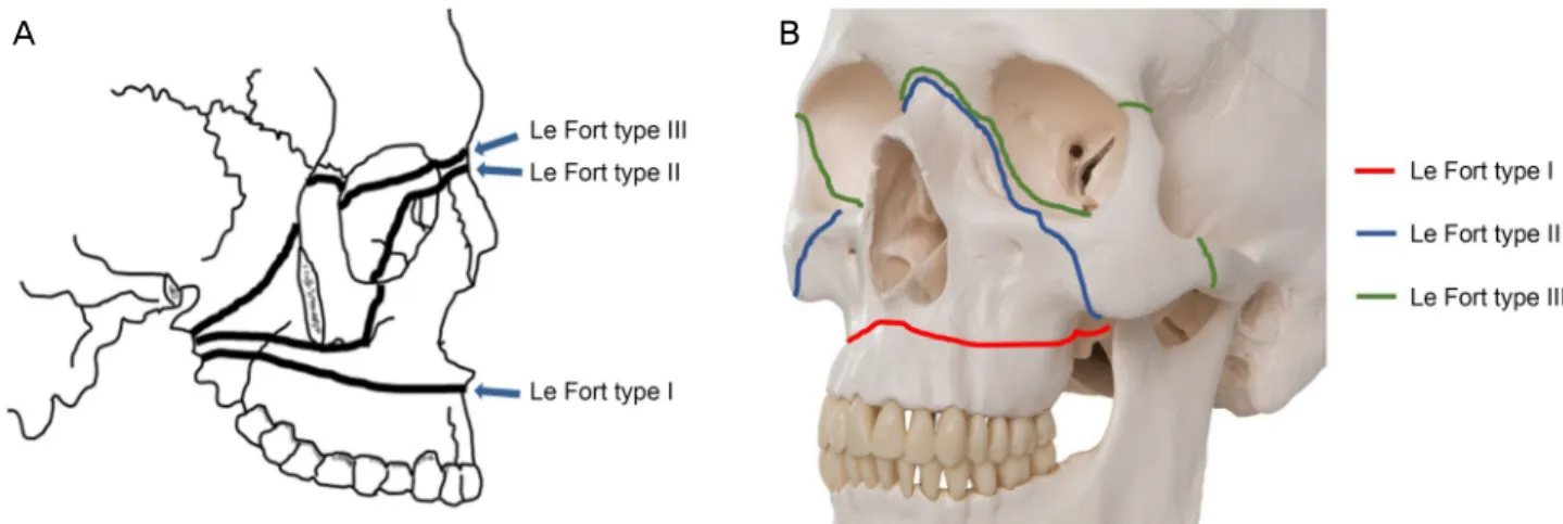
양악수술(double-jaw surgery)은 턱교정수술(orthognathic surgery)의 한 종류로, 상악(maxilla)과 하악(mandible)을 동 시에 절골하는 수술법이다. 양악수술은 부정교합과 같은 생활의 불편함, 안면기형 등 장애를 위한 치료 뿐 아니라 최근에는 단순히 미용을 목적으로도 널리 시행되고 있다. 상악에는 르포절골술(Le Fort osteotomy)을, 하악에는 시상 분할절골술(sagittal split osteotomy)을 시행한다. 르포절골 술은 절골선에 따라 3가지 종류로 나뉘는데(Fig. 1), 그중 제1 르포절골술은 상악의 치아 정렬을 따라 날개위턱유합 (pterygomaxillary junction)을 통과해 절제하는 방식으로, 1864년 Cheever가 비인두암 절제를 위해 처음 고안하였다.1
양악수술은 절골부의 감염이나 출혈,2 괴사,3 수술 후 부 정유합,4 안과적 합병증으로는 눈물분비의 장애,4,5 안와 출
A B
Figure 1. Types of Le Fort osteotomy (A, B). Thick black lines across the skull are osteotomy line of each type.
Figure 2. A coronal section of case patient's facial computed
tomography. The image shows fractured lateral and medial pterygoid plates of the left side (white arrows).혈6 등 여러 술 후 합병증이 발생할 수 있다고 알려져 있으 며 인위적 골절형성과 재건에 따른 신경 손상7 또한 여러 증례가 보고된 바 있다.
국내에서도 양악수술 후 눈물분비 반사의 소실에 대한 2개 의 증례가 보고되어 있으나, 이는 증례의 보고 자체에 중점 을 두고 있으므로,8 저자들은 이러한 합병증이 발생하는 날 개입천장오목(pterygopalatine fossa)의 해부학적 모식도를 통하여 발생 기전 및 신경손상의 원리를 보여줌으로써 발 생기전을 이해하는 데 도움을 주고 주위 해부학적 구조에 대한 지식을 제공하고자 한다.
증례보고
33세 여자 환자가 약 3년 전 부정교합의 교정을 위해 양 악수술을 받은 후 좌안에서는 눈물이 흐르지 않는 것을 주 소로 내원하였다. 환자는 상악의 제1 르포절골술과 하악의 시상분할절골술을 시행받았으며, 통증이나 감정변화 등 반 사적인 눈물분비를 유발하는 모든 상황에서 좌안에서만 눈 물이 흐르지 않고 이와 동시에 좌측 안면부에 국한된 감각 이상을 함께 호소하였다. 환자는 우안에 비하여 좌안에 건 조감이 느껴진다고 하였으나 기존에 건성안으로 치료받은 기왕력은 없었으며, 세극등현미경검사에서 눈물막파괴시간 은 양안 동일하게 약 7초 정도로 감소되어 있었으나 전안 부의 특이 소견은 보이지 않았다. Cochet-Bonnet 각막지각 계검사에서 양안 각막의 감각지각도는 60 mm로 정상이었 다. 양안의 기본적 눈물분비와 자극 시 눈물분비량을 수치 화하기 위해 쉬르머검사(Shirmer test)를 시행하였으며, 무 자극 상태에서 기본 눈물분비량은 우안 5 mm, 좌안 3 mm 측정되었고 눈물흘림 자극 후 분비량은 우안 8 mm, 좌안 5 mm로 좌안에서 약간 감소된 소견을 보였다. 안와 전산화
단층촬영에서 안와 내부의 특이 소견은 없었으며 양악수술 에 기인한 것으로 생각되는 날개판의 골절 소견이 좌측에 국한하여 발견되었다(Fig. 2). 이러한 소견들을 종합적으로 고 려하여 양악수술에 의한 날개판(pterygoid plate)의 골절로, 날개입천장오목에 위치한 날개입천장 신경절(pterygopalatine ganglion)의 손상에 의한 반사적 눈물분비 소실로 진단하였다.
고 찰
반사적 눈물 분비는 부교감신경을 통해 이루어지는데, 눈물분비에 관여하는 부교감신경다발을 포함한 큰바위신 경(greater petrosal nerve)이 중간머리오목(middle cranial fossa)의 기저부에 위치한 관자뼈(temporal bone)에서 기시 하고, 이는 날개관(vidian canal)의 시작점에서 깊은바위신 경(deep petrosal nerve)과 만나 날개신경(vidian nerve)을 이룬다(Fig. 3A, B).9,10 날개신경은 날개관을 통과하여 날개
A B
C D E F
Figure 3. Pterygopalatine ganglion and pterygoid plate anatomy. (A) Greater petrosal nerve originates from temporal bone and joins
deep petrosal nerve to form vidian nerve. Vidian nerve reaches pterygopalatine ganglion. (B) Pterygopalatine fossa (dark blue re- gion, blank arrow) and pterygopalatine ganglion (yellow spot inside dark blue region, black arrow) in real skull image. (C) Axial computed tomography (CT) view of medial pterygoid plate (white arrows) and lateral pterygoid plate (yellow arrows). (D) Coronal CT view of medial pterygoid plate (white arrows) and lateral pterygoid plate (yellow arrows). (E) Medial pterygoid plate (white ar- rows) and lateral pterygoid plate (black arrows) in the skull. (F) Lateral image of lateral pterygoid plate (black arrow) and pter- ygopalatine fossa (yellow arrow, dark blue region). Pterygopalatine ganglion is located in pterygopalatine fossa.입천장오목에 위치한 날개입천장 신경절에 도달하며, 직후 에 상악신경과 함께 아래눈확틈새(inferior orbital fissure)를 따라 안와의 외벽으로 주행하여 눈물샘신경(lacrimal nerve) 과 문합하여 눈물샘을 지배한다. 날개판은 나비뼈의 몸통 과 큰날개의 유합부 사이에서 수직으로 내려오는 얇은 판 과 같은 뼈로, 좌우 대칭으로 존재하며 내측날개판과 바깥 날개판으로 이중벽으로 이루어진 구조이다. 날개입천장 신 경절이 위치한 날개입천장오목은 바깥날개판과 내측날개 판이 유합되어있는 부분과 상악이 이루는 틈새이며, 안와 첨부의 아래, 상악동(maxillary sinus)의 바로 뒤쪽에 위치 하고 있다(Fig. 3C-F). 르포절골술 시 날개위턱유합을 절단 하여 상악을 후방으로 전위시키는 과정에서 날개입천장 신 경절이 손상될 수 있다(Fig. 4).
제1 르포절골술은 날개위턱유합을 절단하여 코 아래범위 의 상악을 3면에서 자유롭게 함으로써 상악의 위치를 재조 정할 수 있는 수술법이다.1 이는 양측 하악의 시상분할절골 술과 동시에 시행함으로써 부정교합, 안면부 기형 등을 교 정할 수 있고 여러 합병증에도 불구하고 미용적인 목적으
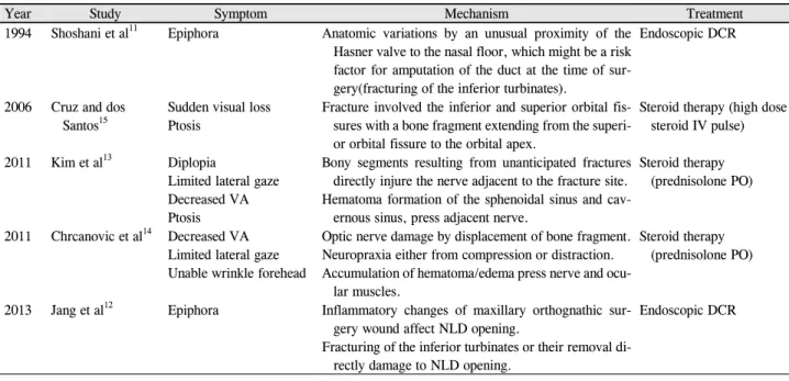
로 국내에서도 그 활용도가 증가하고 있는 추세이다. 여러 연구들에서 제1 르포절골술 시행 후 발생한 합병 증들이 보고되어왔다. 본 증례보고와 비슷한 눈물흘림 반 사의 소실을 보고한 사례들 외에도, Shoshani et al,11 Jang et al12은 코눈물관의 막힘으로 인한 눈물흘림을 보고하기 도 하였으며 수술 후 뇌신경손상 사례,13,14 Cruz and dos Santos15은 실명까지도 보고한 바 있다. 이러한 다른 안과적 합병증들의 증상, 기전, 치료 여부에 대하여 표로 정리하였 다(Table 1).
본 증례와 같이 상악의 양악수술 후 발생한 반사적 눈물 분비의 소실은 흔하지 않은 합병증으로, 국외를 포함하여 현재까지 5예 정도가 보고되어 있다. 1976년에 Tomasetti et al5이 처음으로 제1 르포절골술 후의 반사적 눈물 분비의 소실을 보고한 이후 1993년 Lanigan et al4이 제1 르포절골 술 후 반사적 눈물분비가 감소된 환자의 전산화단층촬영에 서 양측 날개판이 골절되어 있음을 보고하였다. 2014년 Kang et al8은 양악수술을 받은 후 편측의 반사적 눈물분비 가 감소된 2명의 환자들의 전산화단층촬영에서 2명 모두
A B
Figure 4. Pterygoid plate fracture in Le Fort I osteotomy. (A) Pterygoid plate and pterygopalatine fossa may be damaged in the proc-
ess of displacing the maxilla backward by cutting the pterygomaxillary junction. (B) Thick black line across maxilla (white arrows) is osteotomy line, which passes pterygopalatine fossa (blank arrow) and can injure the pterygopalatine ganglion (black arrow).Year Study Symptom Mechanism Treatment
1994 Shoshani et al11 Epiphora Anatomic variations by an unusual proximity of the Hasner valve to the nasal floor, which might be a risk factor for amputation of the duct at the time of sur- gery(fracturing of the inferior turbinates).
Endoscopic DCR
2006 Cruz and dos Santos15
Sudden visual loss Ptosis
Fracture involved the inferior and superior orbital fis- sures with a bone fragment extending from the superi- or orbital fissure to the orbital apex.
Steroid therapy (high dose steroid IV pulse)
2011 Kim et al13 Diplopia
Limited lateral gaze Decreased VA Ptosis
Bony segments resulting from unanticipated fractures directly injure the nerve adjacent to the fracture site.
Hematoma formation of the sphenoidal sinus and cav- ernous sinus, press adjacent nerve.
Steroid therapy (prednisolone PO)
2011 Chrcanovic et al14 Decreased VA Limited lateral gaze Unable wrinkle forehead
Optic nerve damage by displacement of bone fragment.
Neuropraxia either from compression or distraction.
Accumulation of hematoma/edema press nerve and ocu- lar muscles.
Steroid therapy (prednisolone PO)
2013 Jang et al12 Epiphora Inflammatory changes of maxillary orthognathic sur- gery wound affect NLD opening.
Fracturing of the inferior turbinates or their removal di- rectly damage to NLD opening.
Endoscopic DCR
DCR = dacryocystorhinostomy; VA = visual acquity; PO = per oral; NLD = nasolacrimal duct.
Table 1. Other complications of orthognathic surgery than the present case
날개판의 골절이 일어난 것을 확인하였다. 본 증례도 마찬 가지로 좌측의 날개오목의 내측과 외측의 날개판이 모두 골절되어 있는 것을 확인하였는데, 이는 다른 증례 보고들 과의 공통된 소견으로, 일부 환자들에서 해부학적 다양성 으로 인하여 제1 르포절골술의 과정에서 날개판이 골절되 면 날개입천장오목에 위치한 눈물샘을 지배하는 날개입천 장 신경절의 손상으로 반사적 눈물분비가 감소하거나 소실 될 수 있음을 알 수 있다.
또한, 날개입천장오목은 상악동의 바로 뒤쪽에 위치하고 있어 상악동의 감염 혹은 종양, 상악동수술에 의해 직접적
인 영향을 받을 수 있다. 본 증례에서 환자의 전산화단층촬 영에서 양측으로 상악의 부비동염 소견을 보였는데, 제1 르 포절골술시 날개판의 골절에 의한 신경절의 직접적인 손상 외에도 상악동과 직접 연결되는 통로가 생성되어 감염의 전파 혹은 수술 후 염증의 파급에 의해 발생한 날개입천장 신경절의 손상으로 반사적 눈물분비에 영향 미쳤을 가능성 도 있다고 생각한다.
Kang et al8은 모두 증상이 자연적으로 호전되었다고 보 고하였으며, 본 증례의 환자는 인공눈물 사용으로 보존적 치료만을 시행하며 경과 관찰 중으로, 자연 호전에 대해서
는 더 지켜보아야 할 것이다. 날개판의 골절이 더 높은 위 치에서 일어날수록 날개입천장오목의 중앙에 가까워지고, 따라서 내부의 신경절에 더 많은 손상을 야기할 것으로 생 각되므로, 상악교정술 시 수술 전 영상검사를 통해 환자의 해부학적 구조에 대하여 보다 면밀한 평가를 시행하여 절 골 시 더 세심하게 주의를 기울이는 것이 좋겠다.
양악수술 후 발생하는 반사적 눈물분비의 소실은 양악교 정술 후 드물게 발생하는 합병증이며 환자들은 안과적 증 상이 양악수술로 인한 합병증이라고 생각하기 어려워 주로 안과를 먼저 찾게 된다. 안과 전문의는 날개입천장오목과 내부의 신경절의 해부학과 손상 기전에 대하여 잘 이해하 고, 양악수술 후 발생할 수 있는 안과적 합병증에 대해 알 고 있어야 할 것으로 생각된다.
REFERENCES
1) Buchanan EP, Hyman CH. LeFort I osteotomy. Semin Plast Surg 2013;27:149-54.
2) Lanigan DT, Hey JH, West RA. Major vascular complications of orthognathic surgery: hemorrhage associated with Le Fort I osteotomies. J Oral Maxillofac Surg 1990;48:561-73.
3) Kim S, Kim SY, Kim GJ, et al. Partial necrosis of the mandibular proximal segment following transoral vertical ramus osteotomy.
Maxillofac Plast Reconstr Surg 2014;36:131-4.
4) Lanigan DT, Romanchuk K, Olson CK. Ophthalmic complications associated with orthognathic surgery. J Oral Maxillofac Surg
1993;51:480-94.
5) Tomasetti BJ, Broutsas M, Gormley M, Jarrett W. Lack of tearing after Le Fort I osteotomy. J Oral Surg 1976;34:1095-7.
6) Ord RA. Post-operative retrobulbar haemorrhage and blindness complicating trauma surgery. Br J Oral Surg 1981;19:202-7.
7) Agbaje JO, Salem AS, Lambrichts I, et al. Systematic review of the incidence of inferior alveolar nerve injury in bilateral sagittal split osteotomy and the assessment of neurosensory disturbances. Int J Oral Maxillofac Surg 2015;44:447-51.
8) Kang S, Jang SY, Lee A, Jang JW. Loss of reflex tearing after max- illary orthognathic surgery: a report of two cases. BMC Ophthalmol 2014;14:37.
9) Koizumi K, Suda I. Induced modulation in autonomic efferent neu- ron activity. Am J Physiol 1963;205:738-44.
10) Langley JN, Dickinson WL. On the local paralysis of the periph- eral ganglia and on the connection of different classes of nerve fi- bres with them. Proc R Soc Lond 1890;46:423-31.
11) Shoshani Y, Samet N, Ardekian L, Taicher S. Nasolacrimal duct in- jury after Le Fort I osteotomy. J Oral Maxillofac Surg 1994;52:406-7.
12) Jang SY, Kim MK, Choi SM, Jang JW. Nasolacrimal duct ob- struction after maxillary orthognathic surgery. J Oral Maxillofac Surg 2013;71:1085-98.
13) Kim JW, Chin BR, Park HS, et al. Cranial nerve injury after Le Fort I osteotomy. Int J Oral Maxillofac Surg 2011;40:327-9.
14) Chrcanovic BR, Custódio AL. Optic, oculomotor, abducens, and facial nerve palsies after combined maxillary and mandibular os- teotomy: case report. J Oral Maxillofac Surg 2011;69:e234-41.
15) Cruz AA, dos Santos AC. Blindness after Le Fort I osteotomy: a possible complication associated with pterygomaxillary separation.
J Craniomaxillofac Surg 2006;34:210-6.
= 국문초록 =
양악수술 후 발생한 반사적 눈물분비의 소실 증례 보고 및 문헌 고찰
목적: 양악수술 후 편측의 반사적 눈물분비의 소실을 보이는 환자를 경험하였기에 증례와 함께 발생기전, 해부학적 구조에 대하여 보고하고자 한다.
증례요약: 32세 여자 환자가 3년 전 부정교합으로 양악수술을 받은 후 발생한 좌안의 반사적 눈물분비 소실을 주소로 내원하였다.
안와 전산화단층촬영에서 안와 내 특이 소견은 보이지 않았으나, 양악수술로 인한 것으로 보이는 좌측의 날개판(pterygoid plate)의 골절 소견을 보였다. 양안의 눈물막파괴시간은 비슷하였으며 세극등검사에서 양안 각막 모두 특이 소견을 보이지 않았다. 병력과 이 학적 검진, 전산화단층촬영 소견을 바탕으로 날개입천장신경절(pterygopalatine ganglion)의 손상에 의한 반사적 눈물분비 소실로 진 단하였다.
결론: 양악수술 시 인위적 골절형성에 의한 신경손상으로 반사적인 눈물분비의 소실을 보일 수 있으므로 술 전 기본적인 눈물분비와 눈물흘림 반사를 평가해 두는 것이 좋겠다. 신경손상에 의한 반사적 눈물분비의 소실이 초래된 경우 진단을 위해 안와 전산화단층촬 영이 필요하며, 안과의사로서 이러한 반사적 눈물분비 소실의 발생기전, 주위 해부학적 구조에 대하여 숙지하여야 하겠다.
<대한안과학회지 2019;60(12):1295-1300>
방소라 / So Ra Bang
전북대학교 의학전문대학원 안과학교실 Department of Ophthalmology, Chonbuk
National University Medical School