pISSN: 0378-6471 eISSN: 2092-9374
http://dx.doi.org/10.3341/jkos.2012.53.10.1412
= 증례보고 =
이중샤임플러그, 전안부 빛간섭단층촬영계 및 초음파 각막두께측정계를 이용한 각막두께의 비교
김동욱⋅이가영⋅최동규⋅신영주 한림대학교 의과대학 강남성심병원 안과학교실
목적: 이중샤임플러그 전안부사진기(Galilei), 전안부 빛간섭단층촬영계 및 초음파 각막두께측정계를 이용하여 중심각막두께의 측정치 를 비교하고자 한다.
대상과 방법: 정상인 20명 40안을 대상으로 이중샤임플러그, 전안부 빛간섭단층촬영계, 그리고 초음파 각막두께측정계로 측정된 중심 각막두께를 비교하고 상관관계를 알아보았다.
결과: 이중샤임플러그, 전안부 빛간섭단층촬영계, 초음파 각막두께측정계를 이용하여 측정한 중심각막두께는 각각 538.10 ± 31.36 μm, 536.20 ± 31.21 μm, 541.93 ± 34.93 μm였고 초음파 각막두께측정계가 이중샤임플러그, 전안부 빛간섭단층촬영계보다 유의하게 두 껍게 측정되었다(p=0.017, p=0.001). 이중샤임플러그와 전안부 빛간섭단층촬영계에 의해 측정된 중심각막두께는 유의한 차이가 없었 다(p=0.054). 세 검사는 유의한 상관관계를 보였다(r˃0.900, p<0.001).
결론: 세 검사 사이에 유의한 상관관계가 있었지만, 초음파 각막두께측정계에 의해 측정된 중심각막두께가 이중샤임플러그, 전안부 빛간섭단층촬영계의 의한 측정값보다 유의하게 두껍게 측정되어 결과 해석 시 이에 대한 고려가 필요할 것으로 생각한다.
<대한안과학회지 2012;53(10):1412-1418>
■ 접 수 일: 2012년 3월 26일 ■ 심사통과일: 2012년 4월 27일
■ 게재허가일: 2012년 8월 18일
■ 책 임 저 자: 신 영 주
서울특별시 영등포구 신길로 1 한림대학교 강남성심병원 안과 Tel: 02-829-5193, Fax: 02-848-4638 E-mail: [email protected]
중심각막두께의 측정은 굴절교정수술 전후 각막의 평가, 안압의 정확한 측정 및 각막이식술 후 이식실패 평가, 특히 각막내피의 기능을 평가하는 데 매우 중요하다.1-3각막굴 절교정수술을 계획할 때 중심각막의 두께가 중요하여 각막 두께가 실제보다 두껍게 측정될 경우 수술 후 각막의 돌출 등의 심각한 합병증을 일으킬 수 있으며,4녹내장에서는 각 막두께에 따라 안압의 측정치를 보정해야 하므로 정확한 측정이 요구된다.5
현재 각막두께 측정에는 다양한 방법들이 사용되고 있는 데 초음파 각막두께측정계(ultrasound pachymeter)6와 비 접촉형 경면현미경(noncontact specular microscope),7전 안부 빛간섭단층촬영계(anterior segment optical coherence tomography),8 슬릿-스캐닝(slit-scanning)을 이용하는 방 법(Orbscan),9 그리고 회전샤임플러그 카메라(rotating Scheimpflug camera)를 사용하는 방법(Pentacam)10등이 있다. 여러 가지 방법들 중 현재 초음파 각막두께측정계를 이용하여 각막두께를 측정하는 방법이 가장 널리 사용되고
있지만 각막에 직접 접촉해서 측정해야 하므로 각막을 눌 러서 잘못 측정될 수 있고 접촉에 의한 상피 미란 혹은 감 염이 발생할 위험성이 있다.11이에 각막에 직접 접촉하지 않고 각막두께를 측정할 수 있는 방법들이 여럿 대두되었 다. 특히 최근에 도입된 GalileiTM (Ziemer Group; Port, Switzerland)는 이중회전샤임플러그 카메라(dual rotating Scheimpflug camera)를 사용하는 방식으로 정확한 각막두 께 및 전안부 생체계측치를 측정할 수 있다고 제시되었다.12 지금까지 국내에서 이러한 비접촉 방법들과 초음파 각막 두께측정계의 측정치 차이를 보고하고 이를 교정하는 방법 들을 제시한 연구들이 많이 있었지만,13-16이중회전샤임플 러그 전안부사진기를 이용하여 측정한 각막두께와 다른 각 막두께 측정법과의 비교는 아직 보고된 바가 없다. 따라서 본 연구에서는 젊은 정상인을 대상으로 하여 접촉식 각막 두께측정계인 초음파 각막두께측정계와 비접촉식 각막두께 측정계인 전안부 빛간섭단층촬영계, 이중회전샤임플러그 전안부사진기를 이용하여 중심각막두께를 측정하여 비교해 보고자 하였다.
대상과 방법
건강한 성인 지원자 20명 40안을 대상으로 이중회전샤 임플러그(dual rotating Scheimpflug), 전안부 빛간섭단층
Table 1. Central corneal thickness measured with Galilei, an-
terior segment optical coherence tomography, and ultrasound pachymetryMethods Mean ± SD (μm) Range (μm)
Galilei 538.10 ± 31.36 471.00-592.00
AS-OCT 536.20 ± 31.21 476.00-593.00
USP 541.93 ± 34.93 475.00-615.00
SD = standard deviation; AS-OCT = anterior segment optical coherence tomography; USP = ultrasound pachymetry.
Table 2. Intraexaminer repeatability of each method for cen-
tral corneal thicknessMethods ICC (95% CI)
Galilei 0.991 (0.983-0.995)
AS-OCT 0.995 (0.991-0.998)
Ultrasound pachymetry 0.998 (0.996-0.999) ICC = intraclass correlation coefficients; CI = confidence interval;
AS-OCT = anterior segment optical coherence tomography.
Table 3. Pairwise comparison of central corneal thickness
measurementsComparison Mean difference ± SD (μm) p-value*
Galilei and USP - 3.83 ± 10.72 0.017
Galilei and AS-OCT 1.90 ± 6.47 0.054
AS-OCT and USP - 5.73 ± 10.67 0.001
SD = standard deviation; USP = ultrasound pachymetry; AS-OCT
= anterior segment optical coherence tomography.
*Wilcoxon-rank signed test.
촬영계(anterior segment optical coherence tomography), 초음파 각막두께측정계(ultrasound pachymetry)를 이용하 여 중심각막두께를 측정하였다. 안과적 수술력이나 각막 질 환의 과거력, 외상력이 있는 경우는 대상에서 제외하였으며 검사 전 세극등 현미경을 통해 대상자들의 각막 상피에 이 상이 없음을 확인하였다. 검사는 오후 7-8시 사이에 동일 한 한 명의 검사자에 의해 각 검사마다 2회씩 시행되었고 초음파 각막두께측정계의 소식자에 의한 각막 손상을 피하 기 위해 이중회전샤임플러그 전안부촬영검사와 전안부 빛 간섭단층촬영검사를 먼저 시행하고 초음파 각막두께측정검 사를 가장 마지막에 실시하였다. 본 연구는 본원 의학윤리 심의위원회의 심의를 거친 후 지원자들의 동의서를 받은 후 진행되었다.
이중회전샤임플러그 전안부사진기인 GalileiTM (Ziemer Group; Port, Switzerland)를 이용한 각막두께 측정은 우선 검사대의 높이가 적절한지 확인하고 대상자의 머리와 턱을 검사대에 안정된 자세로 고정한 후 눈을 주시점에 고정하 게 하였다. 그 다음 모니터상에서 붉은 십자표시가 네 개의 흰 점 위에 놓이면서 붉은 색의 가로선이 각막의 외측경계 에 닿을 때 스캔을 하도록 하였다. 동공중심이 중앙에 위치 하여 각막의 내외측과 상하측이 각각 대칭이 되고 안구의 움직임이 없으며 눈물층이 마르지 않을 때 측정하였고 결 과지에 나타난 중심각막두께의 값을 측정치로 하였다. 전안 부 빛간섭단층촬영 검사는 Cirrus HD-OCT (Carl Zeiss Meditec Inc., Dublin, CA, USA)의 anterior segment cube 512×124 영상을 찍은 후 Cirrus 소프트웨어(version 5.0) 에서 제공하는 직선측정도구(linear measurement tool)를 이용하여, 각막 중심부의 외측경계에서부터 내측경계까지 수직선을 그어 측정하였다. 초음파 각막두께측정 검사는 가 장 마지막에 시행하였으며 검사 전 proparacaine 0.5%
(Paracaine®, Han Mi Pharm, Korea)로 각막을 마취한 다 음 초음파 각막두께측정계(SP-100, TOMEY®, Japan)의 소식자(probe)를 각막 중심에 수직으로 접촉하여 8회 측정 한 후 평균값을 측정치로 하였다.
이상의 과정을 통해 얻은 중심각막두께 측정치에 대한 검사자내 반복성을 평가하였고 세 검사기기로 측정된 중심 각막두께의 값을 비교하여 검사기계 간의 차이를 분석하였 다. 반복성은 급내상관계수(intraclass correlation co- efficient, ICC)를 계산하여 평가하였고 각 검사의 측정치 차이의 비교는 Wilcoxon-rank signed test를 이용하였으 며 Bland and Altman plots을 이용하여 검사기기 간의 일 치도를 분석하였다. 또한 Pearson correlation test를 이용 해 검사기기 간의 상관관계에 대해서도 알아보았다. 통계학 적 분석은 SPSS 18.0 (SPSS Inc., Chicago, IL, USA)을
이용하였고 통계적 유의수준은 0.05 이하로 하였다.
결 과
전체 대상군은 총 20명 40안이었고 남자 14명, 여자 6명 이었으며 평균 나이는 26.85 ±2.54세(23-31세)였다. 이 중회전샤임플러그 전안부사진기, 전안부 빛간섭단층촬영 계, 초음파 각막두께측정계를 이용하여 측정한 평균 중심각 막두께는 각각 538.10 ±31.36 μm, 536.20 ±31.21 μm, 541.93 ±34.93 μm였다(Table 1). 세 검사기구의 중심각 막두께 측정에 대한 반복성을 평가한 결과 이중회전샤임플 러그 전안부사진기는 ICC가 0.991, 전안부 빛간섭단층촬영 계는 ICC가 0.991, 초음파 각막두께측정계는 ICC가 0.998 로 세 기구 모두 높은 반복성을 보였다(Table 2).
이중회전샤임플러그 전안부사진기와 초음파 각막두께 측정계
초음파 각막두께측정계로 측정된 중심각막두께가 이중
Figure 1. Scattergram showing the correlation of central corneal
thickness measured by Galilei, anterior segment optical coher- ence tomography (AS-OCT), and ultrasound pachtmetry (USP).(A) Correlation between Galilei and USP. (B) Correlation be- tween Galilei and AS-OCT. (C) Correlation between AS-OCT and USP.
회전샤임플러그 전안부사진기로 측정한 값보다 3.83 ± 10.72 μm 더 두껍게 측정되었고 이는 통계적으로 유의하 였다(Wilcoxon-rank signed test, p=0.017) (Table 3).
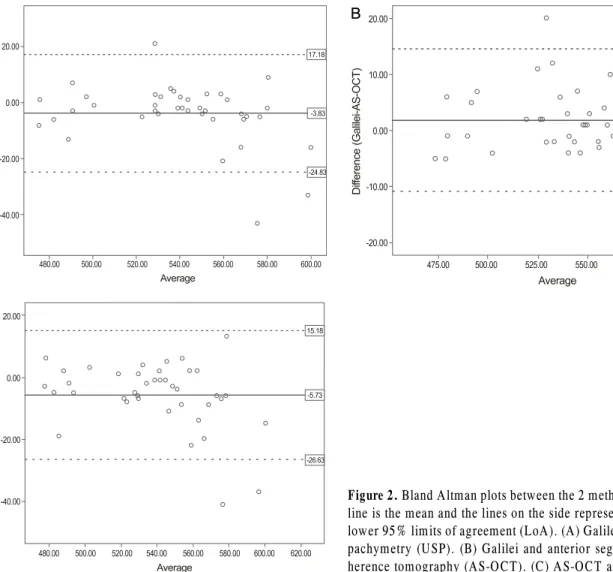
이중회전샤임플러그 전안부사진기와 초음파 각막두께측정 계의 측정값은 높은 양의 상관관계(Pearson correlation, r=0.953, p<0.001)를 보였다(Fig. 1). 또한 Bland and Altman 일치도 분석 결과 두 검사기구 간의 중심각막두께 측정치 의 95% 일치도 범위는 42.01 μm (-24.83~17.18)였다 (Fig. 2).
이중회전샤임플러그 전안부사진기와 전안부 빛간섭단 층촬영계
이중회전샤임플러그 전안부사진기로 측정한 중심각막두 께가 전안부 빛간섭단층촬영계에 의한 측정치보다 1.90 ± 6.47 μm 더 두껍게 측정되었지만 통계적으로 유의하지는 않았다(Wilcoxon-rank signed test, p=0.054) (Table 3).
이중회전샤임플러그 전안부사진기와 전안부 빛간섭단층촬 영계로 측정한 측정치는 높은 상관관계(Pearson correla- tion, r=0.979, p<0.001)를 보였다(Fig. 1). 또한 Bland and Altman 일치도 분석 결과 두 검사기구 간의 중심각막 두께 측정치의 95% 일치도 범위는 25.36 μm (-14.58~
10.78)이었다(Fig. 2).
전안부 빛간섭단층촬영계와 초음파 각막두께측정계
초음파 각막두께측정계는 전안부 빛간섭단층촬영계보다 5.73 ±10.67 μm 더 두껍게 측정되었고 이는 통계적으로 유 의하였다(Wilcoxon-rank signed test, p=0.001) (Table 3).
전안부 빛갑섭단층촬영계와 초음파 각막두께측정계로 측정 한 각막두께는 높은 양의 상관관계(Pearson correlation, r=0.954, p<0.001)를 보였다(Fig. 1). 또한 Bland and Altman 일치도 분석 결과 두 검사기구 간의 중심각막두께 측정치의 95% 일치도 범위는 41.81 μm (-26.63~15.18)
A B
C
Figure 2. Bland Altman plots between the 2 methods. The middle
line is the mean and the lines on the side represent the upper and lower 95% limits of agreement (LoA). (A) Galilei and ultrasound pachymetry (USP). (B) Galilei and anterior segment optical co- herence tomography (AS-OCT). (C) AS-OCT and USP.였으며 높은 양의 상관관계(Pearson correlation, r=0.954, p<0.001)를 보였다(Fig. 2).
고 찰
정확한 각막두께의 측정은 안과 영역에서 필수적인 검사 이며, 특히 각막굴절교정수술이 발달하면서 정확한 측정이 더욱 중요하게 여겨지고 있다.17중심각막두께는 여러 가지 방법으로 측정할 수 있으나 초음파 각막두께측정계가 현재 까지 기준으로 쉽게 사용할 수 있고 상대적으로 저렴한 검 사비용으로 가장 널리 이용되어 왔다.18-20하지만 검사 전 점안 마취가 필요하고 소식자가 각막에 직접 접촉하여 측 정하기 때문에 가해지는 압력의 차이나 측정위치에 따라 측정값의 차이가 있을 수 있으며, 각막상피의 손상, 감염의 전파 등의 위험이 있다.11이런 단점을 보완하기 위해 비접 촉 각막두께 측정법들이 개발되어 사용되고 있는데 그중에 한 가지 방법인 전안부 빛간섭단층촬영계는 비접촉성 영상 기기로 조직 내에서 반사되는 빛의 시간 차이를 광학적 간
섭계를 통해 전안부 및 망막의 고해상도 영상을 나타낼 수 있다.21
한편 이중회전샤임플러그 전안부사진기(Galilei)는 최근 에 도입된 각막지형도 장비로 1대의 샤임플러그 카메라가 360도 회전하여 측정하는 Pentacam과는 달리 2대의 샤임 플러그 카메라가 180도씩 분리되어 회전하면서 측정하기 때문에 보다 더 정확한 각막두께에 대한 정보를 제공할 수 있다.22각막두께 이외에도 홍채, 동공, 전방, 렌즈 등의 영 상과 생체계측치를 제공할 수 있어 그 활용폭이 점차 확대 되고 있다. 또한 비접촉 방식을 사용하기 때문에 마취가 필 요없고 접촉으로 생길 수 있는 오차를 줄일 수 있으며, 감 염의 위험성이 없다는 장점이 있다. 따라서 이중회전샤임플 러그 전안부사진기를 통한 중심각막두께 측정치의 정확성 을 평가하고, 널리 이용되고 있는 다른 각막두께측정 기기 들과 측정치를 비교해 보는 것이 필수적이라 생각한다.
기존의 연구에서 Yeter et al23은 근시 환자 81명 161안 을 대상으로 이중회전샤임플러그 전안부사진기와 초음파 각막두께측정계로 중심각막두께를 측정하여 비교하였는데
A B
C
두 검사기기의 측정값 사이에 유의한 차이는 없었고 유의 한 상관관계를 보였다고 보고하였다. Ladi and Shah24는 정 상안 46명 92안을 대상으로 중심각막두께를 측정하였는데, 이중회전샤임플러그 전안부사진기는 높은 재현성과 반복성 을 보였으며 초음파 각막두께측정계에 의해 측정된 중심각 막두께와 유의한 차이가 없었다고 보고하였다. 본 연구에서 는 이중회전샤임플러그 전안부사진기로 측정한 중심각막두 께 값이 전안부 빛간섭단층촬영계와는 유의한 차이를 보이 지 않았지만, 초음파 각막두께측정계에 비해 평균 3.83 ± 10.72 μm 얇게 측정되었다. 하지만 이중회전샤임플러그 전 안부사진기와 전안부 빛간섭단층촬영계, 이중회전샤임플러 그 전안부사진기와 초음파 각막두께측정계 사이에 상관관 계는 매우 유의하게 나타났으며 세 검사기기 모두 높은 재 현성을 보였다.
전안부 빛간섭단층촬영계와 초음파 각막두께측정계의 경우에는 두 검사기기 사이에 유의한 상관관계가 있었지만 초음파 각막두께측정계의 측정값이 평균 5.73 ±10.67 μm 더 두껍게 측정되었다. 기존의 연구에서 Bechmann et al25 은 정상인에서 초음파 각막두께측정계로 측정한 중심각막 두께가 전안부 빛간섭단층촬영계보다 평균 51 μm 두껍게 측정되었다고 보고하였고, Kim et al16은 초음파 각막두께 측정계가 전안부 빛간섭단층촬영계에 비해 평균 26.3 μm 두껍게 측정되었다고 보고하였다. 반면에 Leung et al26은 정상안을 대상으로 한 연구에서 전안부 빛간섭단층촬영계 로 측정한 중심각막두께가 초음파 각막두께측정계보다 평 균 23 μm 두껍게 측정되었다고 한 보고도 있다.
본 연구에서 이중회전샤임플러그 전안부사진기, 전안부 빛간섭단층촬영계에 비해 초음파 각막두께측정계로 측정한 중심각막두께가 조금 더 두껍게 측정되었으며 이는 통계적 으로 유의하였다. 이러한 차이의 원인은 정확히 알 수 없지 만 다음과 같은 것들을 생각해 볼 수 있다. 우선, 각막의 전 면 및 후면에서 반사되는 빛을 이용하는 이중회전샤임플러 그 전안부사진기와 전안부 빛간섭단층촬영계와는 달리 초 음파 각막두께측정계는 아직 명확하게 밝혀지지 않은 후면 의 어느 점에서 반사된 음파를 이용하기 때문인 것으로 생 각해 볼 수 있다. 한 연구에서는 그 반사점이 아마도 데스 메막과 전방 사이에 위치할 것이라고 추측하고 있다.27또 한 이중회전샤임플러그 전안부사진기는 각막 중심이 컴퓨 터에 의해 자동으로 결정하기 때문에 검사자의 영향을 받 지 않지만, 초음파 각막두께측정계는 각막 중심이 검사자에 의해 수동적으로 결정되기 때문에 검사자의 숙련도에 영향 을 받을 수 있다. 이외에도 이중회전샤임플러그 전안부사진 기와 전안부 빛간섭단층촬영계는 초음파 각막두께측정계와 는 달리 빛의 질과 각막의 투명도에 영향을 받을 수 있다.
이중회전샤임플러그 전안부사진기는 두 개의 샤임플러 그 카메라를 이용하여 측정된 중심각막두께의 값을 평균화 하여 나타내기 때문에 환자가 어느 정도 주시점을 잘 주시 하지 못해도 비교적 정확한 중심각막두께의 측정이 가능하 다고 알려졌다.28 또한 이중회전샤임플러그 전안부사진기 는 여러 연구에서 보고된 것처럼 중심각막두께 측정 시 재 현성이 뛰어나다.28,29하지만 눈꺼풀 틈새가 작거나 건성안 이 심한 환자, 그리고 각막 혼탁이 심하게 있는 환자 등에 서는 검사 결과를 신뢰할 수 없거나 검사 자체를 시행할 수 없는 경우도 있다. 이중회전샤임플러그 전안부사진기는 비 접촉 방식으로 검사 전 마취가 필요없고 자주 측정이 필요 하거나 각막 상피의 질환이 있는 환자에서 유용하며 접촉 으로 인한 감염의 위험성도 없다는 장점이 있다.
결론적으로, 정상인에서 중심각막두께를 측정할 때 이중 회전샤임플러그 전안부사진기는 초음파각막두께 측정계, 빛간섭단층촬영계와 유의한 상관관계가 있으며 높은 재현 성과 안정성의 장점이 있지만 초음파 각막두께측정계를 대 체하여 사용할 경우에는 초음파 각막두께측정계에 비하여 얇게 측정될 수 있기 때문에 향후 이중회전샤임플러그 전 안부사진기를 사용할 때에는 이를 고려해야 할 것으로 생 각한다.
참고문헌
1) Yau CW, Cheng HC. Microkeratome blades and corneal flap thick- ness in LASIK. Ophthalmic Surg Lasers Imaging 2008;39:471-5.
2) Whitacre MM, Stein RA, Hassanein K. The effect of corneal thick- ness on applanation tonometry. Am J Ophthalmol 1993;115:592-6.
3) Kim DH, Kim MS, Kim JH. Early corneal-thickness changes after penetrating keratoplasty. J Korean Ophthalmol Soc 1997;38:1355-61.
4) Wang Z, Chen J, Yang B. Posterior corneal surface topographic changes after laser in situ keratomileusis are related to residual cor- neal bed thickness. Ophthalmology 1999;106:406-9.
5) Doughty MJ, Zaman ML. Human corneal thickness and its impact on intraocular pressure measures: a review and meta-analysis approach. Surv Ophthalmol 2000;44:367-408.
6) Realini T, Lovelace K. Measuring central corneal thickness with ultrasound pachymetry. Optom Vis Sci 2003;80:437-9.
7) Sanchis-Gimeno JA, Lleo-Perez A, Casanova J, et al. Inter-ob- server variability of central corneal thickness measurements using non-contact specular microscopy after laser in situ keratomileusis.
Clin Exp Optom 2004;87:15-8.
8) Yoo C, Eom YS, Suh YW, Kim YY. Central corneal thickness and anterior scleral thickness in Korean patients with open-angle glau- coma: an anterior segment optical coherence tomography study. J Glaucoma 2011;20:95-9.
9) Hashemi H, Roshani M, Mehravaran S, et al. Effect of corneal thickness on the agreement between ultrasound and Orbscan II pachymetry. J Cataract Refract Surg 2007;33:1694-700.
10) Ceylan OM, Turk A, Erdurman C, et al. Comparison of Oculus
Pentacam and Stratus optical coherence tomography for measure- ment of central corneal thickness. Cornea 2011;30:670-4.
11) Li EY, Mohamed S, Leung CK, et al. Agreement among 3 methods to measure corneal thickness: ultrasound pachymetry, Orbscan II, and Visante anterior segment optical coherence tomography.
Ophthalmology 2007;114:1842-7.
12) Wang L, Shirayama M, Koch DD. Repeatability of corneal power and wavefront aberration measurements with a dual-Scheimpflug Placido corneal topographer. J Cataract Refract Surg 2010;36:425-30.
13) Zhao MH, Zou J, Wang WQ, Li J. Comparison of central corneal thickness as measured by non-contact specular microscopy and ul- trasound pachymetry before and post LASIK. Clin Experiment Ophthalmol 2007;35:818-23.
14) Doughty MJ, Jonuscheit S. An assessment of regional differences in corneal thickness in normal human eyes, using the Orbscan II or ultrasound pachymetry. Optometry 2007;78:181-90.
15) Al-Mezaine HS, Al-Amro SA, Kangave D, et al. Comparison of central corneal thickness measurements using Pentacam and ultra- sonic pachymetry in post-LASIK eyes for myopia. Eur J Ophthalmol 2010;20:852-7.
16) Kim HY, Budenz DL, Lee PS, et al. Comparison of central corneal thickness using anterior segment optical coherence tomography vs ultrasound pachymetry. Am J Ophthalmol 2008;145:228-32.
17) Thomas J, Wang J, Rollins AM, Sturm J. Comparison of corneal thickness measured with optical coherence tomography, ultrasonic pachymetry, and a scanning slit method. J Refract Surg 2006;22:
671-8.
18) Ling T, Ho A, Holden BA. Method of evaluating ultrasonic pachometers. Am J Optom Physiol Opt 1986;63:462-6.
19) Copt RP, Thomas R, Mermoud A. Corneal thickness in ocular hy- pertension, primary open-angle glaucoma, and normal tension glaucoma. Arch Ophthalmol 1999;117:14-6.
20) Harper CL, Boulton ME, Bennett D, et al. Diurnal variations in hu-
man corneal thickness. Br J Ophthalmol 1996;80:1068-72.
21) Rodriques EB, Johanson M, Penha FM. Anterior segment tomog- raphy with the cirrus optical coherence tomography. J Ophthalmol 2012;2012:806989. Epub 2012 Jan 24.
22) Savini G, Carbonelli M, Barboni P, Hoffer KJ. Repeatability of au- tomatic measurements performed by a dual Scheimpflug analyzer in unoperated and post-refractive surgery eyes. J Cataract Refract Surg 2011;37:302-9.
23) Yeter V, Sönmez B, Beden U. Comparison of central corneal thick- ness measurements by Galilei Dual-Scheimpflug analyzer® and ultrasound pachymeter in myopic eyes. Ophthalmic Surg Lasers Imaging 2012;43:128-34.
24) Ladi JS, Shah NA. Comparison of central corneal thickness meas- urements with the Galilei dual Scheimpflug analyzer and ultra- sound pachymetry. Indian J Ophthalmol 2010;58:385-8.
25) Bechmann M, Thiel MJ, Neubauer AS, et al. Central corneal thick- ness measurement with a retinal optical coherence tomography de- vice versus standard ultrasonic pachymetry. Cornea 2001;20:50-4.
26) Leung DY, Lam DK, Yeung BY, Lam DS. Comparison between central corneal thickness measurements by ultrasound pachymetry and optical coherence tomography. Clin Experiment Ophthalmol 2006;34:751-4.
27) Azen SP, Burg KA, Smith RE, Maguen E. A comparison of three methods for the measurement of corneal thickness. Invest Ophthalmol Vis Sci 1979;18:535-8.
28) Lee YE, Jun RM. The intra and inter-examiner repeatability of cor- neal parameters obtained by GALILEI(TM) in normal subjects. J Korean Ophthalmol Soc 2009;50:1611-6.
29) Menassa N, Kaufmann C, Goggin M, et al. Comparison and re- producibility of corneal thickness and curvature readings obtained by the Galilei and the Orbscan II analysis systems. J Cataract Refract Surg 2008;34:1742-7.
=ABSTRACT=
Corneal Thickness Measured by Dual Scheimpflug, Anterior Segment Optical Coherence Tomography, and Ultrasound Pachymetry
Dong Wook Kim, MD, Ka Young Yi, MD, PhD, Dong Gyu Choi, MD, PhD, Young Joo Shin, MD, PhD
Department of Ophthalmology, Kangnam Sacred Heart Hospital, Hallym University College of Medicine, Seoul, Korea
Purpose: To compare central corneal thickness (CCT) as measured by dual rotating Scheimpflug camera (Galilei), ante- rior segment optical coherence tomography (AS-OCT), and ultrasound pachymetry (USP).
Methods: The measurements of CCT using a dual rotating Scheimpflug camera, AS-OCT, and USP in 40 eyes of 20 healthy subjects were compared.
Results: The average measurements of CCT by dual rotating Scheimpflug camera, AS-OCT, and USP were 538.10 ± 31.36 μm, 536.20 ± 31.21 μm, and 541.93 ± 34.93 μm, respectively. The CCT measurement by USP was statistically sig- nificantly thicker than by the dual rotating Scheimpflug camera and AS-OCT (p = 0.017, p = 0.001, respectively). There was no significant difference between the dual rotating Scheimpflug camera and AS-OCT (p = 0.054). A significant linear corre- lation was observed between the dual rotating Scheimpflug camera, the AS-OCT, and the USP (r > 0.900, p < 0.001).
Conclusions: The results of the 3 methods have significant correlation with each other, but the measurement by USP was significantly thicker than the dual rotating Scheimpflug camera and AS-OCT. Therefore, CCT should be interpreted in the context of the instrument used.
J Korean Ophthalmol Soc 2012;53(10):1412-1418
Key Words: Anterior segment optical coherence tomography, Central corneal thickness, Dual rotating scheimpflug camera, Galilei, Ultrasound pachymetry
Address reprint requests to Young Joo Shin, MD, PhD Department of Ophthalmology, Kangnam Sacred Heart Hospital
#1 Singil-ro, Yeongdeungpo-gu, Seoul 150-950, Korea
Tel: 82-2-829-5193, Fax: 82-2-848-4638, E-mail: [email protected]