Articles published in Obstet Gynecol Sci are open-access, distributed under the terms of the Creative Commons Attribution Non-Commercial License (http://creativecommons.
org/licenses/by-nc/3.0/) which permits unrestricted non-commercial use, distribution, and reproduction in any medium, provided the original work is properly cited.
Copyright © 2014 Korean Society of Obstetrics and Gynecology
Case Report
Obstet Gynecol Sci 2014;57(6):557-559 http://dx.doi.org/10.5468/ogs.2014.57.6.557 pISSN 2287-8572 · eISSN 2287-8580
www.ogscience.org 557
Introduction
Most cases of inguinal hernia containing an ovary and fallo- pian tubes reported to date occurred in infants and were of- ten accompanied by other congenital anomalies of the genital tract [1]. However, inguinal ovaries can occur rarely in adult women without any other genital anomalies. Moreover, ingui- nal ovary accompanying other ovarian diseases is extremely rare. Ozkan et al. [2] reported the only case of inguinal ovary with other ovarian pathology in which a right sliding indirect inguinal hernia containing a paraovarian cyst and fallopian tube was noted. In addition, inguinal hernia containing endo- metriosis is extremely rare [3,4]. Herein we report a case of an indirect inguinal hernia containing endometriosis, an ovary, and fallopian tube in an adult woman without genital anoma- lies, which was successfully repaired laparoscopically using polypropylene mesh.
Case report
A 44-year-old multiparous patient was scheduled to undergo total laparoscopic hysterectomy for the treatment of micro- invasive cervical cancer. She reported a lump in the left groin that became more apparent during menstruation, movement, and constipation. She had no specific medical or surgical his-
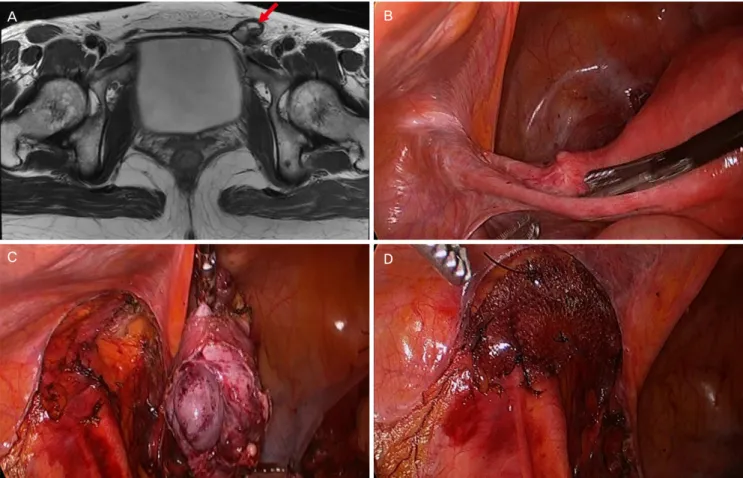
tory. Moreover, no genital anomalies were found on physical examination. A magnetic resonance imaging (MRI) examina- tion of the pelvis demonstrated a 4×3 cm cystic structure in the left inguinal area (Fig. 1A).
The intervention was performed laparoscopically. The uterus and right ovary, fallopian tube, and round ligament appeared normal. However, the left ovary and a portion of the left fal- lopian tube were herniated through the inguinal canal (Fig.
1B). After the left ovary was mobilized, the left adnexa was removed. Furthermore, a defect of the parietal peritoneum in the inguinal internal ring area was identified (Fig. 1C).
Polypropylene mesh was used to repair the hernia and was anchored to the anterior abdominal wall as well as Cooper’s ligament using laparoscopic sutures to prevent migration (Fig.
Received: 2014.5.27. Revised: 2014.7.10. Accepted: 2014.7.15.
Corresponding author: Gun Oh Chong
Gynecologic Cancer Center, Kyungpook National University Medical Center, 807 Hoguk-ro, Buk-gu, Daegu 702-210, Korea Tel: +82-53-200-2684 Fax: +82-53-200-2028
E-mail: [email protected]
Laparoscopic repair of indirect inguinal hernia
containing endometriosis, ovary, and fallopian tube in adult woman without genital anomalies
Ji Hyun Kim
1, Gun Oh Chong
1, Ji Young Lee
1, Yoon Hee Lee
1, Dae Gy Hong
1, Soo Yeun Park
2, Ji Young Park
31Gynecologic Cancer Center, 2Colorectal Cancer Center, 3Department of Pathology, School of Medicine, Kyungpook National University Medical Center, Daegu, Korea
Indirect inguinal hernia containing an ovary is a rare condition, especially in adult women who do not have any other genital tract anomalies. In addition, inguinal hernia containing an ovary and endometriosis is exceedingly rare. In the present report, we describe a case of indirect inguinal hernia containing an ovary, fallopian tube, and endometriosis.
Laparoscopic repair was performed successfully using polypropylene mesh for the treatment of the inguinal hernia.
Keywords: Endometriosis; Hernia, inguinal; Laparoscopic repair; Ovary
www.ogscience.org 558
Vol. 57, No. 6, 2014
1D). Finally, the peritoneal flap was closed over the mesh by suturing to prevent adhesion of the intra-abdominal structures
to the mesh. The patient had an uneventful postoperative course and was discharged on day 3. A histopathological examination confirmed the presence of endometriosis con- taining endometrial glands, with stromal cells and scattered hemosiderin-laden macrophages within fibroadipose tissue (Fig. 2).
Discussion
The inguinal canal in females not as well demarcated as in males. Several different structures normally pass through it, including the round ligament of the uterus, a vein, an artery from the uterus that forms a cruciate anastomosis with the labial arteries, and extra peritoneal fat [5]. Most cases of ingui- nal ovary have been reported in pediatric patients with other genital tract anomalies [1]. Inguinal ovaries in adult women are rarely reported; in fact, only 12 cases were identified in a recent review article [6]. In a retrospective review of 1,950 cas-
Fig. 2. Microscopic findings of the left inguinal ovary: endometrial glands withstromal cells and scattered hemosiderin-laden macrophages in fibroadipose tissue (H&E, ×40).
Fig. 1. (A) Magnetic resonance imaging of the pelvis showing a 4×3 cm cystic structure in the left inguinal area (red arrowhead). (B) Laparoscopic view of herniation of the left ovary and a portion of the left fallopian tube in the inguinal canal. (C) Laparoscopic view of defect of the parietal peritoneum in the inguinal internal ring area. (D) Laparoscopic view of polypropylene mesh used to repair the hernia.
www.ogscience.org 559