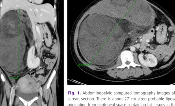
A case of huge retroperitoneal liposarcoma in pregnancySang Eun Oh, Hyun Joo Kim, Suk-Joo Choi, Soo-Young Oh, Cheong-Rae Roh, Jong-Hwa Kim
4
0
0
전체 글
(2)
(3)
(4)
수치
관련 문서
Chang Goo Kang, Ah Hyun Park, Jang Ho Ha, Young Soo Kim, Joon-Ho Oh, Jeong Min Park, Soo Mee Kim, Seung-Jae Lee, Seung Hee Lee, and Han Soo Kim(KAERI). Fabrication
Chang Je Park, Kwoen Ho Kang, Sang Ho Na, Young Hee Kim, Ho Jin Ryu, Geun Il Park, and Kee Chan Song (KAERI). Geun-Suk Choi and
Dong Won Lee, Young Dug Bae, Suk Kwon Kim, Hee Yun Shin, Bong Guen Hong, Hyun Kyu Jung, Yang Il Jung, Jeong Yong Park, Byung Kwon Choi, and Yong Hwan Jeong(KAERI). P07B04
P04G02 Design and Fabrication of Irradiation Testing Capsule for Research Reactor Materials Seong Woo Yang, Bong Goo Kim, Seung Jae Park, Man Soon Cho, Kee Nam Choo, Jong Myeong
11:40 Fabrication of Nitride Coated U-Mo Powders for an Advanced Research Reactor Fuel Jae Soon Park, Yong Jin Jeong, Sang Oh Bae, Sun Chil Kwon, Eung Soo Kim, Se Jung Jang,
Sang-Keun Woo, Yong Jin Lee, WonHo Lee, Min Hwan Kim, Ji Ae Park, In Ok Ko, Jin Su Kim, Jong Guk Kim, Young Hoon Ji, Joo Hyun Kang, Gi Jeong Cheon, Chang Woon Choi, Sang Moo
IL Soon Hwang, Myung Hyun Kim, Han Gyu Joo, Kyung Woo Yi, Bong Yoo, Moo Hwan Kim, Seung Rok Oh, Yoon Jae Kim, Jong Gye Shin, Kwang Myung Lee, Jae Yong
At the end of the study, a reevaluation of each study case was performed with the same questionnaire. The result shows there has been a meaningful result in the group
