Veterinary Science
http://dx.doi.org/10.4142/jvs.2012.13.2.187
Received: 1 Jun. 2011, Revised: 20 Jul. 2011, Accepted: 17 Mar. 2012
Original Article
*Corresponding author: Tel: +82-2-880-1281; Fax: +82-2-880-1281; E-mail: [email protected]
ⓒ 2012 The Korean Society of Veterinary Science.
This is an Open Access article distributed under the terms of the Creative Commons Attribution Non-Commercial License (http://creativecommons.org/licenses/by-nc/3.0) which permits
unrestricted non-commercial use, distribution, and reproduction in any medium, provided the original work is properly cited.
Evaluation of the effect of a 0.0584% hydrocortisone aceponate spray on clinical signs and skin barrier function in dogs with atopic dermatitis
Eui-Hwa Nam, Seol-Hee Park, Ji-Young Jung, Seung-Hee Han, Hwa-Young Youn, Jun-Seok Chae, Cheol-Yong Hwang*
College of Veterinary Medicine and Research Institute for Veterinary Science, Seoul National University, Seoul 151-742, Korea
The purpose of this study was to evaluate the effects of a topical spray containing 0.0584% hydrocortisone aceponate (HCA) on canine atopic dermatitis (CAD) and to evaluate the skin barrier function during the treatment of CAD.
Twenty-one dogs that fulfilled the diagnostic criteria for CAD were included in this study. The HCA spray was applied once a day to the lesions of all dogs for 7 or 14 days.
Clinical assessment was performed before (day 0) and after treatment (day 14), and clinical responses were correlated with changes in skin barrier function. CAD severity significantly decreased after 14 days of HCA treatment based on the lesion scores (p < 0.0001), which were determined using the CAD extent and severity index (CADESI-03) and pruritus scores (p < 0.0001) calculated using a pruritus visual analog scale. Transepidermal water loss, a biomarker of skin barrier function, was significantly reduced compared to baseline (day 0) measurements (p = 0.0011). HCA spray was shown to be effective for significantly improving the condition of dogs suffering from CAD. This treatment also significantly improved cutaneous hydration and skin barrier function in the animals.
Keywords: atopic dermatitis, canine, hydrocortisone aceponate, transepidermal water loss
Introduction
Canine atopic dermatitis (CAD) is a chronic pruritic skin disorder with characteristic clinical features associated with the production of IgE antibodies against environmental allergens [10,16]. The pathogenesis of CAD is not fully understood but this is a complex, multifactorial disease involving interactions between environmental factors,
immunologic abnormalities, and skin barrier defects [7,15,19]. Treatment options include allergen avoidance, allergen-specific immunotherapy, topical and systemic treatments such as administration of glucocorticoids, cyclosporine, anti-histamines, or essential fatty acids, and management of secondary bacterial or yeast infections [23,25,27].
Glucocorticoids are the most commonly used drugs for treating a variety of inflammatory skin diseases including CAD [28]. However, the adverse effects of glucocorticoids such as skin atrophy and iatrogenic Cushing’s syndrome has limited the use of these compounds [2]. Topical glucocorticoids are an attractive treatment option since the drug can be directly absorbed at the site of inflammation, thereby avoiding systemic exposure. However, this mode of application is not recommended in veterinary dermatology due to a lack of penetration into the skin through the hair coat and concerns regarding the ingestion of the product [28,35].
Although ideal topical glucocorticoids with high potency and a low risk of side effects have not yet been synthesized, esterification to a hydrocortisone template molecule was performed to produce new glucocorticoid products with significant anti-inflammatory effects and minimal adverse effects [5,32]. Diester molecules can rapidly penetrate the stratum corneum and undergo metabolism within the dermis, thus mitigating local and systemic side effects [12]. These compounds are widely used for treating atopic dermatitis and other forms of eczema in humans [3,30].
Recently, a new generation of topical glucocorticoids with excellent pharmacological activities and low benefit-risk ratios was developed for use in veterinary dermatology [4,20]. The purpose of this study was to evaluate the effects of a topical diester formula spray containing 0.0584%
hydrocortisone aceponate (HCA) on canines with CAD,
Fig. 1. Changes in lesion scores during treatment with 0.0584%
hydrocortisone aceponate (HCA) spray (n = 21). A statistically significant difference in lesion scores was observed between day 0 and day 14 (p < 0.0001). Box and whisker plots show the median, 25th and 75th percentiles, and range of the scores.
Fig. 2. Changes in pruritus scores during treatment with 0.0584%
HCA spray (n = 21). A statistically significant difference in pruritus scores was observed between day 0 and day 14 (p < 0.0001). The box represents the 25th and 75th percentiles with the bold lines indicating the median. The whiskers indicate the range. Outliers (asterisks) are also indicated.
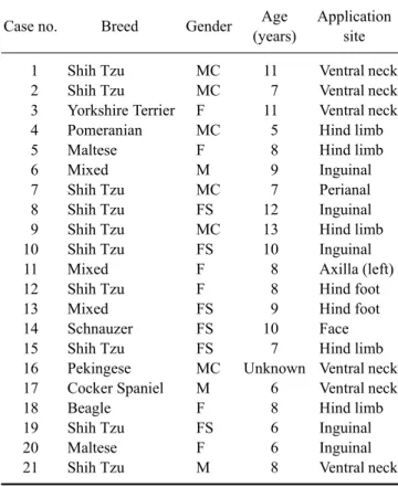
Table 1. Data for the 21 dogs with canine atopic dermatitis enrolled in this study
Case no. Breed Gender Age
(years)
Application site 1
2 3 4 5 6 7 8 9 10 11 12 13 14 15 16 17 18 19 20 21
Shih Tzu Shih Tzu Yorkshire Terrier Pomeranian Maltese Mixed Shih Tzu Shih Tzu Shih Tzu Shih Tzu Mixed Shih Tzu Mixed Schnauzer Shih Tzu Pekingese Cocker Spaniel Beagle Shih Tzu Maltese Shih Tzu
MC MC F MC F M MC FS MC FS F F FS FS FS MC M F FS F M
11 7 11 5 8 9 7 12 13 10 8 8 9 10 7 Unknown
6 8 6 6 8
Ventral neck Ventral neck Ventral neck Hind limb Hind limb Inguinal Perianal Inguinal Hind limb Inguinal Axilla (left) Hind foot Hind foot Face Hind limb Ventral neck Ventral neck Hind limb Inguinal Inguinal Ventral neck
F: female, FS: spayed female, M: male, MC: castrated male.
Fig. 3. Changes in transepidermal water loss (TEWL) during treatment with 0.0584% HCA spray (n = 21). TEWL values differed significantly between day 0 and day 14 (p = 0.0011). Box and whisker plots show the median, 25th and 75th percentiles, and range of the TEWL values. Outlier (asterisk) are also indicated.
compared to day 0 (mean: 6.8 ± 1.5, range: 3∼8; p < 0.0001, Fig. 2). The scores for 16 of the 21 atopic dogs improved by 50% or more following HCA treatment.
TEWL findings
To evaluate the effect of the HCA spray treatment on skin barrier function, TEWL was measured in the lesions of the CAD dogs. Post-treatment TEWL values (mean: 17.5 ± 6.9 g/m
2/h, range: 9.5∼29.8 g/m
2/h) were significantly lower than the pre-treatment values (mean: 48.7 ± 42.4 g/m
2/h, range: 12.9∼211.0 g/m
2/h; p = 0.0011, Fig. 3). Reductions in TEWL values of 50% or more were observed in 13 of the 21 atopic dogs following HCA treatment.
Adverse events
No adverse events were observed in any of the 21 dogs
during the study period by the owners or clinicians.