© 2014 The Korean Ophthalmological Society
This is an Open Access article distributed under the terms of the Creative Commons Attribution Non-Commercial License (http://creativecommons.org/licenses /by-nc/3.0/) which permits unrestricted non-commercial use, distribution, and reproduction in any medium, provided the original work is properly cited.
Original Article
Patient’s Self-recognition of Reduced Visual Acuity Due to Recurrence of Macular Edema and Prompt Visitation to the
Hospital in Retinal Vein Occlusion
Seong Hun Jeong1, Jae Hui Kim1, Jong Woo Kim1, Tae Gon Lee1,2, Chul Gu Kim1, Su Jin Yoo1, Mun Jung Choi1
1Department of Ophthalmology, Kim’s Eye Hospital, Konyang University College of Medicine, Seoul, Korea
2Graduate School of Medicine, Kyung Hee University, Seoul, Korea
Purpose: To evaluate patients’ self-recognition of reduced visual acuity due to recurring macular edema in reti- nal vein occlusion.
Methods: A retrospective review of medical records of patients who were diagnosed with recurring macular edema secondary to retinal vein occlusion was performed. The proportion of patients who recognized re- duced visual acuity due to the recurrence of macular edema and who visited the hospital before the sched- uled follow-up date was determined. Parameters including age, sex, diagnosis, visual acuity before recurrence of macular edema, and extent of visual acuity reduction due to recurrence of macular edema were compared in patients who recognized a reduction in visual acuity and those who did not. The proportion of patients who visited the hospital promptly was also determined.
Results: Forty eyes of 40 patients were included in the analysis. Sixteen and 24 patients were diagnosed with central retinal vein occlusion and branch retinal vein occlusion, respectively. Twenty-one patients (52.5%) rec- ognized reduced visual acuity due to recurring macular edema. These patients were younger (59.2 ± 7.6 vs.
64.8 ± 9.4 years, p = 0.046), had better visual acuity before recurrence of macular edema (0.52 ± 0.48 vs. 1.02
± 0.46, p = 0.002), and exhibited a greater reduction in visual acuity after recurrence of macular edema (0.34
± 0.24 vs. 0.14 ± 0.13, p = 0.003). Only four patients visited the hospital before the scheduled follow-up date, and all of these patients lived relatively close to the hospital.
Conclusions: For prompt treatment of recurring macular edema, more intensive education about the self-esti- mation of visual acuity is necessary, particularly for elderly patients who have relatively poor visual acuity. In addition, a simple and easy way to identify the recurrence of macular edema at the local clinic should be es- tablished for patients who live relatively far from the hospital.
Key Words: Macular edema, Recognition, Retinal vein occlusion, Visual acuity
It is well-known that macular edema secondary to reti- nal vein occlusion (RVO) has a negative influence on visu-
al acuity [1,2]. While spontaneous resolution of macular edema and subsequent recovery of visual function have been observed in branch retinal vein occlusion (BRVO) [1], spontaneous improvement has seldom been reported in the context of central retinal vein occlusion (CRVO) [2]. A va- riety of methods, including intravitreal anti-vascular endo- thelial growth factor injection [3-7], and intravitreal triam- cinolone injection [8-10], have been advocated as effective
Received: June 24, 2013 Accepted: September 23, 2013
Corresponding Author: Jae Hui Kim, MD. Department of Ophthalmolo- gy, Kim’s Eye Hospital, #136 Yeongsin-ro, Yeongdeungpo-gu, Seoul 150- 034, Korea. Tel: 82-2-2639-7664, Fax: 82-2-2639-7824, E-mail: kimoph@
gmail.com
treatments for macular edema secondary to RVO. Prompt treatment for macular edema may minimize deterioration of retinal function.
In clinical practice, patients diagnosed with RVO are usually advised to visit the hospital earlier than their scheduled follow-up date if they recognize a reduction in visual acuity, due to the possibility of recurrence of macu- lar edema or aggravation of vascular occlusion. In our ex- perience, however, patients usually wait for the scheduled follow-up date even if the aforementioned condition is en- countered. Prolonged duration of macular edema results in the accumulation of tissue damage and may have a nega- tive effect on long-term visual prognosis. To date, numer- ous studies have investigated treatment modalities for macular edema secondary to RVO. However, patients’ be- havior following decreased visual acuity due to recurring macular edema has not yet been thoroughly investigated, despite the fact that doing so may provide useful informa- tion for consultation with patients about their treatment plan.
In the present study, we investigated both patients’
self-recognition regarding recurring macular edema second- ary to RVO and early visits to the hospital. We discuss the factors that may influence patients’ recognition of a reduc- tion in visual acuity due to the aforementioned condition.
Materials and Methods
We conducted a retrospective review of the medical re- cords of patients diagnosed with macular edema secondary to RVO between July 2010 and June 2012. Patients who ex- perienced resolution of macular edema after treatment and recurrence of macular edema during regular follow-up were included. Exclusion criteria included severe media opacity, previous vitreoretinal surgery, intraocular inflam- mation, and other disorders that may influence macular function, including exudative age-related macular degen- eration, proliferative diabetic retinopathy, and epiretinal membrane. Patients with visual acuity of counter finger or hand motion before the recurrence of macular edema were also excluded.
Retinal thickness was measured using spectral domain optical coherence tomography (Spectral OCT/SLO; OTI Ophthalmic Technologies Inc., Miami, FL, USA). Because evaluation of macular volume was not routinely performed, central foveal thickness was measured for the analysis. The
vertical distance between the internal limiting membrane and retinal pigment epithelium at the foveal center was measured based on horizontal and vertical optical coher- ence tomography images centered at the foveal center (Fig.
1). The mean values of the two images were used for analy- sis. The resolution of macular edema was defined as less than 250 microns of central foveal thickness. The recur- rence of macular edema was diagnosed when the increase in the central foveal thickness exceeded 100 microns.
Each patient’s sex, age, and residence location were re- corded. To reflect each patient’s subjective recognition of change in visual acuity, visual acuity was measured with eyeglasses in patients who ordinarily wore eyeglasses, and without them in patients who did not. Visual acuities were converted to logMAR (logarithm of minimal angle of res- olution) values for analysis. The distance between the hos- pital and the patient’s residence location was measured us- ing an electronic caliper provided by a web-based map service (http://map.naver.com). Visual acuity was mea- sured before and after the recurrence of macular edema.
The proportion of patients who recognized reduced visual acuity due to recurrence of macular edema and the propor- tion of patients who visited the hospital earlier than the scheduled follow-up were determined. Age, sex, types of RVO, visual acuity before the recurrence of macular ede- ma, and extent of visual acuity reduction following the re- currence of macular edema were compared between pa- tients who recognized a reduction in visual acuity and those who did not. In addition, the difference between the visual acuity of the studied eye before macular edema re- currence and that of the fellow eye was compared between the two groups. Visual acuity measured 3 months after treatment or recurred macular edema was also compared
Fig. 1. Central foveal thickness (arrow) was defined as the vertical distance between the internal limiting membrane and the retinal pigment epithelium at the foveal center, based on optical coher- ence tomography imaging centered at the center of the fovea.
between patients who did and did not recognize a reduction in visual acuity.
Comparisons within the same group were performed us- ing the paired t-test. Comparisons between the different patient groups were performed using the independent sam- ples t-test. Non-parametric analyses were also performed using the chi-squared test or Fisher’s exact test. Statistical analyses were performed with the commercially-available software package SPSS ver. 12.0 (SPSS Inc., Chicago, IL, USA). A p-value of <0.05 was considered significant.
Results
Forty patients (40 eyes, 17 men and 23 women) were in- cluded in the analysis. The mean (±standard deviation) age was 61.6 ± 8.8 years. Fifteen patients wore eyeglasses and 25 patients did not. CRVO and BRVO were diagnosed in 16 and 24 patients, respectively. All patients were adminis- tered at least one intravitreal anti-vascular endothelial growth factor injection to treat macular edema. Eight pa- tients were also administered intravitreal triamcinolone in- jections or posterior sub-Tenon triamcinolone injections.
Macular edema resolution was confirmed via optical co-
herence tomography. Mean central foveal thickness before recurrence of macular edema was 195.9 ± 26.6 microns and the mean visual acuity was 0.73 ± 0.53 (Snellen equiv- alent, 20 / 107). The patients were scheduled to visit the hospital a mean of 2.2 ± 1.1 months (range, 1 to 5 months) later. The mean central foveal thickness after recurrence of macular edema was 557.8 ± 157.1 microns and the mean visual acuity was deteriorated to 0.99 ± 0.49 (Snellen equivalent, 20 / 195). The central foveal thickness was sig- nificantly increased (p < 0.001), and visual acuity was sig- nificantly decreased (p < 0.001), following recurrence of macular edema.
Twenty-one (52.5%, 9 men and 12 women) patients rec- ognized reduced visual acuity, whereas the remaining 19 (47.5%, 8 men and 11 women) did not. Table 1 compares the characteristics of these patients. Eight patients were di- agnosed with CRVO and 13 with BRVO, among the pa- tients that recognized visual changes. The mean age, visual acuity before the recurrence of macular edema, extent of reduction in visual acuity after recurrence of macular ede- ma, visual acuity of the fellow eye, and difference between the visual acuity of the studied eye before macular edema recurrence and that of the fellow eye were 59.2 ± 7.6 years, 0.52 ± 0.48 (Snellen equivalent, 20 / 66), 0.34 ± 0.24, 0.12 ±
Table 1. Comparison between characteristics of patients who recognized reduced visual acuity due to recurring macular edema secondary to retinal vein occlusion and patients who did not
Characteristics Recognition (+)
n = 21 Recognition (-)
n = 19 p-value
Age (yr) 59.2 ± 7.6 64.8 ± 9.4 0 . 046*
Sex 0 . 701†
Male 9 (42.9) 8 (42.1)
Female 12 (57.1) 11 (57.9)
Diagnosis 0 . 721†
Central retinal vein occlusion 8 (38.1) 7 (35.0)
Branch retinal vein occlusion 13 (61.9) 13 (65.0)
BCVA (logMAR)
At previous FU 0.52 ± 0.48 1.02 ± 0.46 0 . 002*
At recurrence 0.86 ± 0.51 1.15 ± 0.42 0 . 065*
Change in visual acuity 0.34 ± 0.24 0.14 ± 0.13 0.003*
Fellow-eye BCVA (logMAR) 0.12 ± 0.22 0.22 ± 0.26 0 . 184*
Difference in BCVA at previous FU and that of the fellow-eye 0.38 ± 0.43 0.79 ± 0.48 0 . 010* Values are presented as mean ± SD or no. (%).
BCVA = best-corrected visual acuity; logMAR = logarithm of the minimal angle of resolution; FU = follow-up.
*Statistical significance was determined using the independent samples t-test; †Statistical significance was determined using the chi- square test.
0.22 (Snellen equivalent, 20 / 26), and 0.38 ± 0.43, respec- tively. Of the patients who did not recognize a reduction in
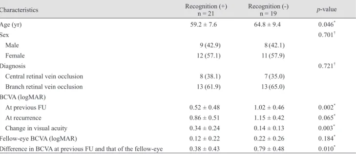
visual acuity, seven were diagnosed with CRVO and 13 with BRVO. The mean age, visual acuity before the recur- Fig. 2. Optical coherence tomography findings before (A,B) and after (C,D) recurrence of macular edema in 2 patients with retinal vein occlusion who recognized reduced visual acuity. Left column: visual acuity deteriorated from 20 / 40 (A) to 20 / 200 (C). The patient visited the hospital 3 weeks earlier than the scheduled follow-up date. The distance between the hospital and this patient’s residence was approximately 8 km. Right column: visual acuity had deteriorated from 20 / 30 (B) to 20 / 100 (D). The patient visited the hospital on the scheduled follow-up date despite recognizing a definite reduction in visual acuity. The distance between the hospital and this patient’s residence was approximately 295 km.
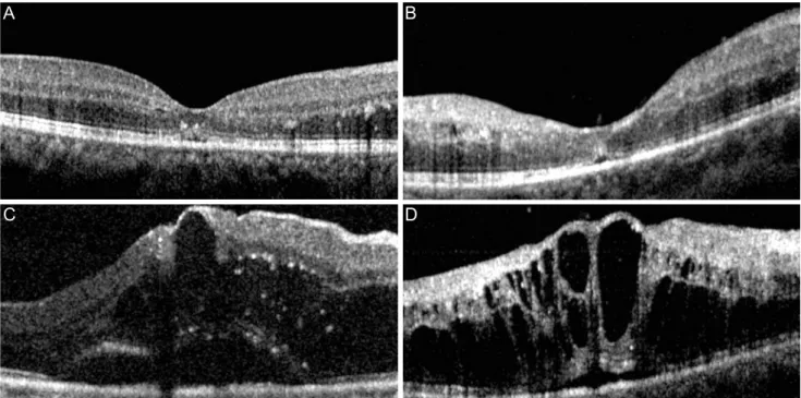
Fig. 3. Optical coherence tomography findings before (A,B) and after (C,D) recurrence of macular edema in 2 patients with retinal vein occlusion who did not recognize a reduction in visual acuity. Left column: visual acuities before and after recurrence were 20 / 100 (A) and 20 / 200 (C), respectively. Right column: visual acuities before and after recurrence were 20 / 63 (B) and 20 / 100 (D), respectively.
A
C
A
C
B
D
B
D
rence of macular edema, the extent of reduction in visual acuity after recurrence of macular edema, visual acuity of the fellow eye, and difference between the visual acuity of the studied eye before macular edema recurrence and that of the fellow eye were 64.8 ± 9.4 years, 1.02 ± 0.46 (Snellen equivalent, 20 / 209), 0.14 ± 0.13, 0.22 ± 0.26 (Snellen equivalent, 20 / 33), and 0.79 ± 0.48 respectively. Patients who recognized reduced visual acuity were younger (p = 0.046), had better visual acuity before the recurrence of macular edema (p = 0.002), experienced greater reductions in visual acuity after recurrence of macular edema (p = 0.003), and showed smaller differences between the visual acuity score of the studied eye before macular edema re- currence and that of the fellow eye (p = 0.010), than pa- tients who did not recognize reduced visual acuity. Sex (p
= 0.701), types of RVO (p = 0.721), and fellow eye visual acuity (p = 0.184) did not differ significantly between the two groups. Figs. 2 and 3 show representative cases of pa- tients who recognized and did not recognize reduced visu- al acuity, respectively.
Only four patients visited the hospital before the sched- uled follow-up date. The mean age of these four patients was 63.5 ± 9.3 years, mean visual acuity before the recur- rence was 0.51 ± 0.42 (Snellen equivalent, 20 / 64), and the mean extent of reduction in visual acuity following the re- currence was 0.59 ± 0.33. The mean distance between the hospital and the location of the four patients’ residences was 9.5 ± 6.2 km. The hospital was within 20 km for all four patients (100%). The mean distance for the remaining 17 patients who recognized reduced visual acuity, but did not visit the hospital prior to the scheduled date, was 50.5
± 88.1 km. The hospital was within 20 km for 10 patients (58.8%) and beyond 20 km for seven patients (41.2%). The proportion of patients residing within 20 km from the hos- pital was greater in the early-visit group (p = 0.035).
All patients underwent additional treatment for macular edema recurrence. After macular edema recurrence, 34 patients were administered 1 to 2 intravitreal anti-vascular endothelial growth factor injections alone for 6 months, while five were also administered intravitreal triamcino- lone injections. Five patients were administered intravitre- al triamcinolone injections alone and one was treated with a posterior sub-tenon triamcinolone injection only. The mean visual acuity at 3 months after treatment was 0.91 ± 0.53 (Snellen equivalent, 20 / 162). The mean visual acuity in patients who did and did not recognize a reduction in
visual acuity was 0.75 ± 0.45 and 1.12 ± 0.53, respectively (p = 0.032). The mean visual acuity at 3 months after treat- ment of patients who visited the hospital before the sched- uled follow-up date was 0.65 ± 0.48.
Discussion
In this study, we first investigated patients’ recognition and behavior regarding the recurrence of macular edema in RVO. Approximately half of the patients recognized reduced visual acuity due to recurrence of macular edema. Younger patients with relatively good visual acuity and patients who had experienced greater reduction in visual acuity were more likely to recognize reduced visual acuity. However, only a few patients visited the hospital promptly, and all of these few patients were living relatively close to the hospital.
In the present study, the exact time of macular edema recurrence could not be accurately determined. However, considering the approximately 2-month period between the follow-ups before and after recurrence of macular ede- ma, treatment may have been delayed by at least several weeks in many of the included patients. Generally, macu- lar edema secondary to RVO was not regarded as urgent.
In the present study, the effect of prompt hospital visitation on treatment outcome was not demonstrated. However, given that the results of some previous studies have sug- gested that prompt treatment may improve visual progno- sis [10-13], delayed treatment due to a delay in visiting the hospital may have a negative impact on visual prognosis.
In particular, one previous study revealed a definite difference in the treatment outcomes of eyes with symp- toms for ≤3 months and those with symptoms for ≥3 months [10]. Significantly improved visual acuity and decreased foveal thickness was maintained for 6 months after treatment in eyes with shorter symptom duration, whereas the values at 3 months and 6 months after treatment were not different from the baseline values in eyes with longer symptom duration. These results indicate the importance of early treatment after visual and anato- mical prognosis. Recognition of reduced visual acuity is thus an extremely important issue with regard to prompt treatment for recurring macular edema. In this study, older patients and patients with relatively poor visual acuity tended not to recognize reduced visual acuity. This result suggests that more intensive patient education with regard
to the recurrence of macular edema as indicated by chang- es in vision, and the fact that it necessitates a prompt visit to the hospital, is very important for these vulnerable pa- tients. Frequent, regular self-examination, such as that which is usually recommended for patients with exudative age-related macular degeneration, should be recommended to these patients [14]. This patient education is particularly pertinent when the scheduled period between follow-up visits is relatively long. In addition, considering the rela- tively older age of patients who did not recognize visual changes in this study, educating the relevant caretakers may be as important as educating the patients themselves.
The significantly smaller difference in the visual acuity scores of the studied eye before macular edema recurrence and that of the fellow eye observed in the present study was interesting. It is possible that patients may depend on the vision in the fellow eye during daily life when there is a relatively greater difference between the visual acuities of the two eyes. In this respect, a more specific and sensi- tive method, such as occluding the fellow eye and measur- ing the visual acuity of the affected eye once every several days, may be required to determine a decrease in the visual acuity of the affected eye.
In this study, we could not definitively determine why a majority of patients that recognized reduced visual acuity did not visit the hospital promptly. The mean age of the four patients who did visit the hospital promptly was slightly older than that of the remaining patients who rec- ognized reduced visual acuity. Visual acuity before the re- currence was comparable between the patients with and without prompt hospital visitation. While various personal reasons may contribute to a decision not to visit the hospi- tal early despite changes in visual acuity, we suspect that the distance between the hospital and the patient’s resi- dence is one of the primary reasons. All four of the pa- tients who visited the hospital promptly lived relatively close to it. Although statistical analysis was not feasible due to the very small number of cases, the mean distance between the hospital and the patients’ residences in these four cases was approximately 1 / 5 of that of the other pa- tients who recognized reduced visual acuity but waited until the scheduled follow-up date to visit the hospital.
This result may highlight the need for a simple and easy way to verify macular edema recurrence in these patients, as well as a need for more intensive patient education.
Considering potential problems related to a patient’s time
and expense, an initial eye check at a clinic located close to the patient’s residence may be an option. If the recurrence of macular edema is confirmed at the clinic, the patient could be promptly referred to the hospital before the scheduled follow-up date. For this method to operate effec- tively, information about the location of a local clinic with optical coherence tomography facilities is needed, because optical coherence tomography exhibits superior sensitivity with regard to the detection of macular edema, as com- pared to standard fundus examination [15,16].
The present study has several limitations. The study was retrospective. The exact time when visual acuity was reduced was not known. The patient may not think that a prompt hos- pital visit is necessary if the reduction in visual acuity occurs just few days before the scheduled follow-up date. Also, the results were all derived from a single treatment facility. So- cio-economic status and level of education may be different among patients at different hospitals. The relative proportions of patients who live close to the hospital and far from it will also differ for each given hospital. Thus, some of our results may not be directly applicable to other hospitals.
In summary, approximately 1 / 2 of the patients investi- gated recognized recurrence of macular edema in RVO.
These patients were relatively younger, had better visual acuity, and experienced greater reductions in visual acuity.
Among them, only a few patients visited the hospital promptly, and all of them lived relatively close to the hos- pital. These results underscore the need for intensive pa- tient education for elderly patients with poor visual acuity, and the importance of establishing simple and easy meth- ods to verify the recurrence of macular edema for patients who live far from the hospital. We hope further studies may reveal the influence of prompt hospital visitation on the long-term visual prognosis in these patients.
Conflict of Interest
No potential conflict of interest relevant to this article was reported.
Acknowledgements
This study was supported by Kim’s Eye Hospital Re- search Center.
References
1. Rogers SL, McIntosh RL, Lim L, et al. Natural history of branch retinal vein occlusion: an evidence-based systemat- ic review. Ophthalmology 2010;117:1094-101.
2. McIntosh RL, Rogers SL, Lim L, et al. Natural history of central retinal vein occlusion: an evidence-based systemat- ic review. Ophthalmology 2010;117:1113-23.
3. Campochiaro PA, Hafiz G, Shah SM, et al. Ranibizumab for macular edema due to retinal vein occlusions: implication of VEGF as a critical stimulator. Mol Ther 2008;16:791-9.
4. Brown DM, Campochiaro PA, Singh RP, et al. Ranibizum- ab for macular edema following central retinal vein occlu- sion: six-month primary end point results of a phase III study. Ophthalmology 2010;117:1124-33.
5. Campochiaro PA, Heier JS, Feiner L, et al. Ranibizumab for macular edema following branch retinal vein occlusion:
six-month primary end point results of a phase III study.
Ophthalmology 2010;117:1102-12.
6. Shin HY, Jee DH. The short-term efficacy of intravitreal ranibizumab for macular edema in central retinal vein oc- clusion. J Korean Ophthalmol Soc 2011;52:1048-54.
7. Kim JY, Park SP. Comparison between intravitreal bevaci- zumab and triamcinolone for macular edema secondary to branch retinal vein occlusion. Korean J Ophthalmol 2009;23:
259-65.
8. Chen SD, Sundaram V, Lochhead J, Patel CK. Intravitreal tri- amcinolone for the treatment of ischemic macular edema as- sociated with branch retinal vein occlusion. Am J Ophthalmol 2006;141:876-83.
9. Gregori NZ, Rosenfeld PJ, Puliafito CA, et al. One-year safety and efficacy of intravitreal triamcinolone acetonide for the management of macular edema secondary to cen- tral retinal vein occlusion. Retina 2006;26:889-95.
10. Oh JY, Seo JH, Ahn JK, et al. Early versus late intravitreal triamcinolone acetonide for macular edema associated with branch retinal vein occlusion. Korean J Ophthalmol 2007; 21:
18-20.
11. Haller JA, Bandello F, Belfort R Jr, et al. Randomized, sh- am-controlled trial of dexamethasone intravitreal implant in patients with macular edema due to retinal vein occlu- sion. Ophthalmology 2010;117:1134-46.
12. Scott IU, Ip MS, VanVeldhuisen PC, et al. A randomized trial comparing the efficacy and safety of intravitreal tri- amcinolone with standard care to treat vision loss associat- ed with macular edema secondary to branch retinal vein occlusion: the Standard Care vs Corticosteroid for Retinal Vein Occlusion (SCORE) study report 6. Arch Ophthalmol 2009;127:1115-28.
13. Ip MS, Scott IU, VanVeldhuisen PC, et al. A randomized trial comparing the efficacy and safety of intravitreal tri- amcinolone with observation to treat vision loss associated with macular edema secondary to central retinal vein oc- clusion: the Standard Care vs Corticosteroid for Retinal Vein Occlusion (SCORE) study report 5. Arch Ophthalmol 2009;127:1101-14.
14. Fine SL. Early detection of extrafoveal neovascular mem- branes by daily central field evaluation. Ophthalmology 1985;92:603-9.
15. Yang CS, Cheng CY, Lee FL, et al. Quantitative assessment of retinal thickness in diabetic patients with and without clinically significant macular edema using optical coher- ence tomography. Acta Ophthalmol Scand 2001;79:266-70.
16. Schaudig UH, Glaefke C, Scholz F, Richard G. Optical co- herence tomography for retinal thickness measurement in diabetic patients without clinically significant macular ede- ma. Ophthalmic Surg Lasers 2000;31:182-6.