Case Report pISSN: 1011-8942 eISSN: 2092-9382
Korean J Ophthalmol 2014;28(1):83-85 http://dx.doi.org/10.3341/kjo.2014.28.1.83
© 2014 The Korean Ophthalmological Society
This is an Open Access article distributed under the terms of the Creative Commons Attribution Non-Commercial License (http://creativecommons.org/licenses /by-nc/3.0/) which permits unrestricted non-commercial use, distribution, and reproduction in any medium, provided the original work is properly cited.
83
A Korean Patient with Lattice Corneal Dystrophy Type IV with Leu527Arg Mutation in the TGFBI Gene
Jinsun Kim1, Kyung A Lee2, Eung Kweon Kim1,3, Hyung Keun Lee1,3
1Institute of Vision Research, Department of Ophthalmology, Yonsei University College of Medicine, Seoul, Korea
2Department of Clinical Diagnosis, Yonsei University College of Medicine, Seoul, Korea
3Corneal Dystrophy Research Institute, Yonsei University, Seoul, Korea
Recent advances have allowed us to understand corneal dystrophies on a basic molecular level, and several studies have reported specific mutations in the TGFBI gene show- ing various phenotypes [1]. Among these are lattice corneal dystrophy (LCD), granular corneal dystrophy, Thiel-Behn- ke corneal dystrophy, Reis-Bucklers corneal dystrophy, and Cogan’s dystrophy [2,3].
LCD is one of the most common corneal disorders and is characterized by lineal stromal amyloid depositions that usually present bilaterally and symmetrically. Among the LCD subtypes, type IV is extremely rare, and only a few cases of this subtype have been discovered in the Japanese [4] and Indian populations [3], and one case in the Italian
population [2]. We recently discovered a Korean LCD type IV patient, and therefore we are hereby reporting the first Korean case of LCD type IV, confirmed by DNA sequenc- ing and clinical examination.
Case Report
An 87-year-old woman visited our clinic for a scheduled cataract surgery. At the time of preoperative evaluation, mild localized semitransparent opacity was noted in both corneas. The opacity showed lattice-shaped, granular de- posits with asymmetrical patterns in the stroma (Fig. 1).
The right eye was more severely affected, showing nodulo- linear amyloid deposits located mainly in the anterior stro- ma; the left eye showed less linear and macular opacity. The patient demonstrated a good visual outcome after the surgery, with a best-corrected visual acuity of 0.8 in both eyes.
Genomic DNA samples from the patient were extracted from peripheral leukocytes using the Easy-DNA Kit (Invi- trogen, Carlsbad, CA, USA). Polymerase chain reaction An 87-year-old woman visited our clinic for a scheduled cataract surgery. At the time of preoperative evaluation, slit lamp examination showed lattice-shaped and granular deposits with asymmetrical patterns in the stroma of both corneas. Genomic DNA samples of the patient, amplified by polymerase chain reaction, showed a single nucleotide substitution, c. 1580T>G (p.L527R), in the transforming growth factor-β-induced TGFBI gene. We also found two additional SNP mutations, c.1620T>C (p.F540F) and c.1678+23G>A, along with the well-known L527R mutation. This is the first report of lattice corneal dystrophy type IV with an L527R mutation outside of Japan, and could challenge the idea that L527R is caused by a mutation from a single Japanese ancestor.
Key Words: Hereditary corneal dystrophies, Transforming growth factor beta
Received: December 1, 2012 Accepted: December 13, 2012
Corresponding Author: Hyung Keun Lee, MD. Department of Ophthal- mology, Gangnam Severance Hospital, #211 Eonju-ro, Gangnam-gu, Seoul 135-720, Korea. Tel: 82-2-2019-3444, Fax: 82-2-3463-1049, E-mail:
This study was previously presented as a poster at the 105th Annual Meeting of The Korean Ophthalmological Society.
84
Korean J Ophthalmol Vol.28, No.1, 2014
was performed on this genomic DNA with specific prim- ers for the TGFBI gene (Table 1); the results showed a Leu527Arg mutation in the TGFBI gene (Fig. 2A). In addi- tion to the Leu527Arg mutation, two more single-nucleo- tide polymorphisms (SNPs) were also identified: a T>C variation at cDNA position 1620 resulting in Phe540Phe and a G>A variation at cDNA position 1678+23 (Fig. 2B and 2C).
The patient’s only family members, her son and grand- daughter, did not show any corneal stromal manifestations.
They also did not have any known mutations in the TGFBI gene specific for LCD type IV, though both showed the T>C variation at cDNA position 1620 and the G>A varia- tion at cDNA position 1678+23, just as in the present case.
Discussion
LCD type IV was first reported in 1998 [4]. The LCD type IV patients with L527R are characterized by asym- metric corneal opacities, sporadic occurrences, and late onset [5]. In the present case, because visual impairment was not directly related to the cornea, visual acuity was improved after cataract surgery. We could not identify the etiology of the condition because the patient’s lesion did not cause her visual symptoms.
Similar to previous LCD type IV cases, the patient in this report also showed only mild clinical manifestation without subjective symptoms. We assert that the novelty of this case is two-fold. Firstly, we found two novel SNP mu-
tations in addition to the well-known L527R mutation;
these were also found in the patient’s family members.
Table 1. Primers for TGFBI gene
Exon Primer sequence (5’→3’) Product size (bp)
12 TGFBI ex12 F AGCCTGGAATCACTC CCTCT 396
TGFBI ex12 R GATGTGCCAACTGTTTGCTG 396
Fig. 2. Open-frame sequencing of the TGFBI gene from the pa- tient. Direct sequencing of polymerase chain reaction products corresponding to exon 12 of TGFBI. (A) Heterozygous mutation, c.1580T>G (arrow) was detected in the TGFBI gene. (B) Hetero- zygous c.1620T>C p.Phe540Phe (rs4669) was detected (arrow). (C) Heterozygous c.1678+23G>A (rs2072239) was detected (arrow).
B
C A
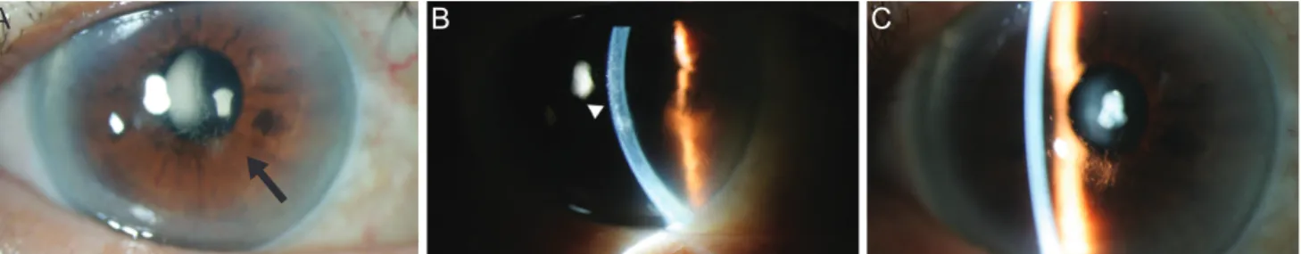
Fig. 1. Corneal photographs of lattice corneal dystrophy type IV patients. (A) The right eye was the more severely affected eye, display- ing nodulolinear amyloid deposits (arrow). (B) The deposits are mainly located in anterior stroma (arrowhead). (C) The left eye showed less linear and macular opacity than the right.
A B C
85 J Kim, et al. Korean Lattice Corneal Dystrophy Type IV Patient
Since the effects of these SNP mutations on the develop- ment and progression of LCD type IV remain uncertain, the role of these mutations should be investigated in future studies.
Secondly, since the initial report of the L527R mutation in the BIGH3 gene [4], all LCD type IV patients with an L527R mutation have been found exclusively in the Japa- nese population. This has led Fukuoka et al. [5] to report that the L527R SNP mutation might have been caused by a founder mutation in a single Japanese ancestor. To our knowledge, present case is the first report of an L527R mu- tation outside Japan, and it could challenge the idea that L527R is the product of a Japanese founder effect [5].
Lastly, because the clinical manifestations of LCD type IV are sporadic and extremely mild with subtle subjective symptoms, only a few cases have been detected and re- ported.
In summary, the patient in this report showed typical asymmetric nodulolinear stromal opacity in both corneas, and an L527R mutation in the TGFBI gene with two novel SNP mutations. This is the first report of the L527R muta- tion in Korea, as well as outside Japan, challenging the idea that L527R was caused by a founder mutation in a single Japanese ancestor.
Conflict of Interest
No potential conflict of interest relevant to this article was reported.
References
1. Munier FL, Korvatska E, Djemai A, et al. Kerato-epithelin mutations in four 5q31-linked corneal dystrophies. Nat Genet 1997;15:247-51.
2. Munier FL, Frueh BE, Othenin-Girard P, et al. BIGH3 mu- tation spectrum in corneal dystrophies. Invest Ophthalmol Vis Sci 2002;43:949-54.
3. Chakravarthi SV, Kannabiran C, Sridhar MS, Vemuganti GK. TGFBI gene mutations causing lattice and granular corneal dystrophies in Indian patients. Invest Ophthalmol Vis Sci 2005;46:121-5.
4. Fujiki K, Hotta Y, Nakayasu K, et al. A new L527R muta- tion of the betaIGH3 gene in patients with lattice corneal dystrophy with deep stromal opacities. Hum Genet 1998;
103:286-9.
5. Fukuoka H, Kawasaki S, Yamasaki K, et al. Lattice corneal dystrophy type IV (p.Leu527Arg) is caused by a founder mutation of the TGFBI gene in a single Japanese ancestor.
Invest Ophthalmol Vis Sci 2010;51:4523-30.