INTRODUCTION
Non-small cell lung cancer (NSCLC) is the most common type of lung cancer. Until now, the main therapeutic method for
NSCLC has been surgical resection with adjuvant chemother- apy, and for patients with unresectable, recurrent, or metastat- ic tumors, chemotherapy is the predominant treatment meth- od.1 Unfortunately, the use of traditional chemotherapy in the treatment of NSCLC is greatly limited by primary or secondary multi-drug resistance (MDR).2 MDR, which is defined as a re- sistance to one drug that is accompanied by a resistance to many other drugs, is believed to be a major reason for the pri- mary drug resistance of NSCLC and can lead to the failure of traditional chemotherapy.3,4 Therefore, determining how to treat NSCLC patients with primary drug resistance has be- come an urgent problem. Tyrosine kinase inhibitors targeting epidermal growth factor receptor (EGFR-TKIs) comprise one class of molecular target drugs and have achieved remarkable
Concomitance of P-gp/LRP Expression with EGFR Mutations in Exons 19 and 21 in Non-Small Cell Lung Cancers
Hong Wei1, Weipeng Lu1, Mei Li2, Qiuping Zhang3, and Shen Lu2
1MD Candidate in Pathology, Dalian Medical University, Dalian;
2Central Laboratory, The Second Hospital of Dalian Medical University, Dalian;
3Department of Pathology, The First Hospital of Dalian Medical University, Dalian, China.
Purpose: Traditional chemotherapy is the main adjuvant therapy for the treatment of non-small cell lung cancer (NSCLC). How- ever, the emergence of multi-drug resistance (MDR) has greatly restricted the curative effect of chemotherapy. Therefore, it is necessary to find a method to treat MDR NSCLC clinically. It is worth investigating whether NSCLCs that are resistant to tradi- tional chemotherapy can be effectively treated with tyrosine kinase inhibitors targeting epidermal growth factor receptor (EGFR).
Materials and Methods: The expression of P-glycoprotein (P-gp) and lung resistance-related protein (LRP) was detected by im- munohistochemistry, and mutations in EGFR (exons 19 and 21) and Kirsten rat sarcoma viral oncogene homolog (KRAS) (exon 2) were detected by high-resolution melting analysis (HRMA) of surgical NSCLC specimens from 127 patients who did not undergo traditional chemotherapy or radiotherapy. A Pearson chi-square test was performed to analyze the correlations between the ex- pression of P-gp and LRP and mutations in EGFR and KRAS.
Results: The expression frequencies of P-gp and LRP were significantly higher in adenocarcinomas from non-smoking patients;
the expression frequency of LRP was significantly higher in cancer tissue from female patients. The frequency of EGFR mutations was significantly higher in well to moderately differentiated adenocarcinomas from non-smoking female patients. The frequency of EGFR mutations in the cancers that expressed P-gp, LRP, or both P-gp and LRP was significantly higher than that in cancers that did not express P-gp or LRP.
Conclusion: NSCLCs expressing P-gp/LRP bear the EGFR mutation in exon 19 or 21 easily.
Key Words: Chemotherapy, multi-drug resistance, epidermal growth factor receptor-tyrosine kinase inhibitor, non-small cell lung cancer
Yonsei Med J 2016 Jan;57(1):50-57
http://dx.doi.org/10.3349/ymj.2016.57.1.50 pISSN: 0513-5796 · eISSN: 1976-2437
Received: October 8, 2014 Revised: February 10, 2015 Accepted: March 10, 2015
Corresponding author: Dr. Shen Lu, Central Laboratory, The Second Hospital of Dalian Medical University, Dalian 116027, Liaoning Province, China.
Tel: 86-411-84687554-601, Fax: 86-411-84672130, E-mail: [email protected]
•The authors have no financial conflicts of interest.
© Copyright: Yonsei University College of Medicine 2016
This is an Open Access article distributed under the terms of the Creative Com- mons Attribution Non-Commercial License (http://creativecommons.org/ licenses/
by-nc/3.0) which permits unrestricted non-commercial use, distribution, and repro- duction in any medium, provided the original work is properly cited.
success in the treatment of patients with NSCLCs bearing ac- tivating mutations of EGFR in exons 19 and 21.5,6 Moreover, these activating EGFR mutations appear more frequently in adenocarcinomas from Asian, female, and non-smoking pa- tients.7-10 Primary drug resistance to EGFR-TKIs still exists in NSCLC patients bearing the activating mutations in exon 2 of the Kirsten rat sarcoma viral oncogene homolog (KRAS) gene.11 Activating mutations in EGFR exons 19 and 21 have been used as biomarkers of NSCLC sensitivity to EGFR-TKIs, while activating mutations in KRAS exon 2 have been used as bio- markers of resistance to EGFR-TKIs. Clinically, many NSCLCs with MDR are well differentiated carcinomas, and well differ- entiated adenocarcinomas are generally sensitive to EGFR- TKIs. Therefore, it could be hypothesized that some NSCLCs with primary MDR may be sensitive to EGFR-TKIs or, in other words, that NSCLC patients with primary MDR may benefit from EGFR-TKIs. Both P-glycoprotein (P-gp) and lung resis- tance-related protein (LRP) are drug transporters in cells, and the expression of these factors is often observed in cancers that have developed MDR.4,12-16 Recently, the expression levels of these factors have been recognized as major biomarkers of MDR in NSCLC.
In this study, the expression of P-gp and LRP as biomarkers of MDR was detected in NSCLCs by immunohistochemistry, and mutations in EGFR exons 19 and 21 and KRAS exon 2 (as biomarkers of whether the patients can benefit from EGFR- TKIs, respectively) were analyzed by high-resolution melting analysis (HRMA) in 127 NSCLCs. By comparing the expres- sion of P-gp/LRP and EGFR mutation status, we investigated whether patients with NSCLC resistant to traditional chemo- therapy would benefit from treatment with EGFR-TKIs.
MATERIALS AND METHODS
Patients and tissue samples
The NSCLC samples were collected from 127 patients who underwent surgical procedures without any traditional che- motherapy or radiotherapy at the affiliated hospitals of Dalian Medical University from July 2007 to May 2009. Seventy-five of these patients were male and 52 were female. The patients had a mean±SD age of 62.1±9.1 years (range 36–80 years); 47 patients had a history of smoking, and 80 patients had no his- tory of smoking. Histologically, the tumor samples consisted of 42 squamous cell carcinomas and 85 adenocarcinomas. Lymph node metastases were found in 64 of the patients. There were 74 patients with well to moderately differentiated tumors and 53 patients with poorly differentiated tumors. All of the tissue samples were collected with the approval of the Ethics Com- mittee of the affiliated hospitals of Dalian Medical University and with the informed consent of the patients or families be- fore the surgical procedures.
Immunohistochemistry
Monoclonal antibodies (Abs) against human P-gp (1:200; clone F4; Sigma-Aldrich Co., LLC, St. Louis, MO, USA) and LRP (1:100; clone 1032; Beijing Golden Bridge Biotechnology Com- pany Ltd., Beijing, China) were used as primary antibodies.
Biotin-streptavidin-peroxidase staining with 3, 3’-diamino- benzidine-tetrahydrochloride detection was used. Tumor cells with staining in the cell membrane or cytoplasm were consid- ered to be positive. Each slide was graded blindly according to the percentage of positive tumor cells. The immunoreactivity of P-gp and LRP was scored as negative (-) when the proportion of positive tumor cells was less than 10% or as positive (+) when the number of positive tumor cells was between 10% and 100%.
DNA extraction and gene mutation detection
Tumor-enriched areas were selected and cut from the stained frozen sections that were marked by two pathologists. Genom- ic DNA was extracted from these areas and purified using the TIANamp Genomic DNA kit according to the manufacturer’s protocol (Tiangen, Beijing, China). EGFR exons 19 and 21 and KRAS exon 2 were amplified in triplicate from each sample in a 10-μL reaction volume with a 15-μL mineral oil overlay in each well of a 96-well PCR plate on a Mastercycler thermal cy- cler (Eppendorf, Hamburg, Germany). The primers were 5’-TG GATCCCAGAAGGTGAGAA-3’ and 5’-AGCAGAAACTCACAT CGAGGA-3’ (EGFR exon 19); 5’-CGCAGCATGTCAAGATCA-3’
and 5’-CCTCCTTACTTTGCCTCC-3’ (EGFR exon 21); and 5’-A GGCCTGCTGAAAATGACT-3’ and 5’-AATGGTCCTGCACCA GTAA-3’ (KRAS exon 2). The reaction conditions were described previously. The mutations were detected with HRMA on a LightScanner® 96 (Biofire Diagnostics, LLC, Salt Lake City, UT, USA). Melting curves were acquired at temperatures ranging from 60°C to 95°C and analyzed using LightScanner software (version 2.0, Biofire Diagnostics, LLC, Salt Lake City, UT, USA) according to the manufacturer’s instructions.17,18
Statistics
Pearson chi-square tests were used to analyze correlations for the expression of P-gp and LRP and mutations in EGFR and KRAS with the clinical and pathological characteristics of the patients; similar tests evaluated the correlation between the expression of P-gp and LRP and mutations in EGFR and KRAS.
Statistical significance was defined as p<0.05. Data analysis was performed using the SPSS 11.5 software package (SPSS Inc., Chicago, IL, USA).
RESULTS
P-gp and LRP expressions and their correlations with the clinicopathological characteristics of NSCLC patients
Of the 127 NSCLC samples analyzed by immunohistochemis-
try, the expression of P-gp was detected in 56 samples, corre- sponding to a positive expression frequency of 44.1%. LRP was expressed in 84 samples, corresponding to a positive ex- pression frequency of 66.1% of the tested NSCLCs.
The frequency of positive P-gp expression in patients aged
>60 years (52.1%, 37/71) was significantly higher than that in the patients aged ≤60 years (33.9%, 19/56) (p=0.004), and the frequency of positive P-gp expression in non-smokers was 51.3%
(41/80), which was significantly higher than that in smokers (31.9%, 15/47) (p=0.034). The frequency of positive P-gp ex- pression in adenocarcinomas was 57.6% (49/85), significantly higher than that in squamous cell carcinomas (16.7%, 7/42) (p=0.000). Significant relationships were observed between protein expression and patient age, smoking history, and tu- mor histology, but no significant relationships were found with patient gender, tumor differentiation, or lymph node metasta- sis (Table 1).
The frequency of positive LRP expression in female patients was 80.8% (42/52), significantly higher than that observed in male patients (56.0%, 42/75) (p=0.004), and the frequency of positive LRP expression in non-smokers was 80.0% (64/80), sig- nificantly higher than that observed in smokers (42.6%, 20/47) (p=0.000). The frequency of positive LRP expression in adeno- carcinomas was 85.9% (73/85), significantly higher than that in squamous cell carcinomas (26.2%, 11/42) (p=0.000). We con- cluded that significant relationships exist between protein ex- pression and patient gender, smoking history, and tumor his- tology, but not between protein expression and patient age,
tumor differentiation, or lymph node metastasis (Table 1).
EGFR and KRAS mutations detected by HRMA in NSCLC
Of the 127 NSCLC samples analyzed by HRMA, EGFR muta- tions were detected in 52 samples, corresponding to a muta- tion frequency of 40.9%; 19 samples contained mutations in exon 19 (Fig. 1) and 33 samples contained mutations in exon 21 (Fig. 2). However, mutations in exon 2 of KRAS were detect- ed in seven samples, corresponding to a mutation frequency of 5.5%.
Correlations between EGFR and KRAS mutations and the clinicopathological characteristics of NSCLC patients
The EGFR mutation frequency in female patients (59.6%, 31/52) was significantly higher than that in male patients (28.0%, 21/75) (p=0.000), and the frequency of EGFR mutations in non-smokers (55.0%, 44/80) was significantly higher than that in smokers (17.0%, 8/47) (p=0.000). The frequency of EGFR mutations in adenocarcinomas was 58.8% (50/85), which was significantly higher than that in squamous cell carcinomas (4.8%, 2/42) (p=0.000); and the frequency of EGFR mutations in well to moderately differentiated tumors (50.0%, 37/74) was significantly higher than that in poorly differentiated tumors (28.3%, 15/53) (p=0.014). We concluded that significant rela- tionships exist between EGFR mutation status and patient gen- der, smoking history, tumor histology, and degree of differen-
Table 1. The Associations between Clinicopathological Characteristics and P-gp or LRP Expression in NSCLC Clinicopathological
characteristics No.
P-gp LRP
Positive number
Positive
frequency (%) p value Positive number
Positive
frequency (%) p value
Gender 0.264 0.004
Male 75 30 40.0 42 56.0
Female 52 26 50.0 42 80.8
Age 0.004 0.695
≤60 56 19 33.9 36 64.3
>60 71 37 52.1 48 67.6
Smoking history 0.034 0.000
Non-smokers 80 41 51.3 64 80.0
Smokers 47 15 31.9 20 42.6
Lymph node metastasis 0.131 0.901
Absent 63 32 50.8 42 66.7
Present 64 24 37.5 42 65.6
Histology 0.000 0.000
Adc 85 49 57.6 73 85.9
SCC 42 7 16.7 11 26.2
Differentiation grade 0.390 0.435
Well-moderate 74 35 47.3 51 68.9
Poor 53 21 39.6 33 62.3
Adc, adenocarcinoma; SCC, squamous cell carcinoma; P-gp, P-glycoprotein; LRP, lung resistance-related protein; NSCLC, non-small cell lung cancer.
tiation, but not between EGFR mutation status and patient age or lymph node metastasis (Table 2).
The KRAS mutation frequency in male patients was 9.3%
(7/75), significantly higher than that in female patients (0.0%, 0/52) (p=0.041). We concluded that a significant relationship exists between KRAS mutation status and patient gender, but not between KRAS mutation status and patient age, smoking history, lymph node metastasis, histological type, or differen- tiation (Table 2).
Correlations of P-gp and LRP expression with EGFR mutation
The EGFR mutation frequency in P-gp-positive samples was 57.1% (32/56); this was significantly higher than that in the P- gp-negative samples (28.2%, 20/71) (p=0.001). The EGFR muta- tion frequency in the LRP-positive samples was 53.6% (45/84), significantly higher than that in the LRP-negative samples (16.3%, 7/43) (p=0.000). We then selected samples that were positive for P-gp or LRP and analyzed the correlation between the expression in these samples and the presence of EGFR
mutations. We found that the EGFR mutation frequency in these samples was 52.1% (49/94), significantly higher than that in the negative samples (9.1%, 3/33) (p=0.000). We then select- ed the samples that coexpressed both P-gp and LRP and ana- lyzed the correlation between the coexpression of these pro- teins and EGFR mutations. We found that the EGFR mutation frequency in these samples was 60.9% (28/46), significantly higher than that in the P-gp- and LRP-negative samples (29.6%, 24/81) (p=0.001). Because the samples with positive expres- sion of either P-gp or LRP could be considered to have MDR, we concluded that the NSCLCs with MDR always bear the EGFR mutations (Table 3, Fig. 3).
Correlations between P-gp and LRP expression and KRAS mutations
The KRAS mutation frequency in the P-gp-positive samples was 5.4% (3/56), lower than that in the negative samples (5.6%, 4/71), although this difference was not significant (p=1.000).
The KRAS mutation frequency in the LRP-positive samples was 6.0% (5/84), higher than that in the LRP-negative samples
Fig. 1. High-resolution melting curves for exon 19 of the EGFR gene. (A) Temperature shift observed in the melting curves of a NSCLC sample with a muta- tion in exon 19 of the EGFR and a NSCLC sample with a wild-type EGFR. Each sample was analysed in triplicate. (B) Fluorescence difference curves of the same samples depicted in A. The NSCLC sample with a mutation in EGFR exon 19 is a well differentiated adenocarcinoma from a non-smoking woman.
NSCLC, non-small cell lung cancer; EGFR, epidermal growth factor receptor.
1 0.8 0.6 0.4 0.2 0
Temperature (°C) Shifted melting curves
77 78 79 80 81 82 83 84 85
Temperature normalized fluorescence
A
wtmt
0.3 0.25 0.2 0.15 0.1 0.05 0
Temperature (°C) Difference curves
77 78 79 80 81 82 83 84 85
∆ Fluorescence
B
wtmt
Fig. 2. High-resolution melting curves for exon 21 of the EGFR gene. (A) Temperature shift observed in the melting curves of a NSCLC sample with a muta- tion in exon 21 of the EGFR and a NSCLC sample with a wild-type EGFR. Each sample was analysed in triplicate. (B) Fluorescence difference curves of the same samples depicted in A. The NSCLC sample with a mutation in EGFR exon 21 is a moderately differentiated adenocarcinoma from a non-smoking woman. NSCLC, non-small cell lung cancer; EGFR, epidermal growth factor receptor.
1 0.8 0.6 0.4 0.2 0
Temperature (°C) Shifted melting curves
81 82 83 84 85 86 87 88
Temperature normalized fluorescence
A
wtmt
0.15
0.1
0.05
0
Temperature (°C) Difference curves
81 82 83 84 85 86 87 88
∆ Fluorescence
B
wtmt
(4.7%, 2/43), although this difference was also not significant (p=1.000). Moreover, we selected the samples that were posi- tive for P-gp or LRP and analyzed the correlation between the expression of these proteins and KRAS mutations. We found that the KRAS mutation frequency in these samples was 5.3%
(5/94), lower than that in the negative samples (6.1%, 2/33), al- though this difference was not significant (p=1.000). We then selected the samples that coexpressed both P-gp and LRP and analyzed the correlation between the coexpression of these
proteins and the presence of KRAS mutations. We found that the KRAS mutation frequency in these samples was 6.5%
(3/46), higher than that in the P-gp- and LRP-negative samples (4.9%, 4/81), although this difference was not significant (p=
1.000). Thus, we concluded that no significant relationship ex- ists between MDR and the presence of KRAS mutations in NSCLC (Table 4).
Table 3. The Association of P-gp or LRP Expression with EGFR Mutations in NSCLC
MDR protein No. EGFR mutation
Number Frequency (%) p value
P-gp 0.001
Positive 56 32 57.1
Negative 71 20 28.2
LRP 0.000
Positive 84 45 53.6
Negative 43 7 16.3
P-gp/LRP 0.000
Positive* 94 49 52.1
Negative 33 3 9.1
P-gp and LRP 0.001
Positive† 46 28 60.9
Negative 81 24 29.6
P-gp, P-glycoprotein; LRP, lung resistance-related protein; EGFR, epidermal growth factor receptor; NSCLC, non-small cell lung cancer.
*The positive expression of P-gp/LRP was defined as the expression of P-gp or LRP in a sample, †The positive expression of P-gp and LRP was defined as the co- expression of both P-gp and LRP in a sample.
Table 2. The Associations between Clinicopathological Characteristics and EGFR or KRAS Mutations in NSCLC Clinicopathological
characteristics No.
EGFR status KRAS status
Positive number
Positive
frequency (%) p value Positive number
Positive
frequency (%) p value
Gender 0.000 0.041
Male 75 21 28.0 7 9.3
Female 52 31 59.6 0 0.0
Age 0.979 0.746
≤60 56 23 41.1 4 7.1
>60 71 29 40.8 3 4.2
Smoking history 0.000 0.124
Non-smokers 80 44 55.0 2 2.5
Smokers 47 8 17.0 5 10.6
Lymph node metastasis 0.517 0.424
Absent 63 24 38.1 5 7.9
Present 64 28 43.8 2 3.1
Histology 0.000 0.327
Adc 85 50 58.8 3 3.5
SCC 42 2 4.8 4 9.5
Differentiation grade 0.014 0.648
Well-moderate 74 37 50.0 3 4.1
Poor 53 15 28.3 4 7.5
Adc, adenocarcinoma; SCC, squamous cell carcinoma; EGFR, epidermal growth factor receptor; KRAS, Kirsten rat sarcoma viral oncogene homolog; NSCLC, non- small cell lung cancer.
Table 4. The Association between the Expression of P-gp or LRP and Mutations in KRAS in NSCLC
MDR protein No. KRAS mutation
Nnumber Frequency (%) p value
P-gp 1.000
Positive 56 3 5.4
Negative 71 4 5.6
LRP 1.000
Positive 84 5 6
Negative 43 2 4.7
P-gp/LRP 1.000
Positive* 94 5 5.3
Negative 33 2 6.1
P-gp and LRP 1.000
Positive† 46 3 6.5
Negative 81 4 4.9
P-gp, P-glycoprotein; LRP, lung resistance-related protein; KRAS, Kirsten rat sarcoma viral oncogene homolog; NSCLC, non-small cell lung cancer.
*The positive expression of P-gp/LRP was defined as the expression of P-gp or LRP in a sample, †The positive expression of P-gp and LRP was defined as the co- expression of both P-gp and LRP in a sample.
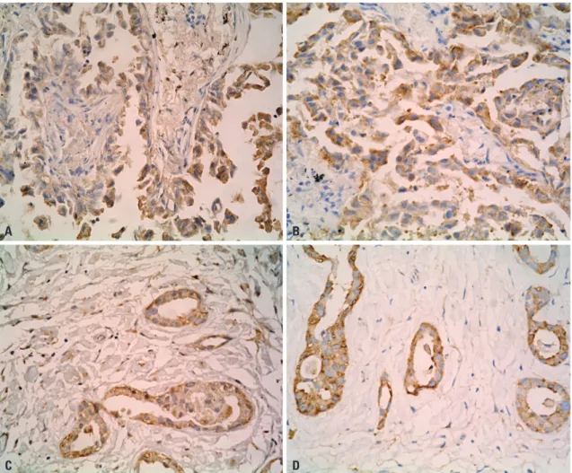
Fig. 3. Immunohistochemical staining of P-glycoprotein and lung resistance-related protein expression in NSCLC samples. Immunohistochemical staining of P-glycoprotein expression (A) and lung resistance-related protein expression (B) in the same sample (a well differentiated adenocarcinoma from a fe- male non-smoker). This was the NSCLC sample with a mutation in exon 19 of the epidermal growth factor receptor, as detected by high-resolution melt- ing analysis, shown in Fig. 1. Immunohistochemical staining of P-glycoprotein expression (C) and lung resistance-related protein expression (D) in the same sample (a moderately differentiated adenocarcinoma from a non-smoking woman). This sample was the NSCLC sample that carried a mutation in exon 21 of the epithemal growth factor receptor, as detected by high-resolution melting, shown in Fig. 2. Original magnification: ×400. NSCLC, non-small cell lung cancer.
A
C
B
D
DISCUSSION
NSCLC accounts for approximately 85% of lung cancers.19 Tra- ditional chemotherapy has played an important role in the treat- ment of NSCLC, although its curative effect has been greatly lim- ited by MDR. It is well known that the occurrence of MDR is closely related to the expression of P-gp/LRP in NSCLCs. P-gp is a 170-kDa transmembrane protein that was first discovered in 1976. P-gp can decrease the concentration of various drugs in the cytoplasm by extruding them out of cells and promot- ing the development of MDR.20-23 LRP, which was initially iden- tified in a non-P-gp MDR lung cancer cell line, is a major com- ponent of the human major vault protein.24-28 LRP can decrease the concentration of chemical drugs in the nucleus by regulat- ing nucleocytoplasmic transport and the drugs that are trans- ported to the cytoplasm can then be extruded from the cancer cell by exocytosis.16,29-31 Previous studies have reported that P-gp is expressed in 35–52% of NSCLCs and LRP is expressed in 65–88% of NSCLCs.29,32,33 The expression of P-gp/LRP could be regarded as a biomarker of primary MDR in NSCLCs. In this study, we found that the frequencies of positive expression of P-gp and LRP detected by immunohistochemistry were 44.1% and 66.1% respectively, which was consistent with the frequencies reported previously, thus the expressions of them were reliable for used as biomarkers to indicate primary MDR of the patients in this study.
In this research, clinicopathological characteristics of the patients with NSCLCs expressing P-gp/LRP positively were similar to those bearing EGFR mutation in exon 19 or 21. Gen- erally, patients with NSCLCs bearing these mutations could benefit from EGFR-TKI. Therefore, one could speculate that patients with NSCLCs expressing P-gp/LRP would also bene- fit from EGFR-TKIs. Recently, mutation in EGFR exon 19 or 21 has been used as a biomarker for sensitivity of NSCLCs to EG- FR-TKIs.34-37 Thus, we analyzed correlations of P-gp/LRP ex- pression with EGFR mutation status to investigate whether pa- tients with NSCLCs expressing P-gp/LRP would bear the EGFR mutation in exon 19 or 21. We found that the EGFR mutation frequency in cancers expressing P-gp/LRP was significantly higher than in those without. These results suggest that NSCLCs expressing P-gp/LRP bear the EGFR mutation in exon 19 or 21. As P-gp/LRP expression was found to be a biomarker for MDR, we discerned that NSCLC patients who are not fit for traditional chemotherapy due to cancers with MDR may benefit from EGFR-TKIs.
ACKNOWLEDGEMENTS
This work was supported by the National Natural Science Foun- dation of China (81071805).
REFERENCES
1. Okamoto T, Ichinose Y. [Adjuvant chemotherapy for non-small cell lung cancer]. Gan To Kagaku Ryoho 2006;33:1985-90.
2. Monzo M, Rosell R, Taron M. Drug resistance in non-small cell lung cancer. Lung Cancer 2001;34 Suppl 2:S91-4.
3. Fojo A, Hamilton TC, Young RC, Ozols RF. Multidrug resistance in ovarian cancer. Cancer 1987;60(8 Suppl):2075-80.
4. Baguley BC. Multiple drug resistance mechanisms in cancer. Mol Biotechnol 2010;46:308-16.
5. Lynch TJ, Bell DW, Sordella R, Gurubhagavatula S, Okimoto RA, Brannigan BW, et al. Activating mutations in the epidermal growth factor receptor underlying responsiveness of non-small- cell lung cancer to gefitinib. N Engl J Med 2004;350:2129-39.
6. Paez JG, Jänne PA, Lee JC, Tracy S, Greulich H, Gabriel S, et al.
EGFR mutations in lung cancer: correlation with clinical response to gefitinib therapy. Science 2004;304:1497-500.
7. Kaneda H, Tamura K, Kurata T, Uejima H, Nakagawa K, Fukuoka M. Retrospective analysis of the predictive factors associated with the response and survival benefit of gefitinib in patients with ad- vanced non-small-cell lung cancer. Lung Cancer 2004;46:247-54.
8. Takano T, Ohe Y, Kusumoto M, Tateishi U, Yamamoto S, Nokihara H, et al. Risk factors for interstitial lung disease and predictive fac- tors for tumor response in patients with advanced non-small cell lung cancer treated with gefitinib. Lung Cancer 2004;45:93-104.
9. Tamura K, Fukuoka M. Gefitinib in non-small cell lung cancer. Ex- pert Opin Pharmacother 2005;6:985-93.
10. Ando M, Okamoto I, Yamamoto N, Takeda K, Tamura K, Seto T, et al. Predictive factors for interstitial lung disease, antitumor re- sponse, and survival in non-small-cell lung cancer patients treated with gefitinib. J Clin Oncol 2006;24:2549-56.
11. Massarelli E, Varella-Garcia M, Tang X, Xavier AC, Ozburn NC, Liu DD, et al. KRAS mutation is an important predictor of resis- tance to therapy with epidermal growth factor receptor tyrosine kinase inhibitors in non-small-cell lung cancer. Clin Cancer Res 2007;13:2890-6.
12. Sandor V, Fojo T, Bates SE. Future perspectives for the develop- ment of P-glycoprotein modulators. Drug Resist Updat 1998;1:
190-200.
13. Szakács G, Paterson JK, Ludwig JA, Booth-Genthe C, Gottesman MM. Targeting multidrug resistance in cancer. Nat Rev Drug Dis- cov 2006;5:219-34.
14. Kitazono M, Sumizawa T, Takebayashi Y, Chen ZS, Furukawa T, Nagayama S, et al. Multidrug resistance and the lung resistance- related protein in human colon carcinoma SW-620 cells. J Natl Cancer Inst 1999;91:1647-53.
15. Scheffer GL, Schroeijers AB, Izquierdo MA, Wiemer EA, Scheper RJ. Lung resistance-related protein/major vault protein and vaults in multidrug-resistant cancer. Curr Opin Oncol 2000;12:550-6.
16. Meschini S, Marra M, Calcabrini A, Monti E, Gariboldi M, Dolfini E, et al. Role of the lung resistance-related protein (LRP) in the drug sensitivity of cultured tumor cells. Toxicol In Vitro 2002;16:
389-98.
17. Li M, Liu L, Liu Z, Yue S, Zhou L, Zhang Q, et al. The status of KRAS mutations in patients with non-small cell lung cancers from mainland China. Oncol Rep 2009;22:1013-20.
18. Li M, Zhang Q, Liu L, Liu Z, Zhou L, Wang Z, et al. The different clinical significance of EGFR mutations in exon 19 and 21 in non- small cell lung cancer patients of China. Neoplasma 2011;58:74- 81.
19. Hayes DN, Monti S, Parmigiani G, Gilks CB, Naoki K, Bhattacha- rjee A, et al. Gene expression profiling reveals reproducible hu-
man lung adenocarcinoma subtypes in multiple independent patient cohorts. J Clin Oncol 2006;24:5079-90.
20. Juliano RL, Ling V. A surface glycoprotein modulating drug per- meability in Chinese hamster ovary cell mutants. Biochim Bio- phys Acta 1976;455:152-62.
21. Riordan JR, Ling V. Purification of P-glycoprotein from plasma membrane vesicles of Chinese hamster ovary cell mutants with reduced colchicine permeability. J Biol Chem 1979;254:12701-5.
22. Debenham PG, Kartner N, Siminovitch L, Riordan JR, Ling V.
DNA-mediated transfer of multiple drug resistance and plasma membrane glycoprotein expression. Mol Cell Biol 1982;2:881-9.
23. Riordan JR, Deuchars K, Kartner N, Alon N, Trent J, Ling V. Ampli- fication of P-glycoprotein genes in multidrug-resistant mammali- an cell lines. Nature 1985;316:817-9.
24. Kedersha NL, Rome LH. Isolation and characterization of a novel ribonucleoprotein particle: large structures contain a single spe- cies of small RNA. J Cell Biol 1986;103:699-709.
25. Rome L, Kedersha N, Chugani D. Unlocking vaults: organelles in search of a function. Trends Cell Biol 1991;1:47-50.
26. Scheper RJ, Broxterman HJ, Scheffer GL, Kaaijk P, Dalton WS, van Heijningen TH, et al. Overexpression of a M(r) 110,000 vesicular protein in non-P-glycoprotein-mediated multidrug resistance.
Cancer Res 1993;53:1475-9.
27. Scheffer GL, Wijngaard PL, Flens MJ, Izquierdo MA, Slovak ML, Pinedo HM, et al. The drug resistance-related protein LRP is the human major vault protein. Nat Med 1995;1:578-82.
28. Izquierdo MA, Shoemaker RH, Flens MJ, Scheffer GL, Wu L, Prather TR, et al. Overlapping phenotypes of multidrug resistance among panels of human cancer-cell lines. Int J Cancer 1996;65:
230-7.
29. Scagliotti GV, Novello S, Selvaggi G. Multidrug resistance in non-
small-cell lung cancer. Ann Oncol 1999;10 Suppl 5:S83-6.
30. Chugani DC, Rome LH, Kedersha NL. Evidence that vault ribo- nucleoprotein particles localize to the nuclear pore complex. J Cell Sci 1993;106(Pt 1):23-9.
31. Schuurhuis GJ, Broxterman HJ, de Lange JH, Pinedo HM, van Heijningen TH, Kuiper CM, et al. Early multidrug resistance, de- fined by changes in intracellular doxorubicin distribution, inde- pendent of P-glycoprotein. Br J Cancer 1991;64:857-61.
32. Paredes Lario A, Blanco García C, Echenique Elizondo M, Lobo C.
[Expression of proteins associated with multidrug resistance and resistance to chemotherapy in lung cancer]. Arch Bronconeumol 2007;43:479-84.
33. Zuo Y, Huang J, Mu C, Shen D. The expression and significance of the multidrug resistance-related proteins P-gp, MRP and LRP in human non-small cell lung cancer tissues. The Chinese-German Journal of Clinical Oncology 2007;6:432-6.
34. Fiala O, Pešek M, Fínek J, Bru˚ha F, Bortlícˇek Z, Krejcˇí J, et al. [EGFR mutations in patients with advanced NSCLC]. Klin Onkol 2012;
25:267-73.
35. Fukihara J, Watanabe N, Taniguchi H, Kondoh Y, Kimura T, Kata- oka K, et al. Clinical predictors of response to EGFR tyrosine ki- nase inhibitors in patients with EGFR-mutant non-small cell lung cancer. Oncology 2014;86:86-93.
36. Wang J, Nong J, Jia H, Qin N, Li X, Zhang H, et al. Efficacy and pre- dictors of EGFR tyrosine kinase inhibitors in Chinese advanced lung adenocarcinoma: analyses of 253 cases from a single insti- tute. Oncol Res 2014;21:237-46.
37. Pallis AG, Fennell DA, Szutowicz E, Leighl NB, Greillier L, Dzi- adziuszko R. Biomarkers of clinical benefit for anti-epidermal growth factor receptor agents in patients with non-small-cell lung cancer. Br J Cancer 2011;105:1-8.