INTRODUCTION
Although there are no generally accepted criteria for surgical treatment of partial anterior cruciate ligament (ACL) tears, in-
terest on rupture pattern with respect to individual bundles based on the double bundle (DB) anatomy of the ACL and its biomechanical properties has increased recently,1-12 and these studies suggest that partial ruptures, depending on injury mechanism, can exclusively affect individual bundles. For ex- ample, an anteriorly directed force has the potential to damage the anteromedial (AM) bundle, whereas a pivoting action can injure the posterolateral (PL) bundle.12 In addition, rupture pat- terns of AM and PL bundles vary widely, and depend on wheth- er injuries result from mild elongation or severe stretching.
Based on this anatomically and biomechanically orientated DB concept, researchers have focused recently on anatomical DB ACL reconstruction7,13-17 and the treatment of symptomatic par- tial ACL tears using individual bundle reconstruction techni- ques.5,18,19 However, surgeons are posed with a dilemma as to
A Comparison between Clinical Results of Selective Bundle and Double Bundle Anterior Cruciate
Ligament Reconstruction
Yon-Sik Yoo
1, Si Young Song
1, Cheol Jung Yang
1, Jong Mun Ha
1, Yoon Sang Kim
2, and Young-Jin Seo
11Department of Orthopedic Surgery, Hallym University Dongtan Sacred Heart Hospital, Hwaseong;
2HiLab, Korea University of Technology and Education, Cheonan, Korea.
Purpose: The purpose of this study was to compare the clinical outcomes of arthroscopic anatomical double bundle (DB) anteri- or cruciate ligament (ACL) reconstruction with either selective anteromedial (AM) or posterolateral (PL) bundle reconstruction while preserving a relatively healthy ACL bundle.
Materials and Methods: The authors evaluated 98 patients with a mean follow-up of 30.8±4.0 months who had undergone DB or selective bundle ACL reconstructions. Of these, 34 cases underwent DB ACL reconstruction (group A), 34 underwent selective AM bundle reconstruction (group B), and 30 underwent selective PL bundle reconstructions (group C). These groups were com- pared with respect to Lysholm and International Knee Documentation Committee (IKDC) score, side-to-side differences of anterior laxity measured by KT-2000 arthrometer at 30 lbs, and stress radiography and Lachman and pivot shift test results. Pre- and post- operative data were objectively evaluated using a statistical approach.
Results: The preoperative anterior instability measured by manual stress radiography at 90° of knee flexion in group A was signifi- cantly greater than that in groups B and C (all p<0.001). At last follow-up, mean side-to-side instrumented laxities measured by the KT-2000 and manual stress radiography were significantly improved from preoperative data in all groups (all p<0.001). There were no significant differences between the three groups in anterior instability measured by KT-2000 arthrometer, pivot shift, or functional scores.
Conclusion: Selective bundle reconstruction in partial ACL tears offers comparable clinical results to DB reconstruction in com- plete ACL tears.
Key Words: Anterior cruciate ligament, double bundle, selective bundle, reconstruction Yonsei Med J 2016 Sep;57(5):1199-1208
http://dx.doi.org/10.3349/ymj.2016.57.5.1199 pISSN: 0513-5796 · eISSN: 1976-2437
Received: July 27, 2015 Revised: December 12, 2015 Accepted: February 17, 2016
Corresponding author: Dr. Young-Jin Seo, Department of Orthopedic Surgery, Hallym University Dongtan Sacred Heart Hospital, 7 Keunjaebong-gil, Hwaseong 18450, Korea.
Tel: 82-31-8086-2410, Fax: 82-31-8086-2429, E-mail: [email protected]
•The authors have no financial conflicts of interest.
© Copyright: Yonsei University College of Medicine 2016
This is an Open Access article distributed under the terms of the Creative Com- mons Attribution Non-Commercial License (http://creativecommons.org/licenses/
by-nc/3.0) which permits unrestricted non-commercial use, distribution, and repro- duction in any medium, provided the original work is properly cited.
how to evaluate the viabilities and functionalities of remaining bundles and how to determine which bundle should be pre- served or sacrificed. As a result, surgeons must rely on subjec- tive feelings of the relative laxity of bundles intraoperatively.19
Recent studies reported 10–25% of ACL surgery provides a good indication of ACL augmentation.5 In the present retro- spective study, we performed selective bundle reconstruction in a more active manner, that is, we tried to preserve moderate- ly attenuated bundles, and only reconstructed bundles that were considered completely torn or remained tensionless during arthroscopic probing. Also, we conducted DB ACL reconstruc- tion for cases with completely torn both bundles.
The purpose of this study was to compare the clinical out- comes of arthroscopic anatomic DB ACL reconstruction with selective AM or PL bundle reconstruction while preserving a relatively healthy ACL bundle. The study hypothesis was that postoperative antero-posterior, rotational, and knee scores in three groups would significantly improve compared to preop- erative data. We also hypothesized that there would be no post- operative clinical differences between DB ACL reconstruction and selective bundle ACL reconstruction.
MATERIALS AND METHODS
Demographic data
Herein, 98 patients who underwent DB ACL reconstruction or selective AM or PL bundle reconstructions between September 2007 and February 2011 with a mean follow-up of 30.8±4.0 months were included in this study. The study protocol of this retrospective comparative study was approved by our Institu- tional Review Board. The final decision to proceed to DB ACL reconstruction or selective bundle reconstruction was made during arthroscopy. Patients with remnant ACL fibers corre- sponding to the AM or PL bundles were surgically indicated for selective bundle reconstruction.
Remnant fibers corresponding to the AM or PL bundles were preserved when their thickness was at least one third of its orig- inal diameter, remnant fibers were still bridging the femur and tibia,5 and when there was acceptable tension under arthroscop- ic probing while applying an anterior drawer force (less than 5 mm laxity). Using this protocol, 34 individuals underwent se- lective AM bundle reconstruction (group B), and another 30 re- ceived selective PL bundle reconstruction (group C). The re- maining 34 patients, in which ACL fibers were completely torn from the intercondylar notch or reattached to PCL fibers, un- derwent DB ACL reconstruction (group A). Mean follow-up pe- riods of each group were 29.0±19.7 months in group A, 31.9±3.8 months in group B, and 31.8±3.6 months in group C. The inclu- sion criteria were as follows: patient was diagnosed as having ACL injury by Lachman test, pivot shift test, and magnetic reso- nance imaging (MRI); patient underwent selective bundle re- construction or DB ACL reconstruction. Patients who received
single-bundle ACL reconstruction due to a small femoral foot- print (smaller than 14 mm in diameter)20 or revision surgery were excluded. In addition, we also excluded patients with mul- tiple ligament injury, such as a concomitant posterior cruciate ligament rupture or PL corner injury, and patients who under- went bilateral ACL reconstruction.
There were 91 men and seven women. Mean age at time of surgery was 31±10.2 years (16–53 years). Injuries were sports related in 72 cases, a fall from height in 17 cases, and a traffic accident in 9 cases.
The average delay between injury and operation was 23 weeks (range 1 week to 250 weeks). The surgical procedures were performed by the senior author, and a double looped tibi- alis anterior allograft was used in all cases. Patient demographic data are summarized in Table 1.
Operative procedure
All patients were operated in the supine position using a leg- holding device under tourniquet control. Standard AM and an- terolateral portals were used for arthroscopic intraarticular in- spection, and the AM portal was used to inspect the ACL femoral attachment. This portal enables visualization of the entire me- dial wall of lateral femoral condyle and the identification of the various ACL injury patterns. An accessory anteromedial (AAM) portal was then established to remove the torn bundle while preserving the remnant. This portal was also used later to es- tablish the femoral tunnel (Fig. 1). The advantage of the AAM portal has been well described previously.21-25 We decided to perform DB ACL reconstruction or selective bundle reconstruc- tion according to the status of the bundles. Selective ACL re- construction was performed if ACL remnants met the above criteria, which comprised remnant fibers corresponding to the AM or PL bundle still bridging the femur and tibia and relatively good tension under proper probing while applying a force to translate the tibia anteriorly (Fig. 2). Meanwhile, DB ACL recon- struction was performed when both bundles were completely torn or when the ACL was absent, as in chronic cases (Fig. 3).
In case of selective bundle reconstruction, a torn AM or PL bundle was removed. The size of the femoral tunnel was deter- mined by the size of the AM or PL femoral footprint. In case of DB reconstruction, 2-mm bony bridge between two femoral tun- nels was considered, while the center of the tunnel was marked.20 For the femoral PL bone tunnel, the center of the PL footprint was marked with a microfracture awl through the AAM portal under 90° of knee flexion. A guidewire was inserted at the cen- ter of the PL footprint under 110° of knee flexion through the AAM portal, and reaming was performed while maintaining the same degree of knee flexion. For the femoral AM bone tun- nel, we positioned the microfracture awl in the center of the AM footprint through the AAM portal under 90° of knee flexion.26 A guidewire was then positioned at the previously marked center of the AM footprint and over drilled using the reamer, which was inserted through the AAM portal under 130° of knee flex-
ion. For double-bundle ACL reconstruction, the PL femoral tunnel was made first. Graft sizes and tunnel lengths were mea- sured intraoperatively. The diameters of the AM and PL grafts in each group were compared. Tunnel lengths in each group were also compared.
Under visualization from the AM portal, the tibial footprints of the AM and (or) PL bundles were marked using a thermal device (ArthroCare, Sunnyvale, CA, USA). To create the tibial AM bone tunnel, the tip of the ACL guide system (Acufex Smith
& Nephew, Andover, MA, USA) was placed at the AM footprint about 5 mm lateral to the medial tibial spine and 5 mm posteri- or to the anterior rim of the of the native AM bundle, while maintaining an angle of 55° to the tibial plateau.18 A tibial bone tunnel with an equal diameter to corresponding femoral tunnel was then made taking care not to damage the anterior tibial cortex or intermeniscal transverse ligament. To create the tibial PL bone tunnel, the tip of the guide at the PL footprint was po- sitioned 5 mm medial to the lateral tibial spine and 5 mm ante- rior to posterior root of the lateral meniscus at an angle of 45 degrees to the tibial plateau.15
A doubled loop of tibialis anterior allograft over an EndoBut-
ton CL (12 mm in length, Smith & Nephew Endoscopy, Ando- ver, MA, USA) was used as a graft for both selective and DB re- construction, and both ends of tendon grafts were whipstitched with FiberWire sutures. During DB ACL reconstruction, the al- lograft was split to obtain two suitable grafts for the AM and PL bundles with diameters of 6, 7, or 8 mm corresponding to the tunnel diameter. Prepared grafts were then passed from the tib- ial bone tunnel to the femoral bone tunnel. Femoral fixation was performed by using the EndoButton. Grafts were precondi- tioned by pulling tibial sides during 20 knee flexion-extension cycles. Tibial fixation was performed using bioabsorbable in- terference screws of the same diameter as the tunnel, and rein- forced with staples. Both AM and PL grafts were fixed at full knee extension, based on the findings of our previous biome- chanical study.11 Finally, grafts were inspected to confirm a full range of knee motion and the absence of roof or lateral wall im- pingement. An arthroscopic view of reconstruction of both bundles or selective bundles is illustrated in Fig. 4.
Rehabilitation
The rehabilitation protocol used in all patients was similar to
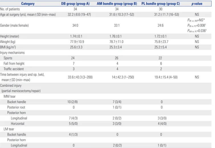
Table 1. Patient Demographics (n=98)
Category DB group (group A) AM bundle group (group B) PL bundle group (group C) p value
No. of patients 34 34 30
Age at surgery (yrs), mean±SD (min–max) 32.2±8.6 (19–47) 31.6±10.3 (17–52) 31.2±11.7 (16–53) NS
Gender (male:female) 34:0 33:1 24:6
PDBvs. AM=NS*
PDB vs. PL=0.008† PAM vs. PL=0.036‡
Height (meter) 1.74±0.1 1.76±0.1 1.72±0.1 NS
Weight (kg) 77.9±10.9 78.7±11.0 75.8±23.7 NS
BMI (kg/m2) 25.6±3.3 25.3±3.4 25.2±5.4 NS
Injury mechanisms
Sports 24 26 22
Fall from height 7 4 6
Traffic accident 3 4 2
Time between injury and op. (wk),
mean±SD (min–max) 33.6±43.3 (3–200) 14±42.3 (1–250) 19.4±15.4 (4–50) NS
Combined injury
(partial meniscectomy/repair) MM tear
Bucket handle 10 (2/8) 7 (3/4) 0
Posterior root 0 1 (0/1) 0
Posterior horn
Longitudinal 7 (4/3) 2 (0/2) 3 (3/0)
Horizontal 5 (5/0) 3 (3/0) 4 (4/0)
LM tear
Bucket handle 4 (1/3) 0 0
Posterior horn
Longitudinal 0 2 (0/2) 1 (0/1)
DB, double bundle; AM, anteromedial; PL, posterolateral; BMI, body mass index; MM, medial meniscus; LM, lateral meniscus; NS, not significant.
*PDB vs. AM=NS (between group A and group B), †PDB vs. PL=0.008 (between group A and group C), ‡PAM vs. PL=0.036 (between group B and group C).
that of standard ACL reconstruction. Full extension and quad- riceps exercises were encouraged during the first postoperative week and heel-gliding exercise was allowed to obtain full range of knee motion thereafter. We recommended partial weight bearing during the 1st postoperative week and full weight bear- ing thereafter. Bracing was permitted without motion restric- tion for 12 weeks. Running was authorized at 12 weeks after surgery, and contact sports were permitted from 6 months post- operatively.
Evaluations
Patients were assessed preoperatively and at final follow-up af- ter the surgery. Every test was performed by the same surgeon (Y.J.S). Degree of translation in stress X-ray was checked with the knee under 90° of flexion. Lachman test and a KT-2000 ar- thrometer (MED metric Corp, San Diego, CA, USA) were checked under 30° of knee flexion.
Anterior instability
Side-to-side anterior instability was assessed using a KT-2000 arthrometer, stress X-ray, and Lachman test. Lachmann test was done under anesthesia on the day of the operation. Objec- tive data using KT-2000 was obtained at 30° of knee flexion un- der an anterior drawer force of 133N (30 lbs). A stress X-ray was checked at 90° of knee flexion under anterior drawer force us- ing a Telos device. Results of the arthrometer measurement and degree of translation on the stress X-ray were recorded as a side- to-side difference between the injured and uninjured knees.
Lachman test was conducted at 30° knee flexion. Lachman test results were classified as grades I (<5 mm), II (5–10 mm), and III (10–15 mm).
Pivot shift test
Intraoperative pivot shift test results under anesthesia were col- lected as preoperative data. Postoperative pivot shift test was performed at final follow up. Both the injured and uninjured knees were assessed. Pivot-shift test results were classified as grades 0 (normal), I (Glide), II (Clunk), and III (Gross).
Functional score
Lysholm knee score was used to evaluate subjective data con- cerning daily living activities. The International Knee Docu- mentation Committee (IKDC) score was obtained to evaluate objective data concerning the affected knee. Preoperatively, the Fig. 1. Anterolateral (AL), anteromedial (AM), and accessory anteromedial
(AAM) portals were established. The AM portal was used as a viewing portal for inspecting femoral ACL insertion sites. Femoral AM and PL bone tunnels were made through the AAM portal. PL, posterolateral; ACL, anterior cruciate ligament.
Fig. 2. Arthroscopic view from anteromedial portal. (A) The PL bundle was elongated, but still bridged tibia and femur (PL), although the AM bundle was completely torn (*). Selective AM bundle reconstruction was performed in this case. (B) A completely torn PL bundle was noted (*) by probing an AM bundle that was moderately attenuated (AM). Selective PL bundle reconstruction was conducted in this case. PL, posterolateral; AM, anteromedial.
A B
two scores were recorded on first day at admission. Postopera- tively, the scores were recorded at final follow up (at a mean of 30.8±4.0 months after the operation).
Statistical analysis
Based on the results of difference in STS difference measured by KT-2000 between pre- & post-op assessment, when the total sample size across the three groups was 98 (DB group: n=34; AM group: n=34; PL group: n=30), an one-way analysis of variance (ANOVA) had 99% power to detect at a 0.05 level a difference in means, assuming that the common standard deviation of 2.0.
The paired t-test was used to compare preoperative and post- operative data in each group, and the differences between the data of each group (including demographic data except gender ratio) were analyzed statistically using one-way ANOVA, with the Tukey honestly significant difference test for pair wise com- parisons. Chi-square test was used to compare the data regard- ing gender ratio, Lachman test, and pivot shift test. Significance was accepted for p-values <0.05. All analyses were performed us- ing SPSS for Windows (version 21.0, SPSS Inc., Chicago, IL, USA).
RESULTS
Assessment of demographic data
No statistical differences were noted preoperatively among the three groups with regard to age, height, weight, body mass in- dex and mean time between injury and operation. Gender ratio was significantly different between group A and groups B and C, respectively. The overall demographic data are illustrated in Table 1.
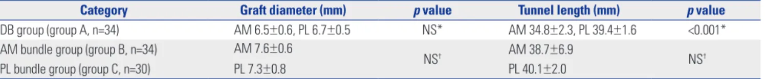
The average AM graft diameter in group A was 6.5±0.6 mm, and PL graft diameter was 6.7±0.5 mm. Average graft diameters were 7.6±0.6 mm in group B and 7.3±0.8 mm in group C. In group A, mean AM and PL tunnel lengths were 34.8±2.3 and 39.4±1.6 mm, respectively. In group B, mean AM tunnel length was 38.7±6.9 mm, and in group C, mean PL tunnel length was 40.1±2.0 mm, without significant difference between the two groups. The mean length of the PL femoral tunnel in group A was significantly longer than that of the AM femoral tunnel in the same group (p<0.001). The data are summarized in Table 2.
Anterior instability
Mean side-to-side instrumented laxities using the KT-2000 de- vice at 30° of knee flexion were 8.3±1.7 mm in group A, 6.6±1.6 mm in group B, and 5.8±1.5 mm in group C preoperatively, and these improved significantly to 1.8±1.4 mm in group A, 2.2±1.4 mm in group B, and 1.9±1.0 mm in group C at last follow-up (all p<0.001). Regarding intergroup differences in terms of preoper- ative KT-2000 values, although there was no significant differ- ences between groups B and C, the preoperative values of group A were found to be significantly greater than those of groups B and C (p=0.001, respectively). No significant intergroup difference was noticed with respect to postoperative side-to- side instrumented laxities (Table 3).
Degree of side to side translation in stress X-ray conducted at 90° of knee flexion were 7.5±2.5 mm in group A, 5.6±2.0 mm in group B, and 5.1±1.6 mm in group C preoperatively, and these improved significantly to 1.1±0.8 mm in group A, 0.9±0.6 mm in group B, and 0.6±0.7 mm in group C at last follow-up (all p<0.001).
The preoperative translation in group A was significantly great- Fig. 3. Arthroscopic view from anterolateral portal. Both AM and PL bun-
dles were absent (arrows). DB ACL reconstruction was conducted in this case. AM, anteromedial; PL, posterolateral; DB, double bundle; ACL, an- terior cruciate ligament.
Fig. 4. Arthroscopic view from anteromedial portal. (A) The AM graft (AM) was passed after PL graft (PL) fixation during DB ACL reconstruction. (B) Re- constructed AM bundle (AM) and (C) reconstructed PL bundle (PL). AM, anteromedial; PL, posterolateral; DB, double bundle; ACL, anterior cruciate ligament.
A B C
er than that in groups B and C (p=0.001, p<0.001, respectively) (Table 2).
In the preoperative Lachman test, all patients showed posi- tive results in all groups. In group A, 7 patients showed grade I, 20 patients showed grade II, and 7 patients showed grade III. In group B, 12 patients showed a grade I, 20 patients showed grade II, and 2 patient showed grade III. In group C, 14 patients dem- onstrated grade I instability, 16 patients demonstrated grade II, and 0 patients demonstrated grade III. A significant difference was found in preoperative Lachman test results between groups A and C. There were no significant differences among three groups postoperatively (Table 4).
Pivot shift
In preoperative pivot-shift test, all of the patients in group A had positive results; grade I (Glide) in 18 cases and grade II (Clunk) in 16 cases. In group B, 17 of 34 patients (50%) showed positive results; grade I (Glide) in 14 cases and grade II (Clunk) in 3 cas- es. In group C, 20 of the 30 patients (67%) showed positive re- sults; grade I (Glide) in 18 cases and grade II (Clunk) in 2 cases.
Significant differences were found in preoperative data between groups A and B and between groups A and C (p<0.001, p<0.001, respectively). There was no significant difference between the data of groups B and C, preoperatively.
The postoperative pivot-shift test at last follow up was posi- tive in 14 cases (grade I) in group A (41%), 13 cases (grade I: 10 cases, grade II: 3 cases) in group B (38%), 11 cases (grade I) in
group C (37%). There were no significant differences among three groups in terms of postoperative pivot shift data. Table 4 shows overall pre- and postoperative pivot shift data.
Functional score
In IKDC score, preoperative test results were abnormal and se- verely abnormal in 23 patients (68%) in group A, 17 patients (50%) in group B, and 13 patients (43%) in group C. Whereas, postoperative test results were normal and nearly normal in 32 patients (94%) in group A, 30 patients (88%) in group B, and 28 patients (93%) in group C (all p<0.001) (Table 4).
Lysholm knee scores improved significantly from 59.4±14.9 to 86.0±6.9 in group A, 56.0±13.3 to 85.9±4.8 in group B, and from 57.4±14.6 to 85.5±5.6 in group C (all p<0.001). No signifi- cant intergroup difference was found with respect to postoper- ative IKDC and Lysholm knee scores (Table 5).
DISCUSSION
This study highlights several interesting aspects. First, it com- pares the clinical results of selective bundle and DB ACL recon- struction. Little information is available in the literature on this subject, although Adachi, et al.27 compared the clinical outcomes of ACL augmentation and ACL reconstruction. Their augmen- tation procedure used single bundle graft with over-the-top femoral tunnel position, whereas both the DB reconstruction Table 2. Graft Diameter and Tunnel Length (n=98)
Category Graft diameter (mm) p value Tunnel length (mm) p value
DB group (group A, n=34) AM 6.5±0.6, PL 6.7±0.5 NS* AM 34.8±2.3, PL 39.4±1.6 <0.001*
AM bundle group (group B, n=34) AM 7.6±0.6
NS† AM 38.7±6.9
NS†
PL bundle group (group C, n=30) PL 7.3±0.8 PL 40.1±2.0
DB, double bundle; AM, anteromedial; PL, posterolateral; NS, not significant.
The AM femoral tunnel length was significantly shorter than PL femoral tunnel length in group A (DB group).
*Between length or diameter of the AM tunnel and PL tunnel in DB group, †Between group B and C.
Table 3. Pre- and Post-Operative Anterior Laxity Measurements (n=98)
Category STS difference measured by KT-2000 (mm) STS difference measured by stress X-ray (mm) Pre-op Post-op p-value (preop vs. postop) Pre-op Post-op p-value (preop vs. postop) DB group
(group A, n=34) 8.3±1.7 1.8±1.4 <0.001 7.5±2.5 1.1±0.8 <0.001
AM bundle group
(group B, n=34) 6.6±1.6 2.2±1.4 <0.001 5.6±2.0 0.9±0.6 <0.001
PL bundle group
(group C, n=30) 5.8±1.5 1.9±1.0 <0.001 5.1±1.6 0.6±0.7 <0.001
p value (intergroup)
<0.001*
PDB vs. AM<0.001† PDB vs. PL<0.001‡ PAM vs. PL=NS§
NS*
<0.001*
PDB vs. AM=0.001† PDB vs. PL<0.001‡ PAM vs. PL=NS§
0.040*
PDB vs. AM=NS† PDB vs. PL=0.031‡ PAM vs. PL=NS§ STS, side to side; DB, double bundle; AM, anteromedial; PL, posterolateral; NS, not significant; HSD, honestly significant difference.
Postoperative data significantly improved in all three study groups from preoperative data (all p<0.001). Regarding intergroup differences, group A (DB group) showed significantly greater preoperative laxity measured by KT-2000 and stress X-ray compared to groups B and C (AM and PL bundle group), respectively.
*p value was derived from ANOVA, †‡§p values were derived from Tukey’s HSD post-hoc test.
and selective procedures in this study have the same purpose in terms of restoring the anatomical DB structure of the ACL.
Postoperative data exhibited significant improvement com- pared to preoperative data in all groups. Also, postoperative clinical outcomes between the three groups showed no signifi- cant differences, except anterior displacement measured by stress X-ray at 90° of knee flexion. These results support our hy- pothesis. Second, group A (DB group) showed significantly greater anterior translation at 30° of knee flexion with KT-2000 and 90° of knee flexion with stress X-ray, compared to groups B (AM bundle group) and C (PL bundle group). These results part- ly concur with a previous study suggesting that remnant ACL fi- bers could contribute to resisting forces to anterior translation loads.28 Furthermore, greater preoperative rotational instability checked by pivot shift test was found in group A (100% posi- tive), compared to groups B (50% positive) and C (67% positive).
We believe precise preoperative clinical evaluation including anteroposterior instability and pivot shift test could provide a clue for operative options for selective bundle reconstruction.
Siebold reported that AM bundle tears show greater instabili- ty at anterior drawer test at 90° of knee flexion and KT-1000 at 30° of knee flexion rather than PL bundle tear. Some have sug- gested that PL bundle tears have a greater effect on rotational stability than AM bundle tears.18 In contrast to these theoretical
concepts, our data revealed that there was no significant differ- ence between groups B (AM bundle group) and C (PL bundle group) in terms of preoperative anterior instability and pivot shift test. The possible explanation for this disparity between the theoretical concept and the present clinical data is that a pure AM or PL bundle tear in clinical situations is very rare. Al- though the tear patterns of groups B and C in the present study varied and the degree of attenuation differed between the AM and PL bundles, both the AM and PL bundles exhibited consid- erable attenuation. Regardless of which bundle was saved, moderate attenuation of the remaining bundle caused greater preoperative side-to-side anterior instability and rotational in- stability than those in previous reports.18
The clinical appearances, imaging characteristics, and treat- ment plans of partial tears are topics of debate. It is difficult to achieve a definite diagnosis of an isolated bundle tear by MRI, because how much intact ligament remains cannot be estimat- ed, and no conclusive criteria are available for the assessment of individual bundle quality. Several treatment options are avail- able for preserving ACL remnants in partial ACL tears. Both conservative and surgically options based on augmentation and selective bundle reconstruction techniques are available.
Good clinical results have been reported after conservative treat- ment due to the intrinsic healing ability of ruptured ACLs.29,30 Table 4. Pre- and Post-Operative Manual Instability Assessment (n=98)
Category
Lachman test Pivot shift test
Pre-op Post-op p value
(preop vs. postop) Pre-op Post-op p value (preop vs. postop)
DB group (group A, n=34) <0.001 <0.001
Normal 0 20 0 20
Grade I 7 14 18 14
Grade II 20 0 16 0
Grade III 7 0 0 0
AM bundle group (group B, n=34) <0.001 NS
Normal 0 22 17 21
Grade I 12 12 14 10
Grade II 20 0 3 3
Grade III 2 0 0 0
PL bundle group (group C, n=30) <0.001 0.039
Normal 0 23 10 19
Grade I 14 7 18 11
Grade II 16 0 2 0
Grade III 0 0 0 0
p value (intergroup)
0.023*
PDB vs. AM=NS† PDB vs. PL=0.008‡
PAM vs. PL=NS§
NS
<0.001*
PDB vs. AM<0.001† PDB vs. PL<0.001‡ PAM vs. PL=NS§
NS
DB, double bundle; AM, anteromedial; PL, posterolateral; NS, not significant; HSD, honestly significant difference.
Group A (DB group) exhibited significantly greater preoperative rotational instability checked by pivot shift test, compared to groups B and C (AM and PL bundle group), respectively (all p<0.001).
*p value was derived from ANOVA, †‡§p values were derived from Tukey’s HSD post-hoc test, †PDB vs. AM=NS (between group A and group B), ‡PDB vs. PL=0.008 (be- tween group A and group C), §PAM vs. PL=NS (between group B and group C), †PDB vs. AM<0.001 (between group A and group B), ‡PDB vs. PL<0.001 (between group A and group C), §PAM vs. PL=NS (between group B and group C).
However, these results are still subjects of debate, and as yet, there is no clear understanding of the indications for conserva- tive treatment.
Meanwhile, the proven biologic and biomechanical advantag- es of preserving the ACL remnant have motivated development of current augmentation and selective bundle reconstruction procedures. Preservation of the ACL remnant during ACL re- construction may contribute to knee function due to its proprio- ceptive and biomechanical properties and because of its vascu- larity, which may facilitate vascularization of grafted tendons, as has been demonstrated recently.1,2,5-7,9,11,12
Adachi, et al.,27 in their clinical study, showed that joint stabil- ity and proprioceptive function in patients that underwent ar- throscopic-assisted ACL augmentation were superior to those of patients that underwent conventional ACL reconstruction.
Ochi, et al.5 in a series of 45 consecutive ACL augmentation cases concluded that the procedure produced improved joint stabili- ty, joint position sense, and Lysholm scores. Also, they found that only 10% of ACL surgeries were indicated for selective bun- dle reconstruction. Furthermore, Siebold and Fu18 reported a frequency of partial ACL tear of between 5 and 10%, and Zan- top, et al.,12 in a report on ACL rupture patterns, found that 25%
of ACL tears involved a partial rupture. In our series of 185 ACL surgeries (except patients with revision surgery and multiple ligament injury), including 87 patients who received single bun-
dle ACL reconstructions because of a small femoral footprint, which was estimated to be less than 14 mm, 64 cases (34.6%) could undergo either AM or PL selective bundle reconstruc- tion. This is much higher than the previously cited 10–25%
which is indicated for selective bundle reconstruction. The pos- sible explanation for this higher percentage of patients with se- lective bundle reconstruction is that we performed selective bundle ACL reconstruction in a more aggressive manner based on broader indications.
Operative treatment of partial ACL tears is technically de- manding due to the difficulties associated with preserving healthy fibers and differentiating between them and a torn bundle. In the present study, a review of the anesthesia record was performed in order to compare operation times between DB ACL reconstruction, selective bundle reconstruction, and typical single bundle ACL reconstruction without remnant preservation. Cases in which the operation was performed with meniscal repair or resection were excluded due to difficulty in defining an accurate ACL reconstruction time. The operative time was defined as beginning with initial portal placement and ending when the skin incisions were closed. The average operation time for DB ACL reconstruction was 102.5±25.7 min- utes (range 84–135 minutes), while selective bundle recon- struction cases averaged 95.7±18.5 minutes (range 64–125 min- utes) with no statistical significance. Meanwhile, the average Table 5. Functional Score Assessment (n=98)
Category
IKDC score Lysholm score
Pre-op Post-op p value
(preop vs. postop) Pre-op Post-op p value (preop vs. postop)
DB group (group A, n=34) <0.001 59.4±14.9 86.0±6.9 <0.001
A (normal) 0 18
B (nearly normal) 11 14
C (abnormal) 19 2
D (severely abnormal) 4 0
AM bundle group (group B, n=34) <0.001 56.0±13.3 85.9±4.8 <0.001
A (normal) 0 18
B (nearly normal) 17 12
C (abnormal) 15 4
D (severely abnormal) 2 0
PL bundle group (group C, n=30) <0.001 57.4±14.6 85.5±5.6 <0.001
A (normal) 0 16
B (nearly normal) 17 12
C (abnormal) 13 2
D (severely abnormal) 0 0
p value (intergroup)
0.163 PDB vs. AM=NS*
PDB vs. PL=0.045† PAM vs. PL=NS‡
NS NS NS
DB, double bundle; AM, anteromedial; PL, posterolateral; NS, not significant.
Pre- and postoperative IKDC and Lysholm scores. Significant postoperative improvements were observed in the three study groups (all p<0.001), although no sig- nificant intergroup difference was found postoperatively.
*PDB vs. AM=NS (between group A and group B), †PDB vs. PL=0.045 (between group A and group C), ‡PAM vs. PL=NS (between group B and group C).
operation time for single bundle reconstruction without rem- nant preservation was significantly shorter than DB ACL and selective bundle reconstruction (p<0.001 respectively, average 78.2±15.3 minutes with a range of 54–107 minutes). During the operation, most decisions to perform selective bundle recon- struction are made based on precise preoperative clinical assess- ment and a surgeon’s subjective feelings on probing the ACL remnants during arthroscopy.19 It is important that surgeons accurately understand the DB anatomy of the ACL and have wide experience with this procedure to avoid erroneous preser- vation of dysfunctional bundles. In particular, an isolated PL bundle tear can be missed because of an overlying intact AM bundle. Sonnery-Cottet and Chambat31 advocated the impor- tance of the figure of four position to prove stretching of the PL bundle. Based on clinical outcomes in the present study, we believe that given a wide indication for a selective bundle re- construction procedure, such as that used in the present study, selective bundle reconstruction produces postoperative results comparable to DB reconstruction. Although postoperative STS antero-posterior translation measured by stress X-ray showed a significant difference between groups A and C, a 0.5 mm differ- ence could be considered to be clinically negligible. Our data support the report by Ochi, et al.5 who postulated that pre- served loose remnant bundles can gradually become tight, re- sulting in good stability postoperatively. This study shows that selective bundle reconstruction is a reasonable treatment op- tion for partial ACL injury patients with appropriate indications.
Despite our results, this study has some limitations that need to be addressed. First, this is a retrospective comparative study rather than a prospective study where the evaluator is blinded to the surgical procedure. Also, the relatively short follow-up period and heterogenous gender ratio could be a bias to affect the outcome analysis obtained in this study. Second, even though the preoperative Lachmann test and pivot shift test were per- formed under anesthesia, other stability evaluations including postoperative test were performed while the patients were awake. Thus, muscle guarding by the patients would be a con- founding variable. Third, the graft size was not controlled re- sulting in heterogenous graft diameter in each case. However, in the present study, the graft diameter was determined accord- ing to the patient’s footprint size based on the concept of indi- vidualized surgery. Fourth, our study compared selective bun- dle reconstruction in partial ACL tears to DB reconstruction in complete ACL tears, rather than DB reconstruction with sacri- ficing the remnant fibers in partial ACL tears. Therefore, the in- jury starting point in terms of rupture patten was not homoge- nous. Fifth, our study did not assess the biological advantages of selective bundle reconstruction with remnant preservation, compared to DB reconstruction. In particular, from a joint posi- tion sense, the potential advantages of a remnant preserving procedure could be included in the future study. A better un- derstanding of proprioception in the context of remnant pres- ervation is required, and could provide additional advantages
for selective bundle reconstruction.
REFERENCES
1. Adachi N, Ochi M, Uchio Y, Iwasa J, Ryoke K, Kuriwaka M. Mech- anoreceptors in the anterior cruciate ligament contribute to the joint position sense. Acta Orthop Scand 2002;73:330-4.
2. Arnoczky SP. Anatomy of the anterior cruciate ligament. Clin Or- thop Relat Res 1983;(172):19-25.
3. Buoncristiani AM, Tjoumakaris FP, Starman JS, Ferretti M, Fu FH.
Anatomic double-bundle anterior cruciate ligament reconstruc- tion. Arthroscopy 2006;22:1000-6.
4. Fu FH, Bennett CH, Ma CB, Menetrey J, Lattermann C. Current trends in anterior cruciate ligament reconstruction. Part II. Oper- ative procedures and clinical correlations. Am J Sports Med 2000;
28:124-30.
5. Ochi M, Adachi N, Uchio Y, Deie M, Kumahashi N, Ishikawa M, et al. A minimum 2-year follow-up after selective anteromedial or posterolateral bundle anterior cruciate ligament reconstruction.
Arthroscopy 2009;25:117-22.
6. Ochi M, Iwasa J, Uchio Y, Adachi N, Kawasaki K. Induction of so- matosensory evoked potentials by mechanical stimulation in re- constructed anterior cruciate ligaments. J Bone Joint Surg Br 2002;84:761-6.
7. Pombo MW, Shen W, Fu FH. Anatomic double-bundle anterior cruciate ligament reconstruction: where are we today? Arthrosco- py 2008;24:1168-77.
8. Woo SL, Debski RE, Withrow JD, Janaushek MA. Biomechanics of knee ligaments. Am J Sports Med 1999;27:533-43.
9. Xerogeanes JW, Fox RJ, Takeda Y, Kim HS, Ishibashi Y, Carlin GJ, et al. A functional comparison of animal anterior cruciate liga- ment models to the human anterior cruciate ligament. Ann Biomed Eng 1998;26:345-52.
10. Yagi M, Wong EK, Kanamori A, Debski RE, Fu FH, Woo SL. Bio- mechanical analysis of an anatomic anterior cruciate ligament reconstruction. Am J Sports Med 2002;30:660-6.
11. Yoo YS, Jeong WS, Shetty NS, Ingham SJ, Smolinski P, Fu F. Chang- es in ACL length at different knee flexion angles: an in vivo bio- mechanical study. Knee Surg Sports Traumatol Arthrosc 2010;18:
292-7.
12. Zantop T, Brucker PU, Vidal A, Zelle BA, Fu FH. Intraarticular rupture pattern of the ACL. Clin Orthop Relat Res 2007;454:48-53.
13. Gadikota HR, Hosseini A, Asnis P, Li G. Kinematic analysis of five different anterior cruciate ligament reconstruction techniques.
Knee Surg Relat Res 2015;27:69-75.
14. Sim JA, Lee YS, Kim KO, Kim JK, Lee BK. Anatomic double-bun- dle anterior cruciate ligament reconstruction using an outside-in technique: two- to six-year clinical and radiological follow-up.
Knee Surg Relat Res 2015;27:34-42.
15. Siebold R, Ellert T, Metz S, Metz J. Tibial insertions of the antero- medial and posterolateral bundles of the anterior cruciate liga- ment: morphometry, arthroscopic landmarks, and orientation model for bone tunnel placement. Arthroscopy 2008;24:154-61.
16. Lee SH, Choi JY, Kim DH, Kang BJ, Nam DC, Yoon HK, et al. Cor- relation between femoral guidewire position and tunnel commu- nication in double bundle anterior cruciate ligament reconstruc- tion. Yonsei Med J 2014;55:1592-9.
17. Kim HS, Seon JK, Jo AR. Current trends in anterior cruciate liga- ment reconstruction. Knee Surg Relat Res 2013;25:165-73.
18. Siebold R, Fu FH. Assessment and augmentation of symptomatic anteromedial or posterolateral bundle tears of the anterior cruci- ate ligament. Arthroscopy 2008;24:1289-98.
19. Sonnery-Cottet B, Lavoie F, Ogassawara R, Scussiato RG, Kidder JF, Chambat P. Selective anteromedial bundle reconstruction in partial ACL tears: a series of 36 patients with mean 24 months fol- low-up. Knee Surg Sports Traumatol Arthrosc 2010;18:47-51.
20. Siebold R. The concept of complete footprint restoration with guidelines for single- and double-bundle ACL reconstruction.
Knee Surg Sports Traumatol Arthrosc 2011;19:699-706.
21. Moon DK, Yoon CH, Park JS, Kang BJ, Cho SH, Jo HS, et al. Effect of anteromedial portal entrance drilling angle during anterior cruciate ligament reconstruction: a three-dimensional computer simulation. Yonsei Med J 2014;55:1584-91.
22. Harner CD, Honkamp NJ, Ranawat AS. Anteromedial portal tech- nique for creating the anterior cruciate ligament femoral tunnel.
Arthroscopy 2008;24:113-5.
23. Lubowitz JH. Anteromedial portal technique for the anterior cru- ciate ligament femoral socket: pitfalls and solutions. Arthroscopy 2009;25:95-101.
24. Lee KW, Hwang YS, Chi YJ, Yang DS, Kim HY, Choy WS. Anatomic single bundle anterior cruciate ligament reconstruction by low accessory anteromedial portal technique: an in vivo 3D CT study.
Knee Surg Relat Res 2014;26:97-105.
25. Sohn OJ, Lee DC, Park KH, Ahn HS. Comparison of the modified transtibial technique, anteromedial portal technique and out-
side-in technique in ACL reconstruction. Knee Surg Relat Res 2014;26:241-8.
26. Siebold R, Ellert T, Metz S, Metz J. Femoral insertions of the an- teromedial and posterolateral bundles of the anterior cruciate lig- ament: morphometry and arthroscopic orientation models for double-bundle bone tunnel placement--a cadaver study. Ar- throscopy 2008;24:585-92.
27. Adachi N, Ochi M, Uchio Y, Sumen Y. Anterior cruciate ligament augmentation under arthroscopy. A minimum 2-year follow-up in 40 patients. Arch Orthop Trauma Surg 2000;120:128-33.
28. Crain EH, Fithian DC, Paxton EW, Luetzow WF. Variation in ante- rior cruciate ligament scar pattern: does the scar pattern affect anterior laxity in anterior cruciate ligament-deficient knees? Ar- throscopy 2005;21:19-24.
29. Deie M, Ochi M, Ikuta Y. High intrinsic healing potential of hu- man anterior cruciate ligament. Organ culture experiments. Acta Orthop Scand 1995;66:28-32.
30. Ihara H, Miwa M, Deya K, Torisu K. MRI of anterior cruciate liga- ment healing. J Comput Assist Tomogr 1996;20:317-21.
31. Sonnery-Cottet B, Chambat P. Arthroscopic identification of the anterior cruciate ligament posterolateral bundle: the figure-of- four position. Arthroscopy 2007;23:1128.e1-3.