• 검색 결과가 없습니다.

Anthocyanins from Clitoria ternatea Attenuate Food-Borne Penicillium expansum and its Potential Application as Food Biopreservative

N/A
N/A
Protected

Academic year: 2022

Share "Anthocyanins from Clitoria ternatea Attenuate Food-Borne Penicillium expansum and its Potential Application as Food Biopreservative"

Copied!
7
0
0

로드 중.... (전체 텍스트 보기)

전체 글

(1)

https://doi.org/10.20307/nps.2017.23.2.125

125

Anthocyanins from Clitoria ternatea Attenuate Food-Borne Penicillium expansum and its Potential Application as Food Biopreservative

Chean-Ring Leong

1

, Muhammad Afif Kamarul Azizi

1

, Md Abu Taher

1

, Suzana Wahidin

1

, Kok-Chang Lee

2

, Wen-Nee Tan

3

, and Woei-Yenn Tong

1,

*

1

Universiti Kuala Lumpur, Malaysian Institute of Chemical and Bioengineering Technology, Lot 1988 Kawasan Perindustrian Bandar Vendor, Taboh Naning, 78000 Alor Gajah, Melaka

2

Universiti Tunku Abdul Rahman, Jalan Universiti, Bandar Barat, 31900 Kampar, Perak, Malaysia

3

School of Distance Education, Universiti Sains Malaysia, 11800 Minden, Penang, Malaysia

Abstract − Clitoria ternatea or Commonly known blue pea, is a perennial climber crop native to Asian countries.

The current study was aimed to evaluate the antimicrobial activity C. ternatea extract on food borne microorganisms and its antifungal effect on Penicillium expansum. The extract showed significant antimicrobial activity against 3 Gram positive bacteria, 2 Gram negative bacteria and 1 filamentous fungus on disc diffusion assay. The extract also showed good biocidal effect on all Gram positive bacteria tested and P. expansum. However, the kill curve analysis revealed that the fungicidal activity of the extract against P. expansum conidia was depend on the concentration of the extract and the time of exposure of the conidia to the extract. The scanning electron micrograph of the extract treated P. expansum culture showed alterations in the morphology of fungal hyphae. The germination of P. expansum conidia was completely inhibited and conidial development was totally suppressed by the extract, suggesting the possible mode of action of anthocyanin. Besides, the extract also exhibited 5.0-log suppression of microbial growth relative to control in the rice model. The results indicate the potential use of the C. ternatea anthocyanin as food biopreservative.

Keywords − Anthocyanin, Antimicrobial activity, Clitoria ternatea, Penicillium expansum

Introduction

Food spoilage is a complex process characterized by any change in a food product that results in the loss of nutritional value, texture, and the flavor of the food.

1

Excessive amount of foods are wasted due to the microbial spoilage. The Food and Agriculture Organization of United Nations reported a waste of 1.3 billion tons of food per year due to food spoilage, which cause a major economic loss of USD 750 billion.

2

Moreover, food- spoilage microorganisms are always associated with food borne illnesses resulted from the ingestion of bacteria, viruses, fungi or parasites in the contaminated food.

Consumption of toxic chemicals produced by the microbial contaminants may lead to common symptoms such as diarrhea, nauseas and vomiting.

3

Despite the improvement of food production with modern technology, food safety is

an increasingly important public health issue. Foodborne outbreaks have accounted 3% of mortality worldwide, which annually cost more than 15.6 billion USD in United States.

4

Penicillum expansum is a broad spectrum pathogen on fruits that has been isolated from a wide range of fruits including tomatoes, strawberries, avocados, mangoes and grapes.

5

P. expansum accounted for up to 90% postharvest decays in apples and pears, even when the most advanced postharvest technologies are applied.

5

It produces high numbers of spores that spread quickly. Furthermore, they are known as producer of patulin, a mycotoxin found in rotting apples. Exposure to this mycotoxin causes adverse health effect to immunological, neurological and gastroin- testinal systems.

6

Up to date, there is no effective preventive measures to control postharvest diseases caused by P.

expansum.

Clitoria ternatea or common known as butterfly pea or blue pea, is a perennial climber crop native to Asian countries.

7

It is a plant species belonging to the Fabaceae family. It has been used for centuries as a memory

*Author for correspondence

Woei-Yenn Tong, Universiti Kuala Lumpur, Malaysian Institute of

Chemical and Bioengineering Technology, Lot 1988 Kawasan Per-

industrian Bandar Vendor, Taboh Naning, 78000 Alor Gajah, Melaka

Tel: +60-16-482-2046; E-mail: [email protected]

(2)

enhancer, nootropic, anti-stress, antidepressant, anticon- vulsant, tranquilizing and sedative agents.

7

This edible flower is also used as a natural colorant for the preparation of various delicacies and being eaten as vegetables in South East Asia. C. ternatea is a flower crops that produce pigments that are mostly anthocyanin compounds.

This compound is a main colorant molecule that are derivatives of basic classes such as pelargonidin for orange-red color, cyanidin for red hues and delphinidin for lilac to blue hues.

8

The anthocyanin pigment is odorless and nearly flavorless, contributing to taste as a moderately astringent sensation.

9

The demands for anthocyanin has been growing steadily in regards of their beneficial health effects and the beautiful colorants to the food processing system. Various groups have also reported the contribution of anthocyanin on treatment of liver dysfunction, hypertension, vision disorders, microbial infections as well as diarrhea.

The current study was aimed to evaluate the antimicrobial activity of C. ternatea extract on food borne microorganisms and its antifungal effect on P. expansum particularly. Besides, the antimicrobial efficiency of the extract was also studied in a food model.

Experimental

Plant materials − The plant samples were collected at Jalan Tengkera (N 2° 12’ 3’’ E 102° 14’ 21’’), Bandaraya Melaka, Malacca, Malaysia. The sample was authenticated by botanist of Universiti Kuala Lumpur. The area of cultivation is free of fungicide and pesticide. The sampling was done by hand picking method and only the flowers with no visible symptoms of any diseases were collected. The collected materials were stored in ziplock plastic bags and process within 24 hours after the collection.

In the laboratory, the samples were cleaned under running tap water. The flowers were dried at 60

o

C until constant weight was obtained. The dried flower was ground using a food blender (Wings). The powdered materials were stored in desiccator until further use.

Extraction of anthocyanin − An acidified ethanol was prepared by adding 1 M acetic acid (Acros) to food grade ethanol until pH 4.5 was reached. The anthocyanin content of the samples was extracted by soaking the powdered plant materials in acidified ethanol for 3 days at ratio of 1:20 (w/v). The mixture was stirred from time to time. The extract was then dried under reduced pressure by using a rotatory evaporator at 50

o

C to obtain the crude extract paste. The anthocyanin test was conducted by adding the extract to equal volume (v/v) of 2N hydrochloric

acid (Acros) and ammonia solution (Acros). The appearance of blue violet solution indicates positive result for anthocyanin.
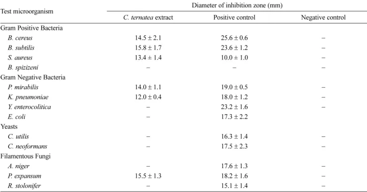
10

Test microorganisms − The test microorganisms used in this study include 4 Gram positive bacteria (Bacillus cereus, Bacillus subtilis, Staphylococcus aureus, Bacillus spizizenii), 4 Gram negative bacteria (Proteus mirabilis, Klebsiella pneumoniae, Yersinia enterocolitica, Escherichia coli), 2 yeasts (Candida utilis, Cryptococcus neoformans) and 3 filamentous fungi (Aspergillus niger, P. expansum, Rhizopus stolonifer). The microorganisms were previously isolated from contaminated food sample and maintained at Upstream Processing Laboratory, Universiti Kuala Lumpur. The colonies of test microorganisms were suspended in sterile distilled water. For test bacteria and yeasts, the turbidity of the suspensions was adjusted to match McFarland 0.5 standard. For test fungi, the density of the spores was determined with hemocytometer (Neu- bauer) under light microscope. Further dilution with sterile distilled water was performed to adjust the inoculums as recommended by Clinical Laboratory Standard Institute.

11

Screening of antimicrobial activity − The antimicrobial activity of the extract was screened by disc diffusion assay.

11

The extract was prepared at concentration of 50 mg/ml by dissolving 0.05 g of extract in 1 ml of sterile distilled water. Nutrient agar (Merck) was used for test bacteria whereas potato dextrose agar (Merck) was used for test yeasts and fungi. Then, 100 µl of the microbial inoculum was spread on the agar plate with sterile hockey stick. Sterile paper disc impregnated with 20 µl of extract was placed on the surface of the inoculated medium. The positive control was included by using 20 µg/ml of chloramphenicol (Sigma) for test bacteria and 20 µg/ml of ketoconazole (Fisher) for test yeasts and fungi. Sterile distilled water was used as negative control. The bacteria and yeasts were incubated at 37

o

C for 24 hours where fungi were incubated at 30

o

C for 72 hours. The test was done in triplicate. After the incubation period, the diameters of the clear zone, including the diameter of the disc were measured to the nearest whole millimeter with a ruler.

Determination of minimal inhibitory concentration −

The minimal inhibitory concentration (MIC) of the extract

was determined via broth microdilution assay in sterile

96-well microtiter plates (Fisher).

12

Only test microor-

ganisms that exhibited susceptibility in the screening test

were selected. The microbial inoculum was prepared by

suspending the microbial colonies in double strength

broth media. Double strength Luria broth (LB) (Merck)

was used for all test bacteria whereas RPMI 1640

medium (Sigma) containing 0.2% dextrose buffered with

(3)

0.165 M 3-(N-morpholino)propanesulfonic acid (MOPS) to a pH of 7.0 at 25

o

C was used for test fungi. The concentration of the extract was prepared at a range from 200 mg/ml to 1.6 mg/ml initially. Then 100 µl of the extract was pipetted into each well. After that, 100 µl of microbial inoculum was added into each well to yield the final concentration of the extract of 100 mg/ml to 0.8 mg/

ml. The sterility control was included with the double strength media and extract at various concentrations.

Next, the growth control was prepared by adding 100 µl of the microbial inoculum and 100 µl of sterile distilled water into each well. The plates were incubated at 30

o

C for 72 hours for fungi, and at 37

o

C for 24 hours for bacteria. After the incubation, 40 µl of Iodonitrotetrazolium violet (INT) (Sigma) at the concentration of 0.2 mg/ml was added into each well to detect the microbial growth.

The color change of the well was observed after 30 min of incubation. The change in color of the broth from yellow to purple indicates microbial growth.

Determination of minimal lethality concentration − To determine minimal lethality concentration (MLC) of the extract, 100 µL of the sample from each well prepared in previous section was taken and suitably diluted before spreading on agar plates to judge the viability. The viable cell count method was performed. The MLC was recorded as the lowest concentration of extract that resulted in 99.9% growth reduction relative to the control.

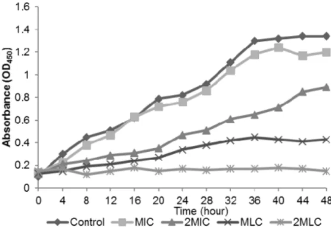
Time kill curve − The kill curve study for P. expansum was performed according to the protocol defined by Dalla Lana et al.

13

The extracts were tested at four concen- trations: the MIC susceptibility breakpoint, twice the MIC, MLC and also twice the MLC. The extract was initially prepared at the concentration 10 times higher than the desired concentration. To achieve a final volume of 50 mL in each flask, 1:10 dilution of the extract was performed by the addition of 25 mL of spore suspension with the inoculum size of 1 × 10

5

and 20 mL of sterile RPMI 1640 medium (Sigma) containing 0.2% dextrose buffered with 0.165M MOPS to a pH of 7.0 at 25

o

C. The flasks were incubated in rotatory shaker for 48 hours at 30

o

C with a rotational speed of 150 rpm. At pre-determined time points (0, 4, 8, 12, 14, …., 44, 48 hours), 1 mL of the sample was withdrawn from each flask. After that, 50 µL of INT salt (Sigma) at concentration 0.5 mg/mL was added to each sample and were incubated at 37

o

C for additional 2 hours. The sample was then centrifuged at 15,000 rpm to pellet the cells. The formazan production in the supernatant was measured in terms of optical density at 450 nm using a spectrophotometer (Sigma). The results were depicted graphically.

Morphological observation of fungal hyphae − The fungus P. expansum was inoculated on PDA plates (Merck).

11

After incubation at 30

o

C for 72 days, 1.0 ml of 50 mg/ml extract was pipetted on the agar surface. The extract was spread by swirling the plate. The plate was then further incubate at 30

o

C for 72 hours. Sterile distilled water was used as negative control. To prepare the sample for scanning electron microscope (SEM) observation, a planchette with tissue-tek was prepared. Then the block of agar with approximately 5 × 5 mm

2

was excised from the periphery of 7-days old culture of fungal culture. The agar block was transferred to the planchette placed on a filter paper lined Petri dish. The vapor fixation was done by wetting the filter paper with a few drops of 2% osmium tetroxide. Once the sample was vapor-fixed for 2 hours, the planchette was plunged into liquid nitrogen ( −210

o

C) and transferred to freeze dryer (Emitech K750) for about 10 hours. After the freeze drying process, the planchette was coated with 5 - 10 nm of gold palladium alloy. The samples were viewed under SEM (Leo Supra 50VP field emission SEM) to observe the morphology of the fungus.

Antimicrobial efficacy in a food model − The pra- boiled rice (Rambutan, Malaysia) was washed thoroughly under running tap water. Then 120 g of rice was cooked with 200 ml of 50 mg/ml extract over a plate heater.

Complete cooking requires 20 minutes. A control was included by replacing the extract with distilled water. The rice was placed on Petri dish, and the thickness of 15 mm was standardized. Then the samples were incubated in laboratory condition for 5 days. On daily basis, the rice sample was collected by using sterile 1 cm cork borer.

The collected sample on each day was processed with food stomacher (Seward) with 10 ml of sterile peptone water (Merck). The microbial load was determined by viable cell count method in plate count agar (Hi-media).

Serial dilutions up to 10

−5

were prepared for each sample.

The plates were incubated for 24 hours at 37

o

C. The microbial load of the rice sample was calculated per gram of sample.

Results and Discussion

Extraction yield of bioactive compound is significantly

affected by the processing conditions. Particularly,

selection of the solvent is crucial to ensure successful

extraction of anthocyanins. Acidified ethanol was used in

this study as Kungsuwan et al. reported that ethanol is the

most efficient solvent to extract anthocyanins.

8

Besides,

ethanol is classified as Q3C guideline solvent recommended

for pharmaceutical and food industry by US Food and

(4)

Administration. The ethanol used in this study was acidified to pH 4.5 as the optimal pH for anthocyanin extraction in purple rice.

8

The anthocyanin test indicates the presence anthocyanin in the extract. A very high content of blue violet color was observed in the test.

The antimicrobial activity of extract was tested against 4 Gram positive, 4 Gram negative, 2 yeast and 3 filamentous fungi. Microbial strains showed different sensitivities against the extract of C. ternatea. As shown in Table 1, the extract showed good antimicrobial activity against 3 Gram-positive bacteria (B. cereus, B subtilis, S.

aureus) and 2 Gram negative bacteria (P. mirabilis, K.

pneumoniae) in disc diffusion assay. Sterile distilled water as the vehicle to dissolve the extract did not exhibit any effect to the growth of all test microorganisms. B. cereus and S. aureus are causative agents of food poisoning by producing toxin in food, followed by toxic symptoms in humans. Thus, these results obtained show that the extract from C. ternatea possess promising potential for application in the food processing industries. In this study, the largest inhibition zone was observed on B. subtilis with a diameter of 15.8 ± 1.7 mm while the smallest inhibition zone was K. pneumoniae with 12.0 ± 0.4 mm. The difference in the diameter of the clear zone indicates different susceptibility of the bacteria to the extract. As indicated by the results, Gram positive bacteria were more

susceptible to the extract compared to Gram negative bacteria.

14

Mechanisms underlying the anthocyanin activity include both membrane and intracellular interactions of these compounds. The similar finding was reported by Tong et al. whereby most medicinal plant extracts are more effective against Gram positive bacteria than Gram negative bacteria.

11

This is due to the lack of outer membrane in cell wall of Gram positive bacteria which cause them to be more permeable to the extract.

The extract did not exhibit inhibitory activity on the test yeasts. The results were in contrast with Kamilla et al.

where they reported the inhibition of C. albicans by C.

ternatea methanolic extract.

15

Among 3 fungal strains tested, only P. expansum was susceptible to the extract.

Ternatin, a penta-acylated anthocyanin was responsible for the antimicrobial activity of C. ternatea.

16

Six major anthocyanins, ternatin A1, A2, B1, B2, D1 and D2 were previously isolated from the flowers of C. ternatea by Kondo et al.

17

Ternatin was previously reported for its excellent gastro-protective and anti-diarrheal properties, which reveals its potential as food preservative.

18

Table 2 shows the MIC and MLC of C. ternatea extract of selected test microorganisms. In general, the results were in agreed with the data obtained from disc diffusion assay. The bacterium that showed the lowest MIC value was K. pneumoniae with the value of 1.6 mg/ml. The Table 1. Antimicrobial activity of C. ternatea extract on disc diffusion assay. The extract showed antimicrobial activity on both Gram positive and Gram negative bacteria

Test microorganism Diameter of inhibition zone (mm)

C. ternatea extract Positive control Negative control Gram Positive Bacteria

B. cereus 14.5 ± 2.1 25.6 ± 0.6 −

B. subtilis 15.8 ± 1.7 23.6 ± 1.2 −

S. aureus 13.4 ± 1.4 10.0 ± 1.0 −

B. spizizeni − − −

Gram Negative Bacteria

P. mirabilis 14.0 ± 1.1 19.0 ± 0.5 −

K. pneumoniae 12.0 ± 0.4 18.0 ± 1.2 −

Y. enterocolitica − 23.2 ± 1.6 −

E. coli − 17.3 ± 2.2

Yeasts

C. utilis − 16.3 ± 1.4 −

C. neoformans − 17.5 ± 2.3 −

Filamentous Fungi

A. niger − 17.6 ± 1.3 −

P. expansum 15.5 ± 1.3 18.2 ± 1.6 −

R. stolonifer − 15.1 ± 1.4 −

( −) = No inhibitory zone

(5)

highest MIC was recorded on S. aureus with the value of 50 mg/ml. Many Gram negative bacteria are difficult food contaminants and pathogens. Therefore, their control in food processing systems is vital. The cells of Gram negative bacteria are surrounded by an outer membrane (OM), which provides the bacterium with a hydrophilic surface.

19

The Gram negative OM functions as a barrier for many external agents and resulting in resistance to hydrophobic antibiotics such as macrolides, novobiocins, rifamycins and actinomycin D.

12

The inhibitory effects C.

ternatea extract on the Gram negative bacteria can be anticipated for the application in food processing and preparation. Antibacterial agents are usually regarded as bactericidal if the MLC is not more than four times of the MIC.

20

The highest MLC was recorded on S. aureus with the concentration of 100 mg/ml. The MLC was signifi- cantly higher than the MIC for all test bacteria. This indicates that the killing activity of the extract was concentration dependent. A higher concentration of the extract was needed in order to kill the microbial cells, than to inhibit the growth. Besides, it is also worthy to note that the extract exhibited bactericidal activity only on Gram positive bacteria, indicates the high susceptibility of Gram positive bacteria to the extract. The extract showed significant fungicidal effect on P. viridicatum, with its noticeably low MLC. Kamilla et al. also reported the inhibitory effect of C. ternatea flower extract on several filamentous fungi.

21

However, there is a lack of literatures on the antifungal activity of ternatin.

For the kill curve study, the formation of formazan was measured as the growth parameter for the kill curve.

Clinical Laboratory Standard Institute recommended formazan detection for measuring fungal growth in kill curve study as the measurement of color intensity for the re-suspended red formazan at 480 nm provides a quanti- tative evidence for the strength of the antifungal agent used.

22

Besides, it is also recommended due to its rapidity, technical simplicity and cost effectiveness compared to the conventional method by using dry mycelia weight as

growth parameter.

23

Figure 1 shows the time kill curve of the extract on P. expansum. The control growth profile exhibited only lag phase and log phase. At concentration of MIC and 2× MIC, the extract did not have a significant effect the growth pattern. However, a noticeable low formazan formation was detected for extract concentration of 2× MIC relative to control. The inhibitory activity of the extract on the fungus was in concentration dependent manner. At the concentration of MLC, the growth curve for P. expansum was totally altered, as no exponential growth was detected. At the concentration of 2× MLC, the kill curve showed prolonged lag phase with no significant growth observed during the experimental period. Thus the fungicidal activity of the extract against P. expansum conidia was dependent on the concentration of the extract and the time of exposure of the conidia to the extract.

Figure 2 shows the SEM micrograph of P. expansum grown on PDA treated with C. ternatea extract. SEM has been successfully used to access the morphological changes of P. expansum induced by the extract. For the fungal culture treated with distilled water control, some coarsely roughed stipes which bear terminal verticillate penicilli were observed on the micrograph. The metulae were cylindrical in whorls of three. The conidia were ellipsoidal in shape, smooth walled and borne in closely packed chain. However, after 3 days of incubation in the treatment, fungal mycelium showed alterations in the morphology of fungal hyphae. Flattened empty hyphae were observed resulted from the cell wall disruption. The hyphae also lost their smoothness. Furthermore, no presence of conidia-bearing conidiophore was observed, suggesting irreversible damage of the conidiophore. At this stage, the germination of P. expansum conidia was completely Fig. 1. The kill curve of C. ternatea extract on P. expansum. The fungicidal activity of the extract was concentration-dependent.

Table 2. The MIC and MLC of C. ternatea extract on test microorganisms. The antimicrobial activity of the extract was con- centration-dependent

Bacteria MIC (mg/ml) MLC (mg/ml)

B. cereus 25.0 50.0

B. subtilis 25.0 50.0

S. aureus 50.0 100.0

P. mirabilis 6.2 50.0

K. pneumoniae 1.6 25.0

P. expansum 12.5 25.0

(6)

inhibited and conidial development was suppressed.

One of the most noticeable role of anthocyanins is to impart colors which linked to the adaptive significance of anthocyanins in plant reproductive organs like flowers that invariably attributed to the attraction of seed dispersers and pollinators. A few studies also suggested that anthocyanins in vegetative tissue might also function in plant defense, although there is absence of solid evidence for such a function. Herein, our results show that the extract of C. ternatea contribute to defense against fruit- rot fungi. P. expansum is a broad spectrum pathogen on fruits that causes postharvest decays on a variety of fruits including apple, grapes and etc. These results indicated that the extract of C. ternatea flowers not only function to attract animal vectors, but might also contribute to the fruit defense against fruit-rot fungi. However, detailed studies are required to clarify the correlation between the risks of fruit-rot in the crops with the anthocyanin contents.

The antimicrobial efficacy of C. ternatea extract was also studied in a food model. Figure 3 shows the microbial load of the rice samples throughout 5 days of incubation period. Based on the results, a high level of microbial contamination was observed for the control sample, with the highest bacterial load recorded on Day 5 with a total of 4.8×10

5

CFU/ml recorded. In the contrary, no significant microbial growth was detected in the rice sample added with C. ternatea extract throughout the 5 days of incubation period. The extract exhibited approxi- mately 5.0-log suppression of microbial growth relative to control from Day 1 to Day 5. The anthocyanin present in the extract showed good antimicrobial stability through the 5 days of test period, thus the results indicate the potential use of the C. ternatea extract as natural food preservative.

In conclusion, the extract of C. ternatea inhibits the growth of food pathogens and may therefore be used as

disinfection compounds against these microorganisms especially in the food processing industries. Further studies of the antimicrobial mechanisms such as disintegration of the outer membrane is required to explain their mechanism as well as their occurrence in plant reproductive organs like flowers.

Acknowledgement

The authors are thankful to Universiti Kuala Lumpur for financial support and Mr Cedric Tan for providing the plant samples.

References

(1) Lima, A. A. N. D.; Sobringho, J. L. S.; Lyra, M. A. M. D. ; Santos F.

L. A. D. ; Figueiredo, C. B. M.; Neto, P. J. R. Int. J. Pharm. Pharm. Sci.

2015, 7, 371-375.

(2) Kagan, C. R. ACS Nano. 2016, 10, 2985-2986.

(3) Kawai, T.; Sekizuka, T.; Yahata, Y.; Kuroda, M.; Kumeda, Y.; Iijima, Y.; Kamata, Y.; Sugita-Konishi, Y.; Ohnishi, T. Clin. Infect. Dis. 2012, 54, 1046-1052.

(4) Abdul-Mutalib, N. A.; Nordin, S. A.; Osman, M.; Ishida, N.;

Tashiro, K.; Sakai, K.; Tashiro, Y.; Maeda, T.; Shirai, Y. Int. J. Food Fig. 2. SEM micrograph of food borne P. expansum treated with (A) sterile distilled water (B) C. ternatea extract (5000× magnification).

The results suggest the irreversible damage on the conidiophore of P. expansum.

Fig. 3. The microbial load of the food model obtained throughout

the 5 days of incubation period. Test substance: ( ◆ ) C. ternatea

extract, ( ■ ) Distilled water control. The extract significantly

inhibits the microbial growth in the rice model.

(7)

Microbiol. 2015, 200, 57-65.

(5) Spadaro, D.; Lorè, A.; Garibaldi, A.; Gullino, M. L. Postharvest Biol. Technol. 2013, 75, 1-8.

(6) Yu, C.; Zhou, T.; Sheng, K.; Zeng, L.; Ye, C.; Yu, T.; Zheng, X. Int.

J. Food Microbiol. 2013, 164, 155-160.

(7) Bhatia, M.; Chahal, J.; Gupta, S. Int. J. Pharm. Sci. Res. 2014, 5, 600-606.

(8) Kungsuwan, K.; Singh, K.; Phetkao, S.; Utama-ang, N. Food Appl.

Biosci. J. 2014, 2, 31-46.

(9) Chong, F. C.; Gwee, X. F. Nat. Prod. Res. 2015, 29, 1485-1487.

(10) Priya, F. J.; Vimala, J. R.; Bama, R. S.; Lavanya, M. World J.

Pharm. Pharm. Sci. 2016, 5, 753-765.

(11) Tong, W. Y.; Ang, S. N.; Darah, I.; Latiffah, Z. World J. Pharm.

Pharm. Sci. 2014, 3, 121-132.

(12) Darah, I.; Chong, C. L.; Tong, W. Y.; Latiffah, Z.; Lim, S. Trop. J.

Pharm. Res. 2015, 14, 2091-2097.

(13) Dalla Lana, D. F.; Donato, R. K.; Bündchen, C.; Guez, C. M.;

Bergamo, V. Z.; de Oliveira, L. F.; Machado, M. M.; Schrekker, H. S.;

Fuentefria, A. M. J. Appl. Microbiol. 2015, 119, 377-388.

(14) Biswas, B.; Rogers, K.; McLaughlin, F.; Daniels, D.; Yadav, A. Int.

J. Microbiol. 2013, 2013, 1-7.

(15) Kamilla, L.; Mnsor, S. M.; Ramanathan, S.; Sasidharan, S. Pharm.

Online. 2009, 1, 731-738.

(16) Nair, V.; Bang, W. Y.; Schreckinger, E.; Andarwulan, N.; Cisneros- Zevallos, L. J. Agric. Food Chem. 2015, 63, 6355-6365.

(17) Kondo, T.; Ueda, M.; Goto, T. Tetrahedron. 1990, 46, 4749-4756.

(18) Rao, V. S.; Santos, F. A.; Sobreira, T. T.; Souza, M. F.; Melo, C. L.;

Silveira, E. R. Planta Med. 1997, 63, 146-149.

(19) Nikaido, H. Microbiol. Mol. Biol. Rev. 2003, 67, 593–656.

(20) Palazzo, I. C. V.; Araujo, M. L. C.; Darini, A. L. C. J. Clin.

Microbiol. 2005, 43, 179-185.

(21) Kamilla, L.; Mansor, S. M.; Ramanathan, S.; Sasidharan, S.

Microsc. Microanal. 2009, 15, 366-372.

(22) Moussa, S. H.; Tayel, A. A.; Al-Hassan, A. A.; Farouk, A. J.

Mycol. 2013, 2013, 753692.

(23) Rafiee, E.; Khodayari, M.; Shahebrahimi, S.; Joshaghani, M. J.

Mol. Catal. A 2011, 351, 204-209.

Received February 15, 2017

Revised April 11, 2017

Accepted April 12, 2017

수치

Table 2 shows the MIC and MLC of C. ternatea extract of selected test microorganisms. In general, the results were in agreed with the data obtained from disc diffusion assay
Figure 2 shows the SEM micrograph of P. expansum grown on PDA treated with C. ternatea extract
Fig. 3. The microbial load of the food model obtained throughout the 5 days of incubation period

참조

관련 문서