Fluoroscopically Guided Three-Tube Insertion for the Treatment of Postoperative Gastroesophageal
Anastomotic Leakage
Guowen Yin, MD
1, Qingyu Xu, MD
1, Shixi Chen, MD
1, Xiangjun Bai, MD
1, Feng Jiang, MD
2, Qin Zhang, MD
2, Lin Xu, MD
2, Weidong Xu, MD
1Departments of 1Interventional Radiology and 2Thoracic Surgery, Cancer Hospital of Jiangsu Province, Cancer Institution of Jiangsu Province, Nanjing 210009, China
Objective: To retrospectively evaluate the feasibility and effectiveness of three-tube insertion for the treatment of postoperative gastroesophageal anastomotic leakage (GEAL).
Materials and Methods: From January 2007 to January 2011, 28 cases of postoperative GEAL after an esophagectomy with intrathoracic esophagogastric anastomotic procedures for esophageal and cardiac carcinoma were treated by the insertion of three tubes under fluoroscopic guidance. The three tubes consisted of a drainage tube through the leak, a nasogastric decompression tube, and a nasojejunum feeding tube. The study population consisted of 28 patients (18 males, 10 females) ranging in their ages from 36 to 72 years (mean: 59 years). We evaluated the feasibility of three-tube insertion to facilitate leakage site closure, and the patients’ nutritional benefit by checking their serum albumin levels between pre- and post- enteral feeding via the feeding tube.
Results: The three tubes were successfully placed under fluoroscopic guidance in all twenty-eight patients (100%). The procedure times for the three tube insertion ranged from 30 to 70 minutes (mean time: 45 minutes). In 27 of 28 patients (96%), leakage site closure after three-tube insertion was achieved, while it was not attained in one patient who received stent implantation as a substitute. All patients showed good tolerance of the three-tube insertion in the nasal cavity. The mean time needed for leakage treatment was 21 ± 3.5 days. The serum albumin level change was significant, increasing from pre-enteral feeding (2.5 ± 0.40 g/dL) to post-enteral feeding (3.7 ± 0.51 g/dL) via the feeding tube (p < 0.001). The duration of follow-up ranged from 7 to 60 months (mean: 28 months).
Conclusion: Based on the results of this study, the insertion of three tubes under fluoroscopic guidance is safe, and also provides effective relief from postesophagectomy GEAL. Moreover, our findings suggest that three-tube insertion may be used as the primary procedure to treat postoperative GEAL.
Index terms: GEAL; Three-tube insertion; Interventional procedure; Esophageal and cardiac carcinoma
Received August 24, 2011; accepted after revision September 22, 2011.
Corresponding author: Qingyu Xu, MD, Department of Interventional Radiology, Cancer Hospital of Jiangsu Province, Cancer Institution of Jiangsu Province, No.42 Baiziting, Nanjing 210009, China.
• Tel: (8625) 83283525 • Fax: (8625) 83283523
• E-mail: [email protected]
This is an Open Access article distributed under the terms of the Creative Commons Attribution Non-Commercial License (http://creativecommons.org/licenses/by-nc/3.0) which permits unrestricted non-commercial use, distribution, and reproduction in any medium, provided the original work is properly cited.
pISSN 1229-6929 · eISSN 2005-8330 Korean J Radiol 2012;13(2):182-188
INTRODUCTION
Although the amounts of postoperative anastomotic leakage can be rather slight, it is a major source of mortality and morbidity (1). In patients with GEAL, quality of life as well as the prognosis is very poor. The leading causes of death in patients with GEAL are infection and nutritional deficiency (2, 3). GEAL is still a therapeutic problem (4, 5) and the most effective treatment option for intra thoracic GEAL is still controversial. Hence, there is no standardized treatment algorithm. Some surgeons recommend aggressive
surgery, while others prefer conservative approaches, such as perianastomotic drainage, total parenteralnutrition, nasogastric decompression, and the use of broad-spectrum antibiotics (6). In this study, we determined the feasibility and effectiveness of using a three-tube insertion for the treatment of postoperative GEAL. The effect on GEAL treatment was evaluated by the time interval of the leakages’ closure, and the effect on nutritional status was evaluated by measuring serum albumin levels.
MATERIALS AND METHODS
The study population included 28 patients suffering from postoperative GEAL and underwent three-tube insertion under fluoroscopic guidance from January 2007 to January 2011. The 28 patients underwent an esophagectomy with intrathoracic esophagogastric anastomotic procedures for esophageal and cardiac carcinoma. The 28 patients consisted of 18 males and 10 females ranging in age from 36 to 72 years (mean age: 59 years). Three of the patients were TNM stage I esophageal carcinoma patients, nine were TNM stage II a, eleven were TNM stage II b, and five were TNM stage III. A frozen-section analysis of resected margins by a competent pathologist was available, and resection margins were found to be cancer- free in all cases. The retrospective study was approved by the Institutional Review Board of our hospital and the Ethics Committee of the Institute for Medical Science. The procedure was explained to all of the patients in detail, and
a written consent was obtained from all patients prior to the procedure. The patients were divided into two groups:
one group contained the patients with leakage within 14 postoperative days, and the other group patients with leakage at more than 14 postoperative days (22). Twenty of the 28 patients experienced acute leakages, and 8 patients had chronic leakages. Signs and symptoms were present in all patients (100%) and included high fever in 26 patients, shortness of breath in 18 patients, chest pain in eight patients, and arrhythmia in six patients. The leakages in the 24 patients (85.7%) were confirmed by first radiographic contrast examination. Four (14.2%) patients initially had negative contrast examination results, but they were all eventually proven to have anastomotic leakage by repeated radiographic contrast examinations 3 to 7 d later. The leakage in all patients was first confirmed by a computed tomography (CT) examination (Fig. 1A). The three tubes were removed after the leak’s closure was testified by CT findings.
Interventional Procedure (Fig. 2)
Nasogastric Placement of the Drainage Tube through the Leak
All the procedures were performed by two interventional radiologists. The patients were in the supine position which allowed an easy performance of interventional maneuvers, and optimal fluoroscopic visualization of the leak. A 0.035- inch guidewire (Terumo, Tokyo, Japan) and a 4F H1 catheter
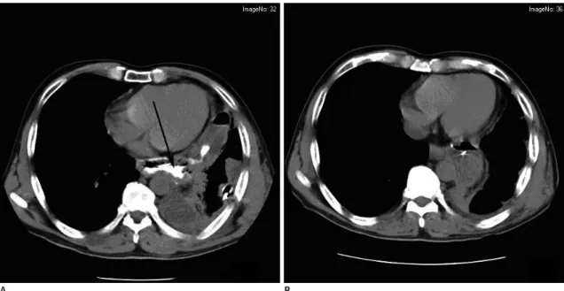
A B
Fig. 1. Intrathoracic anastomotic leak testified by CT at 1 week postoperation.
Fistula was located in pleural cavity (A, black arrow). Fistula closed at 25 days (B).
(Terumo, Japan) were advanced into the esophageal passage through one nasal cavity under fluoroscopic guidance (DSA, Angiostar; Siemens, Germany). The fluid collections aspirated through the catheter were sent for bacterial culture testing. The prosthesis delivery system was then advanced over the guidewire until a 10F Flocare polyurethane un-weighted feeding tube (Nutricia, Export BV, Amsterdam, the Netherlands) was sent into the vomica.
We suggested that the backward distance of the tube should be less than 5 cm. The Flocare tube was connected to a low-pressure continuous vacuum pump whose negative pressure was about 8-10 mm Hg. The fluid collection was examined every day for color and total quantity.
Nasogastric Placement of the Feeding Tube into the Jejunum A 4F H1 catheter (Terumo, Japan) was advanced into the esophageal passage through the ipsilateral nasal cavity.
When gthe H1 catheter entered the stomach, an Amplatz superstiff exchange guidewire (Medi-Tech/Boston Scientific, MA, USA) was then inserted via the catheter into the distal portion of the jejunum. And 10F Flocare polyurethane un- weighted feeding tube (Nutricia, Switzerland) was then inserted into the jejunum using the guidewire. Contrast medium was injected through the Flocare feeding tube to
verify the success of the manipulation. Essential nutrients were poured into the jejunum through the Flocare feeding tube.
A nasogastric decompression tube was placed under fluoroscopic guidance when we applied this treatment to keep the digestive slice from affluxing into the leakage.
When daily volume of the fluid drainage was reduced to 5-10 mL, the drainage tube could be gradually pulled back (Fig. 3) because the adhesion between the lung and the chest wall had already formed at the end of the tube. Under the fluoroscopic guidance, the drainage tube was pulled back from the distal end to the proximal end of the vomica.
Immediate technical success was defined as the successful insertion of the three-tube without any problems. Clinical success was defined as closure of the leakage site (Fig. 4), as confirmed by a follow-up CT examination (Fig. 1B). A Fig. 2. Drain tube (arrow 1) inserted through fistula
with fluoroscopic guidance and distal tip of drain tube was positioned at bottom of abscess cavity (arrow 4).
Feeding nasojejunum tube (arrow 2) and cases of nasogastric decompression tube (arrow 3) were placed during interventional operation for postoperative enteral nutrition supply and digestive slice drainage.
Fig. 3. Eleven days later, fistula (arrow 1) got smaller, after changing position of drainage tube (arrow 2) to keep optimum drainage.
patients’ nutritional benefit was evaluated by the serum albumin level change between the pre- and post-enteral feeding via the feeding tube. The differences in the means were evaluated by t tests.
RESULTS
Immediate technical success was achieved in all patients (28/28 patients). The chronic group (> 14 postoperative day) showed a high incidence of leakage site in the thoracic cavity (8/8 = 100%), but the acute group (< 14 postoperative day) showed a low incidence of leakage site in the thoracic cavity (13/20 = 65%) (Table 1). All patients had a good tolerance of the three-tube in the nasal cavity.
Side effects such as shortness of breath were not seen in any patient. Only slight nasal mucosa diabrosis was seen in
two patients.
The procedure times for three-tube insertion ranged from 30 to 70 min. (mean time: 45 min) and was dependent on the detection of the leakage site. Twenty-seven patients (27/28 = 96.4%) experienced symptomatic relief, while in one patient, though a successful three-tube insertion was achieved, the leakage did not close and the patient suffered from serious sepsis. A covered nitinol stent (Micro-Tech Co, Nanjing, China) was successfully placed to cover the leakage site 31 d after receiving the three-tube treatment.
Sepsis was then under control in 3 weeks. However, the patient died 10 months after placement due to sudden uncontrollable hematemesis. The mean time interval of the leakage treatment was 21 ± 3.5 d. The median follow-up time was 28.2 months (range 7-60 months). No patient was diagnosed with leak recurrences.
The patient’s nutritional benefit was shown by the increasing serum albumin level after enteral feeding via the nasojejunum feeding tube. The mean serum albumin level was 2.5 ± 0.40 g/dL (1.87-3.68 g/dL) in a 14 day of a pre- enteral feeding tube period and 3.7 ± 0.51 g/dL (2.52-4.86 g/dL) in 14 days post-enteral feeding via the feeding tube.
The differences in the mean significantly increased between pre- and post-enteral feeding via the feeding tube (p <
0.001).
DISCUSSION
Although GEAL occurs in less than 10% of patients, it still remains the most feared complication of esophageal resection because of its associated high morbidity and mortality (7, 8). Although several methods such as surgical re-exploration and repair and/or more conservative
approaches like perianastomotic drainage or broad-spectrum antibiotics have been used, no consensus has been reached on the best methods of treatment, and the rate of treatment failure remains high. It may correlate with general nutritive status, anastomotic technique, anastomotic stoma tension, anastomotic manner, blood circulation lesion of the esophageal or gastric wall, digestive tract obstruction, intrathoracic infection, and so on (9-11). HU et al. (12) divides the pathophysiological process of GEAL into 3 stages: diffusion, contained, the elimination of multi-cavity vomicas. The anastomotic leakage closed within durations of drainage tube insertion with hyperalimentation. In our opinion GEAL is a special kind of pyothorax. The key in leak management is adequate and effective drainage.
Fig. 4. Fistula closed on 25 days. Nasojejunum enteral supports tube (arrow 1) was retained for enteral nutrition.
When the leakage was limited in the pleural cavity, routine chest tube could achieve adequate drainage. But previous studies had revealed that most of the contained leaks were limited to the mediastinum (7, 8, 13). It is difficult to deal with this type of leakage because abscess cavities close to the mediastinum are difficult to reach by a conventional chest tube. Nasogastric placement of the drainage tube through the leak in our study may be a helpful treatment for this type of leak. We successfully applied the drainage tube through the leak for the treatment of intrathoracic
anastomotic leaks in 28 patients. Twenty-seven patients (27/28 = 96.4%) experienced symptomatic relief. The mean time interval of the leakage treatment was 21 ± 3.5 d which is much shorter than 32-99 d in the previous study (14, 15).
In our study one patient received placement of self- expanding metallic coated stents after failure of the three- tube treatment. The GEAL was located in the mediastinum and thoracic cavity. The vomicas were huge, coated, and interlinked. Effective drainage could not be achieved. The Table 1. Data in Fluoroscopically Guided by Three-Tube Insertion for Relief of Postoperative Gastroesophageal Anastomotic Leakage
No Age/
Sex Stage Operation Procedures
Leak on POD
Leakage Position
Three-Tube Duration (days)
Further Treatment
Follow-Up Duration (months)
1 44/M IIa Left Ivor–Lewis 10 Mediastinum 7 No 24
2 56/M IIb Left Ivor–Lewis 7 Thoracic cavity 12 No 22 (dead due to recurrence)
3 61/M IIa Left Ivor–Lewis 3 Mediastinum 8 No 21
4 63/F IIb Left Ivor–Lewis 15 Thoracic cavity 17 No 31 (dead due to recurrence)
5 70/F IIb Left Ivor–Lewis 9 Thoracic cavity 21 No 39
6 48/F IIa Left Ivor–Lewis 6 Mediastinum 10 No 10
7 55/M IIb Left Ivor–Lewis 7 Thoracic cavity 20 No 29 (dead due to recurrence)
8 54/M I Left Ivor–Lewis 6 Thoracic cavity 21 No 60
9 60/F IIa Left Ivor–Lewis 10 Thoracic cavity 18 No 18 (dead due to recurrence)
10 61/M IIa Left Ivor–Lewis 8 Thoracic cavity 22 No 8
11 64/M IIb Left Ivor–Lewis 18 Thoracic cavity 24 No 34 (dead due to recurrence) 12 69/M IIa Left Ivor–Lewis 21 Mediastinum-thoracic
cavity 31 No 32 (dead due to recurrence)
13 49/F III Left Ivor–Lewis 5 Mediastinum 8 No 33 (dead due to recurrence)
14 36/M IIb Left Ivor–Lewis 12 Thoracic cavity 15 No 27
15 71/M I Left Ivor–Lewis 19 Thoracic cavity 22 No 49 (dead due to recurrence) 16 67/F IIb Left Ivor–Lewis 20 Thoracic cavity 13 No 34 (dead due to recurrence)
17 51/F IIa Left Ivor–Lewis 11 Mediastinum 12 No 48
18 63/M III Left Ivor–Lewis 5 Thoracic cavity 14 No 9 (dead due to recurrence)
19 66/M IIa Left Ivor–Lewis 4 Thoracic cavity 19 No 21
20 72/F I Left Ivor–Lewis 18 Mediastinum 13 No 60
21 60/F III Left Ivor–Lewis 7 Thoracic cavity 24 No 17 (dead due to recurrence)
22 57/M IIa Left Ivor–Lewis 9 Mediastinum 15 No 22
23 59/M IIb Left Ivor–Lewis 6 Thoracic cavity 21 No 29 (dead due to recurrence) 24 62/F III Left Ivor–Lewis 5 Thoracic cavity 25 No 27 (dead due to recurrence)
25 61/M IIb Left Ivor–Lewis 8 Thoracic cavity 17 No 31
26 64/M III Left Ivor–Lewis 22 Mediastinum-thoracic
cavity Failure Stent 10 (dead due to recurrence) 27 47/M IIb Left Ivor–Lewis 19 Thoracic cavity 23 No 38 (dead due to recurrence)
28 55/M IIb Left Ivor–Lewis 4 Mediastinum 18 No 7
Note.— POD = postoperative days
stent was placed on the 31 d after receiving three-tube treatment. The patient died on 10 months after placement due to uncontrollable hematemesis. Previous studies (16, 17) revealed that certain problems might be associated with stent placement and removal: insufficient closure of the leaks, stent migration and development of strictures after stent removal, severe complications: such as bleeding and food blockage, hard removal because of the growth of granulation tissue, et al. But other studies (18-21) concluded that stent placement has been shown to be an excellent treatment in patients without sepsis and large mediastinal abscess formation. In our view, the leakage in our study can be deemed as optimal and, it is not very suitable to insert the stent leader. Once the stent is placed, it had to be got out which is afflicting for patient. Some patients died of a large amount of haematemesis due to the repeated friction between the stent and fistula (13) over the course of the stents’ dislodgment. Moreover, we know the accurate time of the closure of the leakages while the stent treatment can’t support it. We can monitor the leakage by contrast-medium injection while the stent treatment can’t.
The enteric feeding might contribute to the general nutritional correction from the postsurgical status, and this might contribute to the closure of anastomotic leakage. A routine jejunostomy to nutritional support is a traumatic method. But the nasogastric placement of nutritious tube of jejunum under the fluoroscopic guidance can avoid the injury of the jejunum. Moreover total parenteral nutrition is expensive and it has a high rate of complications.
The enteral feeding allows for a high serum albumin level compared to total parenteral nutrition (22). In our study, the patient nutrition benefit showed a significantly increased serum albumin level after enteral feeding (p
< 0.001). Han et al. (22) reported that they reached a successfull relief of the postoperative gastrointestinal anastomotic obstruction and leakage by fluoroscopically guided feeding tube insertion in thirty-four patients. In our view food intake can irritate the mucosa at the anastomotic site. In addition, the digestive slice can continuously enter the chest space from the fistula. As intrathoracic pleural adhesion has not formed, it is easy to result in diffusion, chemical, and purulent pleuritis; and, eventually, severe sepsis, consumption, and death. In our study, no patient suffered severe sepsis. Ott et al. (23) had reported a success rate for fluoroscopic placement of 90%, with a tube placed into the jejunum in 53% of patients and placed into
the duodenum in 47% of the patients. In the present study, the tubes were placed fluoroscopically in the jejunum in all patients, and the mean procedure time was 45 minutes.
Moreover, the endoscopic and fluoroscopic placement of postpyloric feeding tubes can be performed safely and accurately at the bedside for critically ill patients (24).
Fluoroscopic placement requires less additional sedation, whereas endoscopic placement allows direct visualization of the gastric and duodenal anatomy, and it can be performed without additional X-ray support (24). In our study, fluoroscopic guidance using a catheter and guidewire allowed for the anastomotic sites to be found easily and there were unhindered passages of the catheter into the jejunum because the catheter has a considerably smaller diameter than the endoscopy instrument.
The nasogastric decompression tube was routinely placed when we applied this kind of conservative treatment. Jiang et al. (13) reported that the nasogastric decompression tubes were unnecessary in this management of the
intrathoracic esophagogastric anastomotic leak because the drainage volume of the tube was low. However, our clinical observation revealed that the drainage volume of the nasogastric decompression tube was not low. So we retained the tube.
There are some limitations in our study. One of the limitations was the retrospective and nonrandomized study design. In the coming days, a prospective and randomized study will be designed to evaluate clinical efficacy in three- tube vs stent placement for the treatment of postoperative GEAL. The other one was that some patients feel discomfort.
But in our study all patients had a good tolerance of three- tube in nasal cavity. Shortness of breath was seen in no patient. Only slight nasal mucosa diabrosis was seen in two patients.
In conclusion, three-tube insertion under fluoroscopic guidance was found to provide safe and effective relief of GEAL. We suggest that three-tube insertion should be considered as an alternative therapeutic option for patients having GEAL.
REFERENCES
1. Orringer MB, Marshall B, Iannettoni MD. Transhiatal
esophagectomy: Clinical experience and refinements. Ann Surg 1999;230:392
2. Urschel JD. Esophagogastrectomy anastomotic leaks complicating esophagectomy: a review. Am J Surg 1994;169:634-640
3. Dehn TC, Menon K. Diagnosis and conservative management of intrathoracic leakage after oesophagectomy. Br J Surg 1998;85:1446-1449
4. Wright CD, Kucharczuk JC, O’Brien SM, Grab JD, Allen MS. Predictors of major morbidity and mortality after esophagectomy for esophageal cancer: a Society of Thoracic Surgeons General Thoracic Surgery Database risk adjustment model. J Thorac Cardiovasc Surg 2009;137:587-595
5. Whooley BP, Law S, Alexandrou A, Murthy SC, Wong J. Critical appraisal of the significance of intrathoracic anastomotic leakage after esophagectomy for cancer. Am J Surg 2001;181:198-203
6. Turkyilmaz A, Eroglu A, Aydin Y, Tekinbas C, Muharrem Erol M, Karaoglanoglu N. The management of esophagogastric anastomotic leak after esophagectomyfor esophageal carcinoma. Dis Esophagus 2009;22:119
7. Crestanello JA, Deschamps C, Cassivi SD, Nichols FC, Allen MS, Schleck C, et al. Selective management of intrathoracic anastomotic leak after esophagectomy. J Thorac Cardiovasc Surg 2005;129:254
8. Urschel JD, Blewett CJ, Bennett WF, Miller JD, Young JE.
Handsewn or stapled esophagogastric anastomoses after esophagectomy for cancer: meta-analysis of randomized controlled trials. Dis Esophagus 2001;14:212-217
9. Dan HL, Bai Y, Meng H, Song CL, Zhang J, Zhang Y, et al. A new three-layer-funnel-shaped esophagogastric anastomosis for surgical treatment of esophageal carcinoma. World J Gastroenterol 2003;9:22-25
10. Patil PK, Patel SG, Mistry RC, Deshpande RK, Desai PB. Cancer of the esophagus: esophagogastric anastomotic leak-a retrospective study of predisposing factors. J Surg Oncol 1992;49:163-167
11. Junemann-Ramirez M, Awan MY, Khan ZM, Rahamim JS. Anastomotic leakage post-esophagogastrectomy for esophageal carcinoma: retrospective analysis of predictive factors, management and influence on longterm survival in a high volume centre. Eur J Cardiothorac Surg 2005;27:3-7 12. Hu Z, Yin R, Fan X, Zhang Q, Feng C, Yuan F, et al. Treatment
of intrathoracic anastomotic leak by nose fistula tube drainage after esophagectomy for cancer. Diseases of the Esophagus 2011;24:100-107
13. Jiang F, Yu MF, Ren BH, Yin GW, Zhang Q, Xu L. Nasogastric Placement of Sump Tube Through the Leak for the Treatment of Esophagogastric Anastomotic Leak After Esophagectomy for Esophageal Carcinoma. Journal of Surgical Research 2010;7:1-4
14. Ryan JM, Murphy BL, Boland GW, Mueller PR. Use of transgluteal route for percutaneous abscess drainage in acute diverticulitis to facilitate delayed surgical repair. AJR 1998;170:1189-1193
15. Williams RN, Hall AW, Sutton CD, Ubhi SS, Bowrey DJ.
Management of Esophageal Perforation and Anastomotic Leak by Transluminal Drainage. J Gastrointest Surg 2011;15:777- 781
16. Kanatas AN, Aldouri A, Hayden JD. Anastomotic leak after oesophagectomy and stent implantation: a systematic review.
Oncol Rev 2010;4:159-165
17. Tuebergen D, Rijcken E, Mennigen R, Hopkins AM, Senninger N, Bruewer M. Treatment of thoracic esophageal anastomotic leaks and esophageal perforations with endoluminal stents: efficacy and current limitations. J Gastrointest Surg 2008;12:1168-1176
18. Babor R, Talbot M, Tyndal A. Treatment of upper gastrointestinal leaks with a removable, covered, self- expanding metallic stent. Surg Laparosc Endosc Percutan Tech 2009;19:e1-e4
19. Leers JM, Vivaldi C, Schafer H, Bludau M, Brabender J, Lurje G, et al. Endoscopic therapyfor esophageal perforation or anastomotic leak with a self-expandable metallic stent. Surg Endosc 2009;23:2258
20. Karbowski M, Schembre D, Kozarek R, Ayub K, Low D.
Polyflex self-expanding, removable plastic stents: assessment of treatment efficacy and safety in a variety of benign and malignant conditions of the esophagus. Surg Endosc 2008;22:1326-1333
21. Dai YY, Gretschel S, Dudeck O, Rau B, Schlag PM, Hunerbein M .Treatment of oesophageal anastomotic leaks by temporary stenting with self-expanding plastic stents. Br J Surg 2009;96:887-891
22. Han YM, Kim CY, Yang DH, Kwak HS, Jin GY. Fluoroscopically Guided Feeding Tube Insertion for Relief of Postoperative Gastrointestinal Anastomotic Obstruction and Leakage.
Cardiovasc Intervent Radiol 2006;29:395-400
23. Ott DJ, Mattox HE, Gelfand DW, Chen MY, Wu WC. Enteral feeding tubes: placement by using fluoroscopy and endoscopy.
Am J Roentgenol 1991;157:769-771
24. Foote JA, Kemmeter PR, Prichard PA, Baker RS, Paauw JD, Gawel JC, et al. A randomized trial of endoscopic and fluoroscopic placement of postpyloric feeding tube in critically ill patients. J Parenter Enteral Nutr 2004;28:154-157