Yonsei Med J 48(6):1072 - 1074, 2007 DOI 10.3349/ymj.2007.48.6.1072
Yonsei Med J Vol. 48, No. 6, 2007
We present a case report to show how manganese-enhanced T1- and T2-weighted MR cholangiography could differentiate cystic parenchymal lesions from cystic abnormalities which communicate with the bile ducts.
Key Words: Bile ducts, MR, MR contrast
INTRODUCTION
Recently, several studies reported the useful- ness of manganese-enhanced T1-weighted MR cholangiography in evaluating the biliary system, reflecting the dynamics of bile flow.
1-6We illus- trate here a case report to show how manganese- enhanced T1- and T2-weighted MR cholangio- graphy could differentiate cystic parenchymal lesions from cystic abnormalities which communi- cate with the bile ducts.
CASE REPORT
A 53-year-old-man was referred to our hospital because of abnormal findings on CT scan for health screening showing multiple, nonenhancing, small, hypodense nodules scattered throughout his entire liver. He did not have any malignant
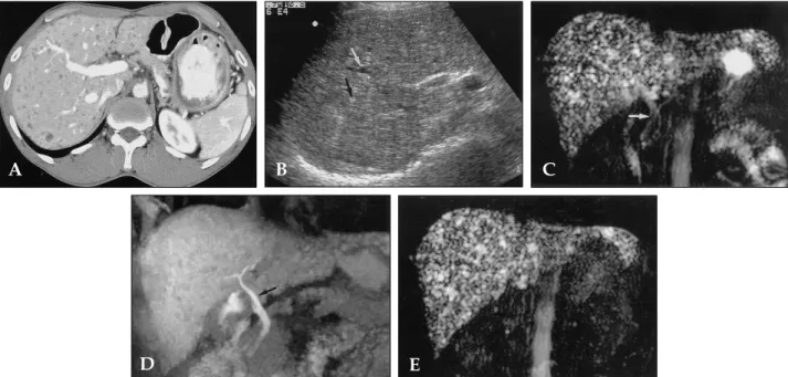
disease. Laboratory findings including liver function and blood chemistry were normal. To differentiate from multiple cysts, ultrasonography was performed. Hepatic US shows an inhomo- geneous echo texture with some hypoechoic and hyperechoic nodules. We could not differentiate cystic lesions from solid nodules on CT and sonography. Bile duct was not dilated. MR cholangiography was performed with a 1.5-T superconducting unit (Magnetom Vision; Siemens Medical Systems, Erlangen, Germany) and a phased-array torso coil. The precontrast T2- weighted MR cholangiography using a half-Fou- rier rapid acquisition with relaxation enhancement (RARE) sequence with breath holds (TR/effective TE, infinite/95 msec; matrix, 240 × 256; field of view, 300 - 350 mm) showed multiple, small, and well-defined high signal intensity foci scattered throughout the liver, suggesting as multiple cystic lesions. And bile ducts appeared as high signal intensity structures. The relationship between the bile ducts and the multiple small cystic lesions could not be clarified in this study. Post-man- ganese 3D T1-weighted fat-saturated volumetric interpolated breath-hold images (TR/TE, 4.2/1.6;
flip angle, 120; matrix 205 × 256; field of view, 300- 350 mm; and 24 partitions interpolated to 48 slices with a thickness of 1.3 mm) showed only bile ducts as high signal intensity structures. Post- manganese enhanced T2-weighted MR cholangio- graphy showed persistent high signals in the multiple small cystic lesions and the loss of bile duct signals. Manganese-enhanced T1- and T2-
Value of Manganese-Enhanced T1- and T2-Weighted MR Cholangiography for Differentiating Cystic Parenchymal Lesions from Cystic Abnormalities which Communicate with Bile Ducts
Mi-Suk Park,
1,2Jeong-Sik Yu,
2Jae Hee Lee,
3and Ki Whang Kim
11