The Clinical Characteristics of Steroid Responsive Nephrotic Syndrome of Children according to the Serum
Immunoglobulin E Levels and Cytokines
You Sook Youn,
1Han Hyuk Lim,
2and Jae Ho Lee
21Department of Pediatrics, Deajeon St. Mary’s Hospital, The Catholic University of Korea, Daejeon;
2Department of Pediatrics, Chungnam National University School of Medicine, Daejeon, Korea.
Received: February 7, 2012 Revised: April 2, 2012 Accepted: April 19, 2012
Corresponding author: Dr. Jae Ho Lee, Department of Pediatrics, Chungnam National University School of Medicine,
282 Munhwa-ro, Jung-gu, Daejeon 301-721, Korea.
Tel: 82-42-280-7247, Fax: 82-42-255-3158 E-mail: [email protected]
∙ The authors have no financial conflicts of interest.
© Copyright:
Yonsei University College of Medicine 2012 This is an Open Access article distributed under the terms of the Creative Commons Attribution Non- Commercial License (http://creativecommons.org/
licenses/by-nc/3.0) which permits unrestricted non- commercial use, distribution, and reproduction in any medium, provided the original work is properly cited.
Purpose: The nephrotic syndrome (NS) is characterized by the favorable response to glucocorticoid therapy and the development of NS may be associated with dys- functional immune systems. In order to investigate the serum immunoglobulin E (IgE) levels and cytokines activity in pediatric NS, the total of 32 steroid responsive NS patients and 5 healthy controls were enrolled in this study. Materials and Meth- ods: All patients were divided into two groups according to the initial serum IgE lev- els, such as normal and high IgE group, and their clinical characteristics were evalu- ated. In addition, serum levels of interleukin (IL)-4, IL-5, IL-10 and transforming growth factor (TGF)-β were compared and correlated with serum albumin, protein- uria by means of disease severity, and cytokines. Results: In the high IgE group, the higher comorbidity of allergic diseases and relapsing rate, the longer duration of ste- roid therapy before initial remission, and the higher serum IL-4 and IL-5 levels were found. In all patients, initially higher serum levels of IL-4 and IL-5 declined to nor- mal levels after steroid therapy, whereas the serum IL-10 levels showed no signifi- cant difference between nephrotic phase (heavy proteinuria) and remission phase (no proteinuria) of NS. The serum TGF-β levels of the nephrotic phase were significant- ly lower than those of remission phase or control group, and returned to normal con- trol levels after steroid therapy. Conclusion: This study indicates that initial IgE level is associated with steroid responsiveness and disease severity, and cytokine activities may also be related to the pathogenesis of pediatric steroid responsive NS.
Key Words: Idiopathic nephrotic syndrome, IgE, TGF-β, cytokines
INTRODUCTION
Idiopathic nephrotic syndrome (NS) is characterized by generalized edema, heavy proteinuria, hypoalbuminemia, and hyperlipidemia, and minimal change disease (MCD) is the most common form of idiopathic NS, accounting for 90% of pa- tients under the age of 10 years and more than 50% of older children.
1,2Although the etiology of idiopathic NS is unknown, several candidate factors
have been identified. After the first report of hypersensitivity and NS by Hard-
national Study of Kidney Disease in Children.
16Five healthy children (1 boy and 4 girls, aged 6 to 8 years) were includ- ed as normal control group.
Study design
The children during their first episode of NS were admitted to the pediatric wards prior to steroid treatment, and clinical data including age, gender, and blood pressure were collect- ed. The blood sample was drawn to evaluate white blood cell count, eosinophil count, platelet, protein, albumin, cho- lesterol, triglyceride, blood urea nitrogen, creatinine, elec- trolytes, and various immune mediators, such as comple- ment 3 and 4, antineutrophil antibody, antistreptolysin O and C-reactive protein.
The proteinuria, hematuria and renal functions were screened by spot and 24-hour urine analysis. When MCD was presumed, prednisolone therapy was initiated at a dose of 60 mg/m
2/day after confirming a negative purified pro- tein derivative skin test.
All of the steroid responsive NS patients were divided into a normal IgE group and a high IgE group according to the initial serum IgE levels. The patients who showed higher IgE levels, measuring more than upper 2 standard deviation score of the age-adjusted reference range, were defined as high IgE group.
17Remission was characterized by a marked reduction in proteinuria (to <4 mg/m
2/hr or urine albumin dipstick of 0 to trace for 3 consecutive days) in association with resolution of edema and normalization of serum albumin to at least 3.5 g/dL. Relapse was defined as recurrence of massive protein- uria (>40 mg/m
2/hr, urine protein/creatinine ratio >2.0 mg/
mg, or urine albumin dipstick ≥2+ on 3 consecutive days), most often in association with recurrence of edema.
2Measurement of the serum immunoglobulin and cytokine levels
During the nephrotic phase prior to steroid treatment and remission phase, the serum was separated from whole blood and was kept at -70°C for immune cytokine assay.
The serum IgE levels were analyzed by Hitachi optigen allergen specific IgE assay system Korean inhaler panel (Hitachi chemical diagnostic, Inc., Mountain View, CA, USA) based on manufacturer’s method. The serum levels of IgG, IgA, and IgM were determined by standard en- zyme immunoassay techniques (Seiken, Denka Seika, Ni- gata-ken, Japan).
The serum IL-4, IL-5 and TGF-β levels were measured by human platinum enzyme-linked immunosorbent assay (ELI- wicke
3in 1959, numerous studies have reported an associa-
tion between NS and allergy.
4,5Several studies have dem- onstrated elevated serum immunoglobulin E (IgE) levels in NS patients and hypothesized its active role in the patho- physiology, however, a direct relationship between IgE and the pathogenesis of NS is controversial.
6,7Meanwhile, it was proposed that abnormal T cell response and Th2 cytokines are strongly related to the pathogenesis of NS.
6,8Various evidences indicate that lymphocytes derived from an abnormal immune system alter the permeability of the glomerular capillary wall, suggesting that immune dys- function plays a key role in the pathogenesis of NS.
8-11Acti- vated macrophages and Th2 lymphocytes may be involved in the pathogenesis of NS, and T-cell dysfunction leads to changes in cytokines, causing a loss of negatively charged glycoproteins within the glomerular capillary wall.
12,13Recently, regulatory T cells (Treg) and T-helper17/Treg imbalance has also been suggested as a potential factor in the pathogenesis of MCD and Treg dysfunction and de- crease of their related cytokines, transforming growth fac- tor (TGF)-β and interleukin (IL)-10, have been reported.
14,15Especially, TGF-β was suggested as a protective cytokine.
14In this study, we evaluated the clinical characteristics of pediatric steroid-responsive NS patients according to the initial serum IgE levels, and several immune mediators such as Th2 cytokines (IL-4, IL-5) and Treg related cyto- kines (TGF-β, IL-10) were compared according to their re- mission status.
MATERIALS AND METHODS
Patients
The study protocol was conducted after approval by the In- stitutional Review Board of Chungnam National University Hospital, Daejeon, Korea and was carried out after informed consent was obtained from the patients or their legitimate guardians.
From September, 2004 to July, 2010, the total of 32 pa-
tients (21 boys and 11 girls, aged 1.8 to 13.9 years) who un-
derwent prednisolone therapy (17 steroid-sensitive and 15
steroid-dependent) for idiopathic nephrotic syndrome at the
Department of Pediatrics, Chungnam National University
Hospital were enrolled in this study (Table 1). The median
duration of follow-up was 4.7 years. The patients with con-
genital or secondary renal diseases were excluded. The di-
agnostic criteria for idiopathic NS were based on the Inter-
and 5 girls) who had normal IgE levels and 20 patients (14 boys and 6 girls) who had high IgE levels. The clinical characteristics among the groups were compared by Mann- Whitney U test and Fisher’s exact test. At the time of initial diagnosis, the data of their age, gender, and the laboratory findings of blood chemistry such as white blood cell count, eosinophil count, serum protein, albumin, cholesterol, IgG, IgA, IgM, immune mediators, and 24-hour urine protein/
creatinine did not show any significant differences between the two groups (Table 2). However, the duration of clinical remission was significantly shorter in the normal IgE group than in the high IgE group (p=0.005). The relapse rate (p=0.003) and the concomitant allergic diseases (p<0.001) were more frequent in the high IgE group. The median (range) time to the initial remission was 8.5 (7.0-12.0) days in the normal IgE group and 12.5 (8.0-32.0) days in the high IgE group (p=0.005) and the median (range) duration of total steroid therapy was 4.0 (2.5-22.0) months in the normal IgE group and 54.0 (3.0-108.0) months in the high IgE group (p=0.006) (Table 2). Total follow-up durations of SA) kit (eBioscience, San Diego, CA, USA). The serum IL-
10 levels were detected by human high sensitivity ELISA kit (eBioscience). Briefly, standard dilutions and samples were pipetted into the wells. And the concentrated streptavidin- horseradish peroxidase solution was added to all the wells.
After a wash to remove any unbound reagent, a TMB sub- strate solution was added to the wells and the intensity of the color was measured by a spectrophotometer at 450 μm.
Statistics analysis
The clinical and laboratory information of the NS patients were expressed as a median and range (minimum to maxi- mum) by frequent analysis. The configuration with NS pa- tients between normal IgE and high IgE levels were ana- lyzed by using Mann-Whitney U test and Fisher’s exact test.
The Kruskall-Wallis test and Scheffe’s post hoc comparison were used to compare the cytokines between NS patients with nephrotic and remission phase and healthy controls.
Significant correlation between circulating cytokines and serum albumin levels was assessed by linear regression analy- sis. The data were analyzed using SPSS version 12.0 for Win- dows (SPSS Inc., Chicago, IL, USA), and a p value<0.05 was considered significant.
RESULTS
Clinical outcomes according to the initial serum IgE levels
Subjects were divided into two groups; 12 patients (7 boys
Table 1. The Profile of Nephrotic Syndrome Patients
Patients (n=32)Age (yrs) 4.8 (1.8-13.9)*
Sex (M : F) 21 : 11
Follow-up duration (yrs) 4.7 (0.1-17.3)*
Steroid-responsiveness
Non-relapse, n (%) 17 (53.1)
Relapse, n (%) 15 (46.9)
*Median (range).
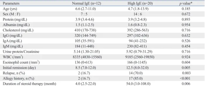
Table 2. The Comparison of Clinical Characteristics of Nephrotic Syndrome according to the Initial Serum IgE Levels
Parameters Normal IgE (n=12) High IgE (n=20) p value*
Age (yrs) 6.6 (2.7-11.0) 4.7 (1.8-13.9) 0.185
Sex (M : F) 7 : 5 14 : 6 0.672
Protein (mg/dL) 3.9 (3.4-4.6) 3.9 (3.2-4.8) 0.893
Albumin (mg/dL) 1.5 (1.1-2.5) 1.6 (0.8-2.3) 0.954
Cholesterol (mg/dL) 410 (170-738) 392 (286-563) 0.716
IgG (mg/dL) 320 (144-749) 297 (102-636) 0.632
IgA (mg/dL) 105 (35-591) 94 (41-232) 0.526
IgM (mg/dL) 184 (11-448) 230 (82-411) 0.454
Urine protein/Creatinine 5.14 (1.30-21.05) 5.92 (0.79-31.29) 0.716
WBC (/mm3) 8335 (4830-15560) 9185 (2560-19850) 0.924
Esoinophil count (/mm3) 136 (0-613) 166 (0-1145) 0.604
Initial remission (day) 8.5 (7.0-12.0) 12.5 (8.0-32.0) 0.005
Relapse, n (%) 2 (16.7) 14 (70.0) 0.003
Allegy history, n (%) 2 (16.7) 17 (85.0) <0.001
Duration of steroid therapy (month) 4.0 (2.5-22.0) 54.0 (3.0-108.0) 0.006
IgG, immunoglobulin G; IgA, immunoglobulin A; IgM, immunoglobulin M; WBC, white blood cell.
*Results of the Mann-Whitney U test and Fisher's exact test.
The serum levels of IL-5 were higher also in the nephrot- ic phase patient than those of remission phase of NS (p=0.051) and controls (p=0.028), but there were no signifi- cant differences between the patients with remission phase and control group (p=0.858) (Fig. 3B). The serum levels of IL-10 did not show any differences between both groups (Fig. 3C).
The serum levels of TGF-β in the nephrotic phase of NS patients were significantly lower than those of remission phase (p<0.001) and control group (p<0.001). However, there was no statistically significant difference between pa- tients with remission phase and control group (p=0.576) (Fig. 3D).
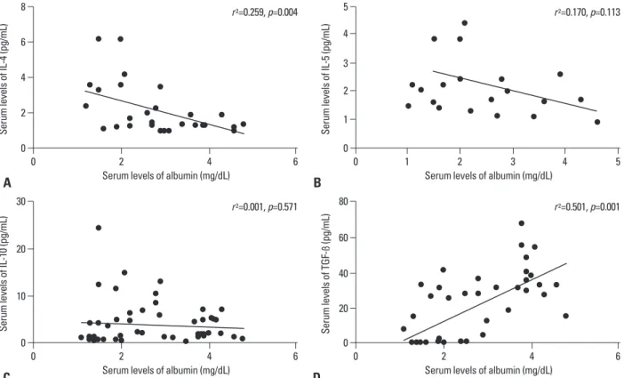
Correlation between serum albumin and cytokines Linear regression analysis showed that the serum levels of IL-4 had a negative correlation with that of albumin (R
2= 0.259, p=0.004) (Fig. 4A), while the serum levels of IL-5 and IL-10 did not show any correlations with that of albu- min (R
2=0.170, p=0.113 and R
2=0.001, p=0.571, respective- ly) (Fig. 4B and C). On the other hand, the serum level of TGF-β had a positive correlation with that of albumin (R
2=0.501, p<0.001) (Fig. 4D).
DISCUSSION
The pathogenesis of idiopathic NS is not thoroughly under- stood. However, the hypothesis was put forward in 1974 that idiopathic NS is an immune disorder with increased levels of lymphocyte-derived permeability factors. Since two groups were same.
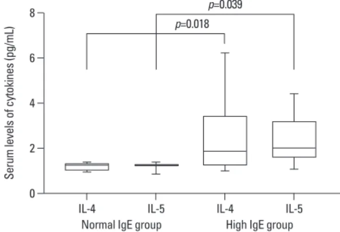
Comparison of serum cytokines according to the serum IgE levels
In the normal IgE group, the median (range) initial serum levels of each IL-4 and IL-5 were 1.3 (1.0-1.4) and 1.3 (0.9- 1.4) pg/mL, respectively. In the high IgE group, the serum levels of IL-4 and IL-5 was 1.9 (1.0-6.2) and 2.0 (1.1-4.4) pg/mL, respectively. Initial serum levels of IL-4 (p=0.018) and IL-5 (p=0.039) were significantly higher in the high IgE group than in the normal IgE group (Fig. 1). However, the serum levels of IL-10 and TGF-β showed no difference between them (data are not shown).
Comparison of serum IgE levels according to the remission state
There were no changes in the median (range) levels of se- rum IgE according to the treatment state (nephrotic or re- mission phase) between the normal and high IgE groups (Fig. 2).
Changes of serum cytokines according to the disease activity
To explore the disease activities and the roles of cytokines, the serum levels of IL-4, IL-5, IL-10, and TGF-β were ex- amined in both NS patients and healthy controls. The se- rum levels of IL-4 were significantly higher in the nephrot- ic phase of NS than in the remission phase (p<0.001) and control group (p<0.001). However no difference was found between remission phase of NS and control group (p=0.991) (Fig. 3A).
Fig. 1. Comparisons of the serum IL-4 and IL-5 levels according to the se- rum IgE levels. Initial serum IL-4 and IL-5 levels were significantly higher in the high IgE group than in the normal IgE group. IgE, immunoglobulin E; IL, interleukin.
Fig. 2. Comparison of serum IgE levels according to the remission state.
There were no changes in the median (range) levels of serum IgE accord- ing to the treatment state (nephrotic or remission phase) between the nor- mal (p=0.667) and high IgE groups (p=0.131). IgE, immunoglobulin.
0 0.00
2 1000.00
4 2000.00
6
3000.00 4000.00
8 5000.00
Serum levels of cytokines (pg/mL) Serum levels of IgE (IU/mL)
IL-4 Nephrotic
phase Nephrotic
phase
IL-5 Remission
phase Remission
phase
IL-4 IL-5
Normal IgE group
Normal IgE group High IgE group
High IgE group p=0.018
p=0.667
p=0.039 p=0.131
*
Fig. 3. Comparisons of the cytokines between nephrotic syndrome patients and healthy controls. (A) The serum levels of IL-4 in nephrotic syndrome patients with nephrotic phase were significantly higher than those of remission phase and controls. (B) The serum levels of IL-5 in nephrotic phase were also higher than those of normal controls. (C) The serum IL-10 levels did not show any significant differences in each group. (D) In the patients with nephrotic phase, the serum levels of TGF-β were significantly lower than those of remission phase or control group. This were calculated by using Kruskal-Wallis test Scheffe’s post hoc comparison. The **means p<0.001, and *indicates p<0.05. IL, interleukin; TGF, transforming growth factor; NS, nephrotic syndrome.
Fig. 4. Correlations between serum cytokines and albumin levels. By linear regression analysis, (A) the serum IL-4 levels had a negative correlation with se- rum albumin (p=0.004), (B) the serum IL-5 levels had no correlation with serum albumin (p=0.113), (C) the serum IL-10 levels did not have a positive correla- tion with serum albumin levels (p=0.571), whereas (D) the serum TGF-β levels had (p<0.001). IL, interleukin; TGF, transforming growth factor.
A
A C
C
B
B D
D
0
0
0 0
0 0
2 1
10 20
40
4 2
3
6 4
20 60
8 5
30 80
0
0 2
20
1
5 4
40
2
10 6
60
4
15 3 8
80
5
20
Serum levels of IL-4 (pg/mL)Serum levels of TGF-ß (pg/mL)Serum levels of IL-4 (pg/mL) Serum levels of IL-5 (pg/mL)
Serum levels of IL-10 (pg/mL) Serum levels of TGF-ß (pg/mL)Serum levels of IL-5 (pg/mL)Serum levels of IL-10 (pg/mL)
Nephrotic phase
Nephrotic phase
0 0
0 0
2 1 2
2 2
4 3 4
4 4
6 5
6 6
Serum levels of albumin (mg/dL) Serum levels of albumin (mg/dL)
Serum levels of albumin (mg/dL) Serum levels of albumin (mg/dL)
r 2=0.259, p=0.004 r 2=0.170, p=0.113
r 2=0.001, p=0.571 r 2=0.501, p=0.001
Nephrotic phase
Nephrotic phase Remission phase
Remission phase
Remission phase
Remission phase Normal controls
Normal controls
Normal controls
Normal controls
**
**
p=0.051
NS
**
**
*
NS NS
NS
NS
NS
pathic NS are driven toward the Th2 phenotype.
13IL-4 is the most important Th2 cytokine which mediates IgE synthesis.
24In NS, IL-4 can act on glomerular visceral epithelial cells by binding IL-4 receptor, and plays a role in glomerular permeability.
25This suggests that, regardless of atopy, IL-4 may be one of the cytokines associated with the pathogenesis of proteinuria and also be a predictive factor for disease severity in NS. In our present study, the serum levels of IL-4 were higher in the nephritic phase of NS than the remission phase and control group and were correlated with serum albumin levels. However, this seems to be vari- ance with the results in many previous studies that serum IL-4 levels decreased during relapse but increased in ne- phritic patients with long-term remission.
26Several authors have reported that serum IL-5 levels in the nephrotic phase were higher than in the remission phase and Ohtomo, et al.
27reported that suplatast tosilate, which suppresses IL-4 and IL-5 production, was effective for ste- roid reduction in idiopathic steroid sensitive NS.
13,23We also found that serum IL-5 levels were higher in the ne- phrotic phase than in the remission phase with steroid treat- ment. However, because other hither-to-uncharacterized factors might have been needed to induce idiopathic NS, further studies should be conducted to clarify the effects of IL-4 and IL-5 on idiopathic NS.
Recently, regulatory T cells (Treg cells) have been de- scribed as subsets distinct from Th1 and Th2 cells. Treg cells expressing the forkhead/winged helix transcription factor (Foxp3) have an anti-inflammatory role and maintain tolerance to self-components through direct contact with cells or by releasing anti-inflammatory cytokines such as IL-10 and TGF-β.
28,29Treg cell dysfunction has been found in several kidney disease animal models.
30-32A reduction in Treg cells induces local tissue inflammation, which may be related to the pathogenesis of proteinuria and the propaga- tion of idiopathic NS.
33In relapsed idiopathic minimal NS, Treg cells have an impaired capacity to suppress T-effector- cell proliferation.
14Liu, et al.
15reported that Th17/Treg ra- tios were increased along with increased proteinuria and decreased albumin levels in adult patients with minimal change NS.
TGF-β has been shown to regulate both immunogenic and immunosuppressive response, depending on the cellular environment.
34Therefore, overall systemic effects of TGF-β must be understood within the context of other collaborative networks involving other immunoregulatory signals.
TGF-β is often associated with the progression of kidney then, numerous lines of evidence have been presented, sug-
gesting that immune mediators play a pivotal role in its pathogenesis.
6-8,10,18Although a pathogenic relationship remains to be con- firmed, the serum levels of IgE show a close relationship to NS, and many studies have shown a strong association be- tween idiopathic NS and atopic disorders.
4,5IgE levels were significantly higher in NS patients with atopy than in non- atopic patients, especially during the relapse phase of NS compared with the remission phase, implicating IgE as a barometer of disease severity and giving it prognostic val- ue.
6,19In our present study, in a comparison of the clinical characteristics of NS patients with normal IgE and those with high IgE, the high-IgE group required a significantly longer time to reach their initial remission, was more sus- ceptible to frequent relapse and comorbid allergic diseases, and required longer steroid therapy to recover from the ne- phrotic phase than the normal-IgE group. Serum IgG levels were lower in NS patients, whereas the levels were not dif- ferent between the normal- and high-IgE groups. It is not certain whether the increased levels of IgE in idiopathic NS are pathogenic or co-incident, nevertheless, our results sug- gest that the regulation of total IgE production correlates with the disease activity and outcome of NS.
20However, it should be noted that part of the nephrotic children had per- sistently normal serum IgE levels, indicating different etiol- ogies in the pathogenesis of NS.
The signals for IgE synthesis are delivered by IL-4 or IL- 13. In relapsed NS, activated T cells increase the secretion of IL-4 and IL-13, which could induce isotype switching in IgE. IL-5 also promotes the production of IgE, whereas IFN-γ inhibits the secretion of IgE.
13,21,22In our study, high- IgE group displayed significantly elevated serum IL-4 and IL-5 levels. Thus, we suggest that the increased production of IL-4 and IL-5 in idiopathic NS may indicate that these cytokines are involved in the enhanced production of serum IgE. Furthermore, allergic treatment alone could not induce the remission of NS, suggesting that the elevated serum IgE in some patients can not cause their NS, but rather reflects allergic responses to the perturbation of their humoral im- munity.
4,23In order to evaluate immunologic pattern and their modu-
lating serum cytokines and to analyze the correlation with
disease activity in children with idiopathic NS, we evaluat-
ed initial serum levels of IL-4, IL-5, IL-10 and TGF- β and
their kinetics during treatment with or without relapse. A
number of reports have shown that activated T cells of idio-
REFERENCES
1. Schachter AD. The pediatric nephrotic syndrome spectrum: clini- cal homogeneity and molecular heterogeneity. Pediatr Transplant 2004;8:344-8.
2. The primary nephrotic syndrome in children. Identification of pa- tients with minimal change nephrotic syndrome from initial re- sponse to prednisone. A report of the International Study of Kid- ney Disease in Children. J Pediatr 1981;98:561-4.
3. Hardwicke J, Soothill JF, Squire JR, Holti G. Nephrotic syndrome with pollen hypersensitivity. Lancet 1959;1:500-2.
4. Abdel-Hafez M, Shimada M, Lee PY, Johnson RJ, Garin EH. Id- iopathic nephrotic syndrome and atopy: is there a common link?
Am J Kidney Dis 2009;54:945-53.
5. Salsano ME, Graziano L, Luongo I, Pilla P, Giordano M, Lama G.
Atopy in childhood idiopathic nephrotic syndrome. Acta Paediatr 2007;96:561-6.
6. van den Berg JG, Weening JJ. Role of the immune system in the pathogenesis of idiopathic nephrotic syndrome. Clin Sci (Lond) 2004;107:125-36.
7. Cheung W, Wei CL, Seah CC, Jordan SC, Yap HK. Atopy, serum IgE, and interleukin-13 in steroid-responsive nephrotic syndrome.
Pediatr Nephrol 2004;19:627-32.
8. Grimbert P, Audard V, Remy P, Lang P, Sahali D. Recent ap- proaches to the pathogenesis of minimal-change nephrotic syn- drome. Nephrol Dial Transplant 2003;18:245-8.
9. Matsumoto K, Kanmatsuse K. Transforming growth factor-beta1 inhibits vascular permeability factor release by T cells in normal subjects and in patients with minimal-change nephrotic syndrome.
Nephron 2001;87:111-7.
10. Souto MF, Teixeira AL, Russo RC, Penido MG, Silveira KD, Teixeira MM, et al. Immune mediators in idiopathic nephrotic syndrome: evidence for a relation between interleukin 8 and pro- teinuria. Pediatr Res 2008;64:637-42.
11. Eddy AA, Symons JM. Nephrotic syndrome in childhood. Lancet 2003;362:629-39.
12. Le Berre L, Hervé C, Buzelin F, Usal C, Soulillou JP, Dantal J.
Renal macrophage activation and Th2 polarization precedes the development of nephrotic syndrome in Buffalo/Mna rats. Kidney Int 2005;68:2079-90.
13. Kanai T, Shiraishi H, Yamagata T, Ito T, Odaka J, Saito T, et al.
Th2 cells predominate in idiopathic steroid-sensitive nephrotic syndrome. Clin Exp Nephrol 2010;14:578-83.
14. Araya C, Diaz L, Wasserfall C, Atkinson M, Mu W, Johnson R, et al. T regulatory cell function in idiopathic minimal lesion nephrot- ic syndrome. Pediatr Nephrol 2009;24:1691-8.
15. Liu LL, Qin Y, Cai JF, Wang HY, Tao JL, Li H, et al. Th17/Treg imbalance in adult patients with minimal change nephrotic syn- drome. Clin Immunol 2011;139:314-20.
16. Nephrotic syndrome in children: prediction of histopathology from clinical and laboratory characteristics at time of diagnosis. A report of the International Study of Kidney Disease in Children.
Kidney Int 1978;13:159-65.
17. Dodig S, Richter D, Benko B, Zivcić J, Raos M, Nogalo B, et al.
Cut-off values for total serum immunoglobulin E between non- atopic and atopic children in north-west Croatia. Clin Chem Lab Med 2006;44:639-47.
18. Shalhoub RJ. Pathogenesis of lipoid nephrosis: a disorder of T-cell function. Lancet 1974;2:556-60.
disease, and exhibits fibrogenic and proinflammatory proper- ties in the kidney.
35-37Proteinuria predominantly involves al- bumin, which triggers the positive feedback loop for TGF-β expression, subsequently inhibiting albumin endocytosis.
38Prolonged proteinuria gives rise to increased TGF-β, which modulates the expression of the extracellular matrix, lead- ing to glomerulosclerosis and interstitial fibrosis, and re- tarding the progression to renal failure.
39On the other hand TGF-β may promote the expression of Foxp3 and induce the differentiation of Treg cells that which originate from CD4+CD25-T cells.
40,41TGF-β is the inhibi- tor of Th17 differentiation in humans,
42and it also inhibits the vascular permeability factor released by T cells in nor- mal subjects and patients with minimal change disease.
9In the present study, we found that TGF-β and IL-10 concen- trations decreased in minimal change NS patients and posi- tively correlated with circulating Treg frequencies, which suggested that TGF-β and IL-10 may play a protective role, partly through inducing the production of Treg cells, and act as one of the effective factors of Treg cells.
15IL-10 is also pleiotropic cytokine that can exert either im- munosuppressive or immunostimulatory effects on a vari- ety of cell types and plays a central role in controlling in- flammatory processes.
43Previous studies have shown that IL-10 is involved in the positive feedback loops with TGF-β and acts at local sites of inflammation, whereas TGF-β is involved in the systemic immune response related to hu- moral lymphoproliferation.
44,45In this study, the initial serum levels of lL-10 and TGF-β did not differ between the normal- and high-IgE groups, whereas the initial serum TGF-β levels in the nephrotic phase of NS were lower than in the remission phase or in the healthy controls, and differed significantly according to the degree of proteinuria and serum albumin level. Howev- er, serum lL-10 levels were not significantly different be- tween the groups. The results of this study suggest that in- sufficient levels of TGF-β in the nephrotic phase of NS may aggravate the inflammatory responses and contribute to the induction of proteinuria. To better understand the nature, regulation, and function of Treg cells and their cytokines in idiopathic NS, further studies should be carried out.
ACKNOWLEDGEMENTS
This study was supported by research fund of Chungnam
National University in 2004.
2006;17:2731-41.
33. Shao XS, Yang XQ, Zhao XD, Li Q, Xie YY, Wang XG, et al. The prevalence of Th17 cells and FOXP3 regulate T cells (Treg) in children with primary nephrotic syndrome. Pediatr Nephrol 2009;24:1683-90.
34. Letterio JJ, Roberts AB. Regulation of immune responses by TGF-beta. Annu Rev Immunol 1998;16:137-61.
35. Harris RC, Neilson EG. Toward a unified theory of renal progres- sion. Annu Rev Med 2006;57:365-80.
36. Liu Y. Renal fibrosis: new insights into the pathogenesis and thera- peutics. Kidney Int 2006;69:213-7.
37. Reidy K, Kaskel FJ. Pathophysiology of focal segmental glomeru- losclerosis. Pediatr Nephrol 2007;22:350-4.
38. Gekle M, Knaus P, Nielsen R, Mildenberger S, Freudinger R, Wohlfarth V, et al. Transforming growth factor-beta1 reduces megalin- and cubilin-mediated endocytosis of albumin in proxi- mal-tubule-derived opossum kidney cells. J Physiol 2003;552(Pt 2):471-81.
39. Diwakar R, Pearson AL, Colville-Nash P, Brunskill NJ, Dockrell ME. The role played by endocytosis in albumin-induced secretion of TGF-beta1 by proximal tubular epithelial cells. Am J Physiol Renal Physiol 2007;292:F1464-70.
40. Chen W, Jin W, Hardegen N, Lei KJ, Li L, Marinos N, et al. Con- version of peripheral CD4+CD25- naive T cells to CD4+CD25+
regulatory T cells by TGF-beta induction of transcription factor Foxp3. J Exp Med 2003;198:1875-86.
41. Mucida D, Park Y, Kim G, Turovskaya O, Scott I, Kronenberg M, et al. Reciprocal TH17 and regulatory T cell differentiation medi- ated by retinoic acid. Science 2007;317:256-60.
42. Wilson NJ, Boniface K, Chan JR, McKenzie BS, Blumenschein WM, Mattson JD, et al. Development, cytokine profile and func- tion of human interleukin 17-producing helper T cells. Nat Immu- nol 2007;8:950-7.
43. Chung F. Anti-inflammatory cytokines in asthma and allergy: in- terleukin-10, interleukin-12, interferon-gamma. Mediators In- flamm 2001;10:51-9.
44. Mottet C, Golshayan D. CD4+CD25+Foxp3+ regulatory T cells:
from basic research to potential therapeutic use. Swiss Med Wkly 2007;137:625-34.
45. Christ M, McCartney-Francis NL, Kulkarni AB, Ward JM, Mizel DE, Mackall CL, et al. Immune dysregulation in TGF-beta 1-defi- cient mice. J Immunol 1994;153:1936-46.
19. Lin CY, Lee BH, Lin CC, Chen WP. A study of the relationship between childhood nephrotic syndrome and allergic diseases.
Chest 1990;97:1408-11.
20. Tain YL, Chen TY, Yang KD. Implication of serum IgE in child- hood nephrotic syndrome. Pediatr Nephrol 2003;18:1211-5.
21. Bacharier LB, Geha RS. Molecular mechanisms of IgE regula- tion. J Allergy Clin Immunol 2000;105(2 Pt 2):S547-58.
22. Hurtado A, Johnson RJ. Hygiene hypothesis and prevalence of glomerulonephritis. Kidney Int Suppl 2005:S62-7.
23. Fuke Y, Endo M, Ohsawa I, Satomura A, Hidaka M, Fujita T, et al. Implication of elevated serum IgE levels in minimal change nephrotic syndrome. Nephron 2002;91:769-70.
24. Finkelman FD, Katona IM, Urban JF Jr, Holmes J, Ohara J, Tung AS, et al. IL-4 is required to generate and sustain in vivo IgE re- sponses. J Immunol 1988;141:2335-41.
25. Van Den Berg JG, Aten J, Chand MA, Claessen N, Dijkink L, Wi- jdenes J, et al. Interleukin-4 and interleukin-13 act on glomerular visceral epithelial cells. J Am Soc Nephrol 2000;11:413-22.
26. Daniel V, Trautmann Y, Konrad M, Nayir A, Schärer K. T-lym- phocyte populations, cytokines and other growth factors in serum and urine of children with idiopathic nephrotic syndrome. Clin Nephrol 1997;47:289-97.
27. Ohtomo Y, Fujinaga S, Hattori M. Suplatast tosilate dimethylsul- fonium treatment for steroid-dependent nephrotic syndrome. Pedi- atr Int 2005;47:230-1.
28. Weaver CT, Hatton RD. Interplay between the TH17 and TReg cell lineages: a (co-)evolutionary perspective. Nat Rev Immunol 2009;9:883-9.
29. Sakaguchi S, Ono M, Setoguchi R, Yagi H, Hori S, Fehervari Z, et al. Foxp3+ CD25+ CD4+ natural regulatory T cells in dominant self-tolerance and autoimmune disease. Immunol Rev 2006;212:
8-27.
30. Wolf D, Hochegger K, Wolf AM, Rumpold HF, Gastl G, Tilg H, et al. CD4+CD25+ regulatory T cells inhibit experimental anti- glomerular basement membrane glomerulonephritis in mice. J Am Soc Nephrol 2005;16:1360-70.
31. Monteiro RM, Camara NO, Rodrigues MM, Tzelepis F, Damião MJ, Cenedeze MA, et al. A role for regulatory T cells in renal acute kidney injury. Transpl Immunol 2009;21:50-5.
32. Mahajan D, Wang Y, Qin X, Wang Y, Zheng G, Wang YM, et al.
CD4+CD25+ regulatory T cells protect against injury in an innate murine model of chronic kidney disease. J Am Soc Nephrol