서론
치근단 방사선촬영은 상의 왜곡이 적고 해상도와 선예도가 우수하며 방사선노출이 비교적 적은 진단영상법으로 치과영 역에서 많이 이용되고 있다. 특히, 고해상도 영상이 요구되는 임플란트 시술 및 근관치료 과정에서 치근단 방사선촬영이 시 행되고 있으며, 최근에는 임플란트 시술 중 이동형 구내방사 선촬영기(portable dental X-ray machine)를 이용한 치근단 방사선 촬영이 많이 이루어지고 있다.
이동형 구내방사선촬영기는 개발 초기에는 촬영실 설치가 불가능한 전쟁지역이나 신원 파악이 필요한 재해지역에서 이 용 되었다.1,2그 이후에는 환자가 움직일 수 없는 수술장 그리고 거동이 불편한 노인이나 장애인의 재택 치료 시 이용되고 있 다.3,4우리나라의 경우에는 무게가 비교적 가볍고 카메라처럼 술자가 들고 촬영할 수 있기에 일반 치과진료실에서도 사용이 증가되고 있으며, 국내업체에서도 여러 종류의 제품을 개발하 여 출시하고 있다.5
전 세계적으로 새로운 진단영상장비의 개발로 인한 이용률 의 증가와 의료영상검사의 빈도가 증가하면서 환자들의 방사
선 노출이 점점 증가하고 있다.6치과영역에서도 장비의 이동 성과 진료의 효율성이 크다는 장점을 가진 이동형 구내방사선 촬영기가 개원가를 중심으로 보급되어 사용되고 있다. 그러나 이러한 장비 사용에 따른 방사선 노출 정도에 대한 자료가 충 분치 않다.
본 연구에서는 이동형 구내 방사선사진촬영기와 벽걸이형 구내 방사선사진촬영기로 치근단 방사선촬영을 할 경우 두경 부 주요 조직 및 기관의 흡수선량을 측정하고 유효선량을 구 하여 환자의 방사선 노출 정도를 알아보고자 하였다.
연구 재료 및 방법 1. 연구 재료
촬영대상은 선량측정용 두경부 마네킨인 ART - 남성 팬텀 (Radiology Support Devices, Inc., Long Beach, USA)을 이용하였 다. 팬텀(phantom)은 인조 두개골에 연조직과 방사선 감쇄계수 가 유사한 재료로 외형을 만들었고, 각 2.5 cm 두께를 갖는 10개 절단면으로 구성되었으며 각 절단면에는 열형광선량계 소자
*교신저자: 한한원원정정
330-716 충청남도 천안시 동남구 단대로 119 단국대학교 치과대학 구강악안면방사선학교실 041-550-1923: e-mail, [email protected] 원고접수일: 2012년 7월 13일 / 원고최종수정일: 2012년 7월 18일 / 원고채택일: 2012년 7월 19일
이동형 구내방사선촬영기와 벽걸이 구내방사선촬영기로 촬영한 치근단 방사선촬영에서 환자의 흡수선량과 유효선량 평가
한원정*
단국대학교 치과대학 구강악안면방사선학교실
연구 목적: 이 연구는 상악 전치부와 하악 구치부 치근단 방사선촬영시 이동형 구내방사선촬영기와 벽걸이 구내방사선촬영기로 촬영한 경우에서 환자의 방사선량을 알 아보고자 하였다.
연구 재료 및 방법: 방사선량 측정은 선량 측정용 두경부 마네킨의 23부위에 열형광선량계 소자를 위치시키고 해당 치근단 방사선촬영을 하였다. 열형광선량계 판독기 로 흡수선량을 구하였고 방사선 조사된 조직의 비율을 곱하여 방사선 가중선량을 구하였다. 국제방사선방호위원회에서 2007년에 공지한 조직 가중계수를 이용하여 각 방 사선촬영의 유효선량을 구하였다.
결과: 환자의 흡수선량은 이동형 구내방사선촬영기로 촬영한 상악 전치부와 이동형 및 벽걸이 구내방사선촬영기로 촬영한 하악 구치부 치근단방사선촬영의 경우 하악 체에서 가장 높았다. 유효선량은 상악 전치부 치근단 방사선촬영에서는 이동형 촬영기로 촬영한 경우 4 μSv, 벽걸이 촬영기로 촬영한 경우 2 μSv 였고 우측 하악 구치부 치 근단 방사선촬영에서는 각각 6 μSv, 2 μSv 였다.
결론: 벽걸이 구내방사선촬영기보다 이동형 구내방사선촬영기로 촬영한 치근단 방사선촬영에서의 유효선량이 더 많기 때문에 술자는 구내방사선촬영기에 따른 방사선 노출 정도를 충분히 인지하고 이를 사용하여야 한다. (대한치과보철학회지 2012;50:184-90)
주요단어: 치근단방사선촬영; 이동형 구내방사선촬영기; 방사선량측정; 열형광선량계
(Thermoluminescent dosimeter chip)를 위치시킬 수 있는 구멍(hole) 들이 있다(Fig. 1).
선량은 1/8" × 1/8" × 0.035"의 열형광선량계 소자(Harshaw Chemical Co., USA)와 열형광선량계 판독기(model 5500 reader, Harshaw Chemical Co., USA)를 이용하여 측정하였다(Fig. 2).
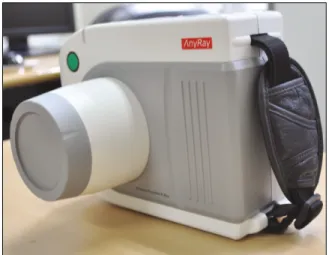
치근단 방사선촬영에 사용된 이동형 구내방사선촬영기 (AnyRay-P, Vatech Co., Hwasung, Korea)의 관전압은 60 kVp, 관전 류는 2 mA, 초점(focal spot) 크기는 0.8 mm, 관구(cone) 모양은 원 형, 관구 직경은 70 mm, 초점-피부간 거리(FSD, focal spot-skin distance)는 110 mm이다(Fig. 3).
벽걸이 구내방사선촬영기(Kodak2200 intraoral X-ray system, Carestream Health, Inc., Rochester, USA)의 관전압 70 kVp, 관전류 8 mA, 초점 크기 0.7 mm, 초점-피부간 거리는 200 mm이다(Fig. 4).
2. 연구 방법
1) 선량 측정 전 준비
방사선량 측정기관(Iljin Radiation Engineering Co., Ltd., Hwasung, Korea)에서 방사선 교정과정을 거쳐 각 소자의 오류정정부호 (ECC, Error correction code) 값을 통상적인 방법으로 구한 후, 그 오
차범위가 5% 미만인 열형광선량계 소자를 방사선량 측정에 사용하였다.
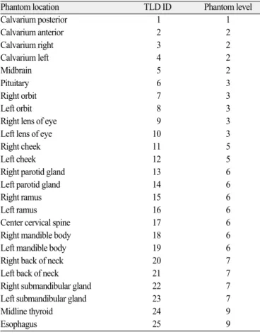
두경부 팬텀의 25부위에 소성된 열형광선량계 소자를 각각 위치시킨 후 치근단 방사선촬영을 하였다. 25부위는 방사선 감 수성이 있는 조직 및 기관이다(Table 1).7
이 부위들 중에서 좌∙우 안구렌즈 부위, 뺨 부위 그리고 뒷 목 부위는 팬텀의 외형 표면에 테이프를 이용하여 소자를 고 정시켰고, 나머지 부위는 팬텀 절단면의 해당 구멍에 소자 홀 더를 이용하여 위치시켰다.
2) 치근단 방사선촬영
이동형 구내방사선촬영기와 벽걸이 구내방사선촬영기로 각각 상악 전치부와 하악 우측 구치부 치근단 방사선촬영을 시행하였다. 이동형 구내방사선촬영에서의 촬영조건은 관전 압 60 kVp, 관전류 2 mA, 관구에서 피부까지의 거리 2 cm, 노출시 간 0.2 초로 구내 CCD센서를 사용하는 조건으로 하였다. 벽걸
Fig. 1. ART - man phantom consisted of ten 2.5 cm horizontal sections which numbered from 1 to 9.
Fig. 2. TLD chips in the container (left) and Harshaw TLD reader system (right).
Fig. 4. Wall-type dental X-ray machine (Kodak2200 intraoral X-ray system, Carestream Health, Inc., Rochester, USA).
Fig. 3. Portable dental X-ray machine (AnyRay-P, Vatech Co., Hwasung, Korea).
이 구내방사선촬영에서는 관전압 70 kVp, 관전류 8 mA, 관구에 서 피부까지의 거리 2 cm, 노출시간 0.074초의 조건으로 촬영하 였다(Table 2).
팬텀의 위치 변동 없이 동일 치아부위에서 치근단 방사선촬 영을 10회 시행하였다. 촬영횟수는 참고문헌들을 참조하여 정 하였다.7-9
3) 흡수선량 측정과 유효선량 계산
방사선에 노출된 25부위의 열형광선량계 소자에서 각 부위 의 흡수선량(μGy)을 구한 후, 해당 조직 및 기관의 평균 흡수선 량을 구하였다. 이때 10개의 소자를 정하고 선량 계산시 배경 방사선량을 감해주었다.
골수의 평균 흡수선량은 하악골, 두개골, 경추에서 각각 구 한 후 합하였다. 하악골은 좌∙우 하악지와 좌∙우 하악체에 서 측정된 흡수선량의 평균값으로 계산하였고 두개골은 전∙
후∙좌∙우 두개골에서 측정된 흡수선량의 평균값으로 계산 하였다. 경추는 경추에서 측정된 흡수선량으로 하였다.
골표면의 평균 흡수선량은 골수의 평균 흡수선량에 4.64를 곱하여 구하였다.10
피부의 평균 흡수선량은 좌∙우 뺨, 좌∙우 안구렌즈 그리고 좌∙우 뒷목에서 측정된 흡수선량의 평균값으로 계산하였다.
타액선의 평균 흡수선량은 좌∙우 이하선과 좌∙우 악하선에 서 측정된 흡수선량의 평균값으로 하였다. 뇌의 평균 흡수선 량은 중간 뇌부위와 뇌하수체에서 측정된 흡수선량의 평균값 으로 구하였다. 기타 조직(remainder)에 lymphatic nodes, muscle, extrathoracic airway, oral mucosa를 포함시켰으며, 흡수선량을 각각 구하였다.7
구해진 각 조직 및 기관의 평균 흡수선량에 전체 조직에 대 한 방사선 조사된 조직의 비율을 곱하여 등가선량(μSv)을 구하 였다(Table 3).7
조사된 두경부 골수의 비율은 하악골은 전체 골수의 1.3%, 두개골은 11.8%, 경추는 3.4%로 계산하여 등가선량을 구하였다.
조사된 식도의 비율은 전체 식도의 10%로 계산하였고, 조사된 피부의 비율은 5%로 계산하여 등가선량을 구하였다.8갑상선, 타액선, 뇌는 조사된 비율을 100%로 계산하여 구하였다. 기타 조직에 대한 등가선량도 구하였다.8
Table 2. Exposure parameters for periapical and panoramic radiography Portable dental Wall-type dental X-ray machine X-ray machine
Image receptor CCD sensor CCD sensor
kVp 60 70
m A 2 8
Exposure time (seconds)
Upper anterior teeth 0.2 0.074
Lower posterior teeth 0.2 0.074
Table 3. Estimated percentage of tissue irradiated and TLDs used to calculated mean dose to a tissue or organ
Fraction irradiated(%) TLD ID
Bone marrow 16.5
Mandible 1.3 15,16,18,19
Calvarium11.8 1 - 4
Cervical spine 3.4 17
Bone surface* 16.5
Mandible 1.3 15,16,18,19
Calvarium11.8 1 - 4
Cervical spine 3.4 17
Brain 100 5,6
Esophagus 10 25
Salivary gland 100
Parotid 100 13,14
Submandible 100 22,23
Skin 5 9 - 12,20,21
Thyroid 100 24
Remainder
Extrathoracic region 100 7,8,13 - 19,22,23,25
Lymphatic nodes 5 13 - 19,22,23,25
Muscle 5 13 - 19,22,23,25
Oral mucosa 100 13 - 19,22,23
Table 1. Locations of TLD chips in phantom
Phantom location TLD ID Phantom level
Calvarium posterior 1 1
Calvarium anterior 2 2
Calvarium right 3 2
Calvarium left 4 2
Midbrain 5 2
Pituitary 6 3
Right orbit 7 3
Left orbit 8 3
Right lens of eye 9 3
Left lens of eye 10 3
Right cheek 11 5
Left cheek 12 5
Right parotid gland 13 6
Left parotid gland 14 6
Right ramus 15 6
Left ramus 16 6
Center cervical spine 17 6
Right mandible body 18 6
Left mandible body 19 6
Right back of neck 20 7
Left back of neck 21 7
Right submandibular gland 22 7
Left submandibular gland 23 7
Midline thyroid 24 9
Esophagus 25 9
유효선량(μSv)은 구해진 각 조직 및 기관의 등가선량에 해당 조직 가중계수를 곱하고 그 합으로 하였다.
조직 가중계수는 전체적인 방사선 위험도에 대한 조직 및 기 관의 상대적인 기여도를 나타내며, 국제방사선방호위원회 (ICRP) 2007년 권고안에 따른 15개 조직 및 기관과 14개의 기타 조직의 계수를 이용하였다(Table 4).
본 연구에서는 치근단 방사선촬영시 방사선에 직접 노출되 지 않는 8개 조직 및 기관인 유선, 결장, 폐, 위, 방광, 생식선, 간, 신장과 9개의 기타 조직은 제외하고 유효선량을 구하였다.
결과
1. 흡수선량
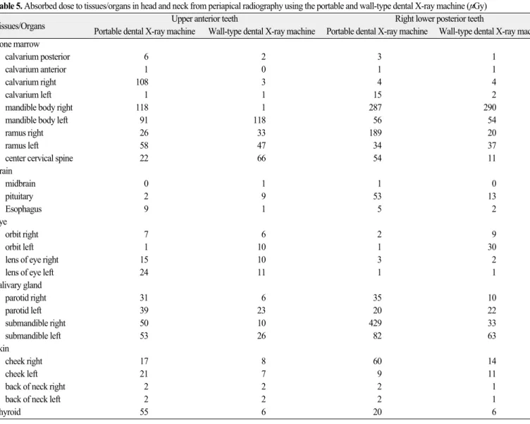
상악 전치부 치근단방사선촬영에서 이동형 구내방사선촬 영기로 촬영한 경우에는 우측 하악체가 118 μGy로 가장 높았으 며, 벽걸이 구내방사선촬영기로 촬영한 경우에도 좌측 하악체 가 118 μGy로 가장 높았다.
우측 하악 구치부 치근단방사선촬영에서는 이동형 구내방 사선촬영기로 촬영한 경우는 우측 악하선이 429 μGy로 가장 높았고 그 다음으로 우측 하악체가 287 μGy 이었다. 벽걸이 구 내 방사선촬영기로 촬영한 경우는 우측 하악체가 290 μGy로 가장 높았다(Table 5).
2. 유효선량
각 조직 및 기관의 평균 흡수선량에 전체 조직에 대한 방사
선 조사된 조직의 비율을 곱한 등가선량을 구하고 기타 조직 및 기관을 제외하고 비교하였다. 상악 전치부 치근단방사선촬 영에서 이동형 구내방사선촬영기로 촬영한 경우는 갑상선이 가장 높았고 벽걸이 구내방사선촬영기로 촬영한 경우에는 타 액선이 가장 높았다. 하악 우측 구치부 치근단방사선촬영에서 는 두 촬영기 모두 타액선이 가장 높았다(Table 6).
유효선량은 상악 전치부 치근단방사선촬영에서는 이동형 구내방사선촬영기로 촬영한 경우 4 μSv, 벽걸이 구내방사선촬 영기로 촬영한 경우 2 μSv 였다. 하악 우측 구치부 치근단방사 선촬영에서는 이동형 구내방사선촬영기로 촬영한 경우 6 μSv, 벽걸이 구내방사선촬영기로 촬영한 경우 2 μSv 였다(Table 7).
고찰
1990년 국제방사선방호위원회(ICRP, International Commission on Radiological Protection)에서는 방사선 위해(radiation detriment) 개념 을 공지하였고 방사선의 확률적 효과가 고려된 유효선량(effec- tive dose)으로 이를 표시하였다.
유효선량은 인체의 일부만 방사선에 노출되었어도 인체 전 신에 대한 효과가 어느 정도인지 나타내주는 선량이며, 단위 는 Sievert (Sv)를 사용한다. 방사선에 노출된 각 조직 및 기관의 등가선량(equivalent dose)과 조직 가중계수(tissue weighted factor) 의 곱을 모두 합하여 구하게 된다. 그러므로 유효선량은 신체 일부에 노출된 방사선으로 전신에 노출되어 야기되는 방사선 위해 정도를 측정할 수 있으며, 다른 촬영법간의 방사선 위해 정도를 비교 할 수 있다.10조직 가중계수는 전체 방사선 위험도 에 대한 조직 또는 기관의 상대적인 기여도를 나타내며, 1990년 국제방사선방호위원회는 방사선에 감수성이 있는 12개 조직 및 기관과 10개 기타조직(remainder)의 조직 가중계수를 공지하 였다. 2007년에는 뇌, 신장, 타액선이 추가된 15개 조직 및 기관 과 14개 기타 조직에 대한 조직 가중계수를 개정하여 공지하였 다.11
이동형 구내방사선촬영기는 1993년 미국에서 처음 제작되 어 군에서 주로 사용되었고 미국 식품의약국(U.S.Food and Drug Administration, FDA)에서 군용으로 승인 받았다.12국내에서는 2000년대부터 임플란트 술식과 연관되어 치과에 보급되기 시 작하였다.
국내에서 시판되고 있는 대부분의 이동형 구내방사선촬영 기는 술자가 직접 들고 촬영할 수 있으며 가볍고 장비의 이동 성이 매우 우수하다. 그러나 벽걸이 구내방사선촬영기보다 더 많은 누설 및 산란선량이 발생되어 술자의 방사선 노출을 증 가시킨다.133종의 이동형 구내방사선촬영기와 1종의 벽걸이 구내방사선촬영기를 이용하여 치근단 필름으로 치근단방사 선촬영을 한 후 환자선량을 비교한 연구에서는 유효선량의 차 이가 크지 않았다.9그러나 이동형 구내방사선촬영기들의 환 자선량에 대한 자료가 아직은 충분하지 않은 상태이다.
본 연구에서는 상악 전치부 치근단 방사선촬영의 경우 이동 Table 4. Tissue-weighting factors for calculation of effective dose - ICRP 2007
recommendation
Tissue/organ 2007 WT
Bone marrow 0.12
Breast 0.12
Colon 0.12
Lung 0.12
Stomach 0.12
Bladder 0.04
Esophagus 0.04
Gonads 0.08
Liver 0.04
Thyroid 0.04
Bone surface 0.01
Brain 0.01
Salivary glands 0.01
Skin 0.01
Remainder tissues* 0.12
*Adrenals, extrathoracic region, gall bladder, heart, kidneys, lymphatic nodes, oral mucosa, pancreas, prostate, small intestine, spleen, thymus and uterus/cervix. Italicized text represents remainder tissues used for calculation of maxillofacial dose.
Table 5. Absorbed dose to tissues/organs in head and neck from periapical radiography using the portable and wall-type dental X-ray machine (μGy)
Tissues/Organs Upper anterior teeth Right lower posterior teeth
Portable dental X-ray machine Wall-type dental X-ray machine Portable dental X-ray machine Wall-type dental X-ray machine Bone marrow
calvarium posterior 6 2 3 1
calvarium anterior 1 0 1 1
calvarium right 108 3 4 4
calvarium left 1 1 15 2
mandible body right 118 1 287 290
mandible body left 91 118 56 54
ramus right 26 33 189 20
ramus left 58 47 34 37
center cervical spine 22 66 54 11
Brain
midbrain 0 1 1 0
pituitary 2 9 53 13
Esophagus 9 1 5 2
Eye
orbit right 7 6 2 9
orbit left 1 10 1 30
lens of eye right 15 10 3 2
lens of eye left 24 11 1 1
Salivary gland
parotid right 31 6 35 10
parotid left 39 23 20 22
submandible right 50 10 429 33
submandible left 53 26 82 63
Skin
cheek right 17 8 60 14
cheek left 21 7 9 11
back of neck right 2 2 2 1
back of neck left 2 2 2 1
Thyroid 55 6 20 6
Table 6. Equivalent dose to tissues/organs in head and neck from full-mouth periapical radiography using the portable and wall-type dental x-ray machine (μSv)
Tissues/Organs Upper anterior teeth Right lower posterior teeth
Portable dental X-ray machine Wall-type dental X-ray machine Portable dental X-ray machine Wall-type dental X-ray machine
Bone marrow 5 3 4 2
Bone surface 24 14 20 9
Brain 1 5 26 6
Esophagus 1 0 0 0
Salivary glands 43 16 142 32
Skin 1 0 1 0
Thyroid 55 6 20 6
Remainder tissues/organs
Lymphatic nodes 2 2 6 3
Extrathoracic region 42 29 99 48
Muscle 2 2 6 3
Oral mucosa 58 33 141 66
Table 7. Effective dose from radiographic examination for periapical images using portable and wall-type dental X-ray machine (μSv)
Upper anterior teeth Right lower posterior teeth
Portable dental X-ray machine Wall-type dental X-ray machine Portable dental X-ray machine Wall-type dental X-ray machine
Effective dose 4 2 6 2
형 구내방사선촬영기와 벽걸이 구내방사선촬영기 모두 하악 체에서 흡수선량이 가장 높았으며, 하악 구치부 치근단방사선 촬영에서도 벽걸이 구내방사선촬영기로 촬영한 경우에 하악 체에서 흡수선량이 가장 높았다. 그러나 이동형 구내방사선촬 영기에서는 악하선에서 흡수선량이 가장 높았다. 이러한 결과 는 이전의 구내방사선촬영기를 이용한 연구들과 유사한 결과
이다.14-16Cho 등의 연구9에서도 하악체, 악하선에서 높은 흡수선
량을 보였다. 하악 구치부 치근단촬영에서 이동형 구내방사선 촬영기로 촬영한 경우에 악하선에서 더 높은 흡수선량이 측정 된 것은 이동형 구내방사선촬영기의 관구의 길이가 벽걸이 구 내방사선촬영기보다 짧아서 환자의 표면 노출의 범위가 커짐 에 따라 악하선의 방사선피폭이 커졌을 것으로 생각된다. 국 내에 시판되고 있는 이동형 구내방사선촬영기의 관구의 길이 가 100 mm 전후인 경우가 대부분이며, 이러한 관구길이는 환자 의 노출량을 증가시키는 주된 요인이다. 조직 및 기관의 등가 선량은 이동형 구내방사선촬영기로 촬영한 경우 타액선, 갑상 선에서 가장 높았다. 이동형 구내방사선촬영기로 전악 치근단 방사선촬영과 파노라마 방사선촬영간의 환자선량을 비교한 연구에서도 갑상선은 파노라마방사선촬영의 6배의 등가선량 을 보여주었으며, 이동형 구내방사선촬영시 갑상선에 대한 방 사선노출을 최소화하기 위한 방법으로 갑상선 보호대 사용을 적극 추천하였다.17이 연구에서 14매 전악 치근단 방사선촬영 에서의 유효선량은 46 μSv였고 파노라마 방사선촬영에서는 38 μSv였다.17
본 연구 결과 유효선량은 이동형 구내방사선촬영기로 촬영 한 치근단 방사선촬영에서 더 높았다. 직접적인 이유는 노출 시간이 0.2초로 벽걸이 구내방사선촬영보다 방사선 노출량이 많았으며, 더 짧은 관구 길이로 인한 환자의 노출범위가 커서 방사선감수성이 높은 갑상선, 타액선등이 노출되었기 때문이 라 생각된다.
그러므로 이동형 구내방사선촬영기로 치근단방사선촬영을 시행할 경우에는 환자의 방사선 노출을 충분히 인지하고 이용 하여야 한다.
결론
상악 전치부와 하악 구치부 치근단 방사선촬영시 벽걸이 구 내방사선촬영기로 촬영한 경우보다 이동형 구내방사선촬영 기로 촬영한 경우가 유효선량이 더 높았으므로, 술자는 이동 형 구내 방사선촬영기로 촬영할 경우 환자의 노출선량을 고려 하여 촬영의 당위성이 인정될 경우에 이용하도록 한다.
참고문헌
1. Coy J, Vandre RH, Davidson WR. Use of the hand-held dental X-ray machine during joint operation, NATO exercise Display Determination-92. Mil Med 1997;162:575-7.
2. Varghese S, Kimmel A, Radmer T, Bradley TG, Bahcall J. In vit- ro evaluation of the XR-15 portable x-ray unit for forensic odontology. J Forensic Odontostomatol 2004;22:5-8.
3. Hermsen KP, Jaeger SS, Jaeger MA. Radiation safety for the NO- MAD portable X-ray system in a temporary morgue setting. J Forensic Sci 2008;53:917-21.
4. Charlton DG. Portable dental equipment: dental units and x-ray equipment. Gen Dent 2009;57:336-41.
5. Kim EK. Effect of the amount of battery charge on tube voltage in different hand-held dental x-ray systems. Imaging Sci Dent 2012;42:1-4.
6. Lee W. Current status of medical radiation exposure and regu- lation efforts. J Korean Med Assoc 2011;54:1248-52.
7. Ludlow JB, Davies-Ludlow LE, Brooks SL, Howerton WB.
Dosimetry of 3 CBCT devices for oral and maxillofacial radiology:
CB Mercuray, NewTom 3G and i-CAT. Dentomaxillofac Radiol 2006;35:219-26.
8. Ludlow JB, Davies-Ludlow LE, Brooks SL. Dosimetry of two extraoral direct digital imaging devices: NewTom cone beam CT and Orthophos Plus DS panoramic unit. Dentomaxillofac Radiol 2003;32:229-34.
9. Cho JY, Han WJ, Kim EK. Absorbed and effective dose from pe- riapical radiography by portable intraoral x-ray machine. Korean J Oral Maxillofac Radiol 2007;37:149-56.
10. White SC, Pharoah MJ. Oral radiology: principles and interpretation.
6thed. Mosby; 2008.
11. ICRP. ICRP Publication 103. The 2007 recommendations of the international commission on radiological protection. Elsevier; 2007.
12. Van Dis ML, Miles DA, Parks ET, Razmus TF. Information yield from a hand-held dental x-ray unit. Oral Surg Oral Med Oral Pathol 1993;76:381-5.
13. Kim EK. Leakage and scattered radiation from hand-held den- tal x-ray unit. Korean J Oral Maxillofac Radiol 2007;37:65-8.
14. Avendanio B, Frederiksen NL, Benson BW, Sokolowski TW.
Effective dose and risk assessment from detailed narrow beam radiography. Oral Surg Oral Med Oral Pathol Oral Radiol Endod 1996;82:713-9.
15. Choi SC, Choi HM. Absorbed dose in the full-mouth periapical radiography, panoramic radiography and zonography. J Korean Acad Oral Maxillofac Radiol 1999;29:255-60.
16. Underhill TE, Chilvarquer I, Kimura K, Langlais RP, McDavid WD, Preece JW, Barnwell G. Radiobiologic risk estimation from dental radiology. Part I. Absorbed doses to critical or- gans. Oral Surg Oral Med Oral Pathol 1988;66:111-20.
17. Han WJ. Absorbed and effective dose from periapical radiography by portable intraoral x-ray machine and panoramic radiography.
J Korean Dent Assoc 2012;50:148-58.
Absorbed and effective dose for periapical radiography using portable and wall type dental X-ray machines
Won-Jeong Han*, DDS, PhD
Department of Oral and Maxillofacial Radiology, College of Dentistry, Dankook University, Cheonan, Korea
Purpose: The purpose of this study was to measure the absorbed dose and to calculate the effective dose for one periapical radiography using the portable and wall type den- tal X-ray machines. Materials and methods: Thermoluminescent chips were placed at 25 sites throughout the layers of the head and neck of a tissue-equivalent human skull phantom. The man phantom was exposed with the portable and wall type dental X-ray machines. For one periapical radiography taken by portable dental X-ray machine, the exposure setting was 60 kVp, 2 mA and 0.2 seconds, while for one periapical radiography taken by wall type dental X-ray machine, exposure setting was 70 kVp, 8 mA and 0.074 seconds. Absorbed dose measurements were performed and equivalent doses to individual organs were summed using ICRP 103 to calculate effective dose. Results:
In the upper anterior periapical radiography using portable dental X-ray machine and in the lower posterior periapical radiography using both machines, the highest absorbed dose was recorded at the mandible body. The effective dose in upper anterior periapical radiography using portable and wall type dental X-ray machines was 4 μSv, 2 μSv, respectively. In the lower posterior periapical radiography, the effective dose for each portable and wall type dental X-ray machines was 6 μSv, 2 μSv. Conclusion: It was recommended that the operator use prudently potable dental X-ray machine because that the effective dose in the periapical radiography using wall type dental X-ray machine was lower than that in the periapical radiography using portable dental X-ray machine. (J Korean Acad Prosthodont 2012;50:184-90)
Key words: Radiography, Dental, Digital; Radiation Protection; Radiation Dosage; Portable Dental X-ray
*Corresponding Author: Won-Jeong Han
Department of Oral and Maxillofacial Radiology, College of Dentistry, Dankook University, 119, Dandae-ro, Dongnam-Gu, Cheonan, Korea +82 041 550 1923: e-mail, [email protected]
Article history
Received July 13, 2012 / Last Revision July 18, 2012 / Accepted July 19, 2012