186 Copyright 2006 by the Korean Society for Clinical Neurophysiology
대한임상신경생리학회지 8(2):186~189, 2006 ISSN 1229-6414Unilateral facial palsy of the peripheral type without other neurologic deficits is mostly diag- nosis as Bell’ s palsy. We present a patient with unilateral peripheral type facial palsy as like as Bell’ s palsy. The blink test was indicative of left facial neuropathy as seen as in the left medullary lesion. So, we must have to check brain image study. Diffusion weighted (DWI) brain MRI demonstrated high signal region in the left dor- solateral ponto-medullary junction. Apperant diffusion coefficient (ADC) brain MRI showed low signal region in the same area. Her peripheral type facial palsy was due to infarction in the left ponto-medullary junction.
CASE
A 69 year old was woman admitted with sudden left facial weakness. She had 4.6 cm ascending aortic aneurysm. Seven years before, she was recommended a surgical resection of aortic aneurysm. However, she refused the operation.
She had had medications during the past 7 years due to the aortic aneurysm, antihypertensive medication for 20 years and anti-diabetic med- ication for 7 years.
On admission day, she could not frown or raise the eyebrow, retract the angle of the mouth or purse the lips, puff out the cheek, tighten the chin, or make wrinkle in her forehead (Fig. 1). In neurologic examination, she had no weakness or sensory loss in all extremities. All the cranial nerves were intact, except for left peripheral type facial nerve palsy. On admission day, nerve con-
말초성 안면신경 마비로 발현된 교뇌-연수 인접 부위 뇌경색 1예
서울보훈병원 신경과
조정선∙김두응∙김정미∙한영수∙하상원∙박상은∙한정호∙조은경
A Case of Peripheral Facial Palsy in Ponto-medullary Junction Area Infarction
Jeong-Seon Cho, M.D., Doo-Eung Kim, M.D., Jung-Mee Kim, M.D., Young-Su Han, M.D., Sang-Won Ha, M.D., Sang-Eun Park, M.D., Jeong-Ho Han, M.D., Eun-Kyoung Cho, M.D.
Department of Neurology, Seoul Veterans Hospital
A 69 year-old woman was admitted with sudden left facial weakness. She had no other neurologic deficit, except for left peripheral type facial palsy. She had a presumptive diagnosis of Bell’s palsy. The blink test was indicative of left facial neuropathy due to left medullary lesion. Diffusion weighted (DWI) brain MRI demonstrated high signal signal lesion in left dorsolateral ponto-medullary junction. Apperant diffusion coefficient (ADC) brain MRI showed low signal lesion in the same area. We present an unusual case of ipsilateral peripheral facial palsy in dosolateral ponto-medullary infarction without other neurologic deficits.
Key Words: Peripheral facial palsy, Ponto-medullary infarction
Address for correspondence Doo-Eung Kim, M.D.
Department of Neurology, Seoul Veterans Hospital Dunchon 2-dong 6-2, Gangdong-gu, Seoul, 134-791, Korea Tel: +82-2-2225-1324 Fax: +82-2-2225-1327
E-mail : [email protected]
duction study, and blink reflex test were done.
Nerve conduction study showed normal terminal latency and compound muscle action potential (CMAP) amplitude in right and left facial nerves.
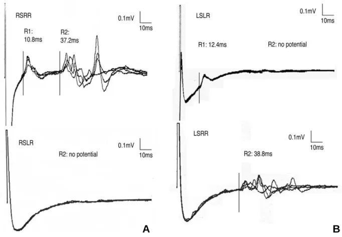
Blink reflex test showed normal R1 responses on both sides. But, ipsilateral R2 and contralateral R2 responses were not obtained by the stimula- tion on the affected side. These findings were indicative of left facial neuropathy, due to left medullary lesion (Fig. 2).
1The diffusion weighted (DWI) brain MRI demonstrated high a signal region in left dorsolateral ponto-medullary junc- tion (Fig. 3). Apperant diffusion coefficient (ADC) brain MRI showed low signal region in the same area.
DISCUSSION
The corticofacial fibers travel within the ven- tromedial base of the pons and cross the midline at the level of the facial nucleus. In some individ- uals, the corticofacial fibers loop down into the ventral part of the upper medulla, cross the mid- line and ascend in the dorsolateral medullary region ipsilaterally to the facial nucleus. The motor nuclei are located in the ventrolateral por- tion of the lower pontine tegmentum near the medulla oblongata. While still in the tegmentum
of the pons, the axons of the neurons first pro- ceed toward the floor of the fourth ventricle near the midline, then loop around the nuclei of the abducens nerves and proceed toward the ponto- cerebellar angle, where they emerge at the ponto-medullary junction just in front of cranial nerve VIII (Fig. 3).
4Within the pons, nuclear and fascicular lesions of the facial nerve result in a peripheral type facial nerve palsy. These lesions usually affect neighboring structures, such as the abducens fascicle or nucleus (lateral rectus paralysis), the paramedian pontine reticular formation (paralysis of conjugate gaze to the ipsilateral side), the cor- ticospinal tract (contralateral hemiplegia), and occasionally the spinal tract and nucleus of the trigeminal nerve and the spinothalamic tract (ipsilateral facial and contralateral body sensory disturbances).
7Occasionally, peripheral type facial palsy was found in pontine infarction. But, isolated periph- eral facial palsy without other neurologic defects is a rare condition in brainstem infarction.
Generally, if a patient presents with peripheral type facial palsy without other neurologic abnor- malities, brainstem lesions were not considered.
In this case, 69 year-old woman developed sud- den left peripheral type facial palsy. She had no
말초성 안면신경 마비로 발현된 교뇌-연수 인접 부위 뇌경색 1예
J Korean Society for Clinical Neurophysiology / Volume 8 / December, 2006 187
Figure 1. The patient shows left all facial muscles paralyzed. The lip was deviated to the right side. Forehead wrinkle is observed in
right side only.
other neurologic deficits. Nerve conduction study showed brainstem lesion. Brain MRI demonstrat-
ed left ponto-medullary infarction.
This case shows us that ponto-medullary
조정선∙김두응∙김정미∙한영수∙하상원∙박상은∙한정호∙조은경
188 J Korean Society for Clinical Neurophysiology / Volume 8 / December, 2006 Figure 2. On blink reflex test R1 and R2 responses are within the normal limits in the non-affected right side (A). R1 response is within normal limit in the affected left-sided, but R2 response is not obtained (B).
A B
Figure 3. Note a high signal lesion in the ponto-medullary junction area on diffusion weighted MR imaging (arrow). Schematic drawing of the voluntary corticofacial projections in the human brainstem. 1 = main ventral pyramidal tract; 2 = ‘aberrant bundle’ in a paralemniscal position at the dorsal base of the pons; 3 = fiber loop into the ventral medullary region, crossing the midline and ascending in the dorsolateral medullary region to the facial nucleus from below. (A) Sagittal view; (B) coronal view.
A B
말초성 안면신경 마비로 발현된 교뇌-연수 인접 부위 뇌경색 1예