• 검색 결과가 없습니다.

Genetic Characteristics of Mitochondrial DNA Was Associated with Colorectal Carcinogenesis and Its Prognosis

N/A
N/A
Protected

Academic year: 2021

Share "Genetic Characteristics of Mitochondrial DNA Was Associated with Colorectal Carcinogenesis and Its Prognosis"

Copied!
12
0
0

로드 중.... (전체 텍스트 보기)

전체 글

(1)

Genetic Characteristics of Mitochondrial DNA Was Associated with Colorectal

Carcinogenesis and Its Prognosis

Jae-Ho Lee

1

, Ilseon Hwang

2

, Yu-Na Kang

2

, In-Jang Choi

1

, Dae-Kwang Kim

3,4

* 1 Department of Anatomy, Keimyung University School of Medicine, Daegu, Republic of Korea, 2 Department of Pathology, Keimyung University School of Medicine, Daegu, Republic of Korea, 3 Department of Medical Genetics, Keimyung University School of Medicine, Daegu, Republic of Korea, 4 Hanvit Institute for Medical Genetics, City Women ’s Clinic, Buk-gu, Daegu, Republic of Korea

* dkkimmd@kmu.ac.kr

Abstract

Clinical value of mitochondrial DNA has been described in colorectal cancer (CRC). To clar- ify its role in colorectal carcinogenesis, mitochondrial microsatellite instability (mtMSI) and other markers were investigated in CRCs and their precancerous lesions, as a multitier ge- netic study. DNA was isolated from paired normal and tumoral tissues in 78 tubular adeno- mas (TAs), 34 serrated polyps (SPs), and 100 CRCs. mtMSI, nucleus microsatellite instability (nMSI), KRAS mutation, and BRAF mutation were investigated in these tumors and their statistical analysis was performed. mtMSI was found in 30% of CRCs and 21.4%

of precancerous lesions. Mitochondrial copy number was higher in SPs than TAs and it was associated with mtMSI in low grade TAs. KRAS and BRAF mutations were mutually exclu- sive in TAs and SPs. CRCs with mtMSI showed shorter overall survival times than the pa- tients without mtMSI. In CRCs without nMSI or BRAF mutation, mtMSI was a more accurate marker for predicting prognosis. The genetic change of mitochondrial DNA is an early and independent event in colorectal precancerous lesions and mtMSI and mitochondrial con- tents are associated with the tubular adenoma-carcinoma sequence, resulting in poor prog- nosis. This result suggested that the genetic change in mitochondrial DNA appears to be a possible prognosis marker in CRC.

Introduction

Colorectal cancer (CRC) is common throughout the world, and two pathways of its carcino- genesis have been identified [1–5]. The first pathways involves chromosomal instability (CIN) characterized by sequential accumulation of genetic alterations, such as APC, KRAS, and p53 mutations [6–11]. The second pathways entails microsatellite instability (MSI or nMSI) charac- terized by longer survival, a strong association with BRAF mutation, and an inverse correlation with KRAS mutation [12–20].

OPEN ACCESS

Citation: Lee J-H, Hwang I, Kang Y-N, Choi I-J, Kim D-K (2015) Genetic Characteristics of Mitochondrial DNA Was Associated with Colorectal Carcinogenesis and Its Prognosis. PLoS ONE 10(3): e0118612.

doi:10.1371/journal.pone.0118612

Academic Editor: Anthony W.I. Lo, Queen Mary Hospital, HONG KONG

Received: November 16, 2014 Accepted: January 21, 2015 Published: March 3, 2015

Copyright: © 2015 Lee et al. This is an open access article distributed under the terms of the Creative Commons Attribution License, which permits unrestricted use, distribution, and reproduction in any medium, provided the original author and source are credited.

Data Availability Statement: All relevant data are within the paper and its Supporting Information files.

Funding: This study was supported by the research promoting grant from the Keimyung University Medical Research Promoting Grant. The funders had no role in study design, data collection and analysis, decision to publish, or preparation of the manuscript.

Competing Interests: The authors have declared

that no competing interests exist.

(2)

For many years, serrated polyps (SP) have been suggested to exhibit no malignant potential [21]. However, recent proposals have suggested that such lesions with nMSI may be precursors of CRC and may progress through a serrated neoplastic pathway characterized by frequent BRAF mutation [22–26]. Due to a lack of studies on these legions, details on the molecular mechanism of such progression remain unclear.

Mitochondrial DNA (mtDNA) differs from nuclear DNA, and multiple copies of mtDNA are present in each mitochondrion. The mutation rate of mtDNA is higher than that of nuclear DNA due to an abundance of reactive oxygen species in the mitochondrial inner membrane, fewer repair mechanisms, and a lack of mtDNA-coating proteins, like histones in the nucleus [27,28]. Previous studies reported a high frequency of mitochondrial microsatellite instability (mtMSI) in various cancers; moreover, mitochondrial and nucleus MSI showed no significant associations [29–31]. In recent studies, researchers have set out to investigate mitochondrial copy number (mtCN) in various cancers [32 – 35]. These studies showed that mtMSI and mtCN in CRCs have been shown to be closely related to various clinical characteristics; howev- er, there was no study to examine both mitochondrial status and other genetic markers via a multitier approach [36–42].

In the present study, mtMSI was evaluated in CRCs with nMSI exhibiting KRAS and BRAF mutations. Additionally, their clinicopathological characteristics and potential for prognostic markers were also discussed. To contribute to a better understanding of mitochondrial status during colorectal carcinogenesis, these markers and mtCN were also studied in precursor le- sions. This study may help in establishing better treatment of CRCs by indentifying the role of mitochondrial DNA in colorectal carcinogenesis.

Materials and Methods Patients and DNA Extraction

All patients who underwent surgical resection for CRCs at Dongsan Medical Center between 1999 and 2003 were initially considered for enrolment in this study. The exclusion criteria in- cluded preoperative chemoradiotherapy, previous history of surgical resection for CRCs, death within 30 postoperative days, and evidence of hereditary non-polyposis colorectal cancer (Am- sterdam criteria) or familial adenomatous polyposis. After excluding patients according to these criteria, the Keimyung Human Bio-Resource Bank at Dongsan Medical Center provided 100 paired normal and tumor samples.

To obtain data on the precancerous lesions, the medical records of colonoscopic polypec- tomies performed between 1999 and 2003 were reviewed retrospectively. All of the pathologic specimens were reviewed by two gastrointestinal pathologists (Hwang and Kang) blinded to knowledge of the clinical data or the results of the molecular assays. The precancerous legions are diagnosed by their microscopic appearance histomorphologically, as a result, it was con- sisted of 78 TAs and 34 SPs. The study was approved by the Institutional Regional Review Board at Dongsan Medical Center (IRB No.10–157), and informed consent was obtained from all individuals involved in the study. IRB center obtained written consent for the approval.

Tumor areas and adjacent normal mucosa were selected by pathologists from slides of he- matoxylin and eosin stained sections. Subsequently, the selected areas from paraffin embedded tissues were used for DNA extraction using a DNA extraction Kit (Absolute DNA extraction Kit, BioSewoom, Korea).

Mitochondrial and nucleus microsatellite instability

MtMSI was analyzed using eight microsatellite markers ((C)n in D-loop, (CA)n in D-loop, (C)

6 in ND1, (A)7 in ND2, (A)7 in COI, (T)7 in COIII, (C)6(A)8 in ND5, and (C)3(A)3 in ND5)

(3)

as previously described [36,43,44]. Recent studies have demonstrated that BAT25 and BAT26 analysis can accurately detect nMSI without the need for additional markers [45,46], therefore, nMSI was analyzed with two microsatellite markers, BAT25 and BAT 26. Polymerase chain re- action (PCR) was performed and PCR products were denatured in formamide loading buffer and electrophoresed through 7.5% and 10% polyacrylamide gels. Silver stain was performed to develop bands. MSI was defined as either a band shift or the appearance of a novel band in DNA from precancerous or cancerous lesions compared to the paired normal tissues. All ex- periments were repeated at least twice to rule out any artifacts. Direct DNA sequencing was performed on those PCR products that showed altered band mobility in the above analysis.

Variations in the sequences between tumor and matched normal tissue were performed using the ABI 3730 DNA sequencer (Bionics Inc, Korea).

KRAS and BRAF Mutations

KRAS mutations in codons 12 and 13, as well as BRAF V600E mutations, were analyzed by pyr- osequencing (PyroMark Q24, Sweden). Primers for amplification and pyrosequencing were de- signed as previously described [47]. The pyrosequencing reaction was performed on a

PyroMark Q24 instrument using Pyro Gold Q24 Reagents (Qiagen, Netherlands). The pyrose- quencing primers were used in a final concentration of 0.3 μmol/L. Resulting data were ana- lyzed and quantified with PyroMark Q24 software, version 2.0.6 (Qiagen, Netherlands).

mtDNA copy number

mtDNA copy number (mtCN) was examined using a real-time PCR assay. For the quantitative determination of mtDNA content relative to nDNA, primers for specific amplification of mtDNA COX1 and nDNA-encoded ß-actin gene were selected according to previous studies [32,42,48]. Real-time PCR was then carried out on an LightCycler 480 II system (Roche Diag- nostics, Germany) with a total volume of 20 μl of reaction mixture containing 10 μl of SYBR Green Master MIX (Takara, Japan), 8 pmol of each primer, and DNA. The PCR conditions were 95°C for 1 min, followed by 40 cycles of 95°C for 15 s, and 60°C for 30 s. The threshold cycle number (Ct) values of the ß-actin gene and the mitochondrial COXI gene were deter- mined. The copy number of mtDNA in each tested specimen was then normalized against that of ß-actin gene to calculate the relative mtDNA copy number. Each measurement was repeated in triplicate and five serially diluted control samples were included in each experiment.

Statistical Analysis

SPSS software for Windows was used to conduct al statistical analyses. Relative mtCN was cal- culated from quantified data and patient subgroups were split based on the median values of T/N ratio (mtCN in tumors divided by that in normal tissue X 100%). Chi-square, Fischer’

exact tests and Student ’s t-test were used to analyze the relationship between variables. Survival curves, estimated with the Kaplan—Meier method (Univariate analysis), were compared by log-rank test. Overall survival (OS) was defined as the time between diagnosis and either death from disease or death from other causes. Disease free survival (DFS) was defined as the time between diagnosis and disease recurrence or development of distant metastasis. All P-values < 0.05 were considered statistically significant.

Result

The precursor lesions of CRC consisted of 78 TAs and 34 SPs. The clinicopathological charac-

teristics of the TAs and SPs are presented in S1 Table. TAs were classified into those of low and

(4)

high grade (LTA and HTA) according to histological features; SPs were categorized as serrated adenomas or hyperplastic polyps. Among SPs, mixed form and traditional serrated adenomas were excluded. The characteristics of the CRCs included in the present study are presented in S2 Table. The clinicopathological characteristics were analyzed according to CRC stage. In doing so, location, N stage, differentiation, and vascular invasion were shown to be significantly different between early and advanced stage CRCs.

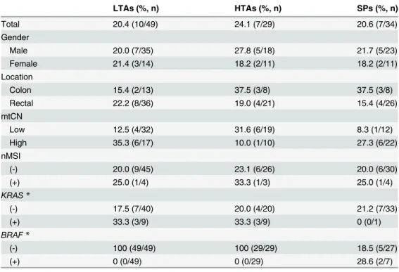
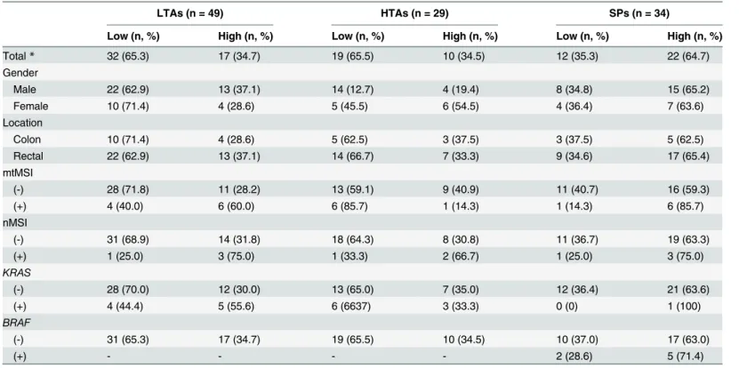
Molecular genetic analysis in Tubular Adenomas and Serrated Polyps The mtMSI results for 78 TAs and 34 SPs are presented in Table 1. In colorectal precancerous legions, mtMSI was found in 21.4% (24/112) of all lesions, and this was similar between TAs and SPs, regardless of their classification. mtMSI was not associated with any clinicopathologi- cal characteristic in both TAs and SPs. Direct sequencing results showed that mtMSI was found in only (C)n (D303 –310) and (CA)n (D514) regions of D-loops. Considering the molec- ular function of D-loops, mtMSI may be associated with mitochondrial content, as a result of mitochondrial replication. Accordingly, subsequent analysis of mtCN revealed significant asso- ciations in these legions (Table 2). Higher mtCN was found in SPs (64.7%, 22/34) than in TAs (34.6%, 27/78), a difference that was statistically significant (p = 0.003). Of note, mtMSI was observed in LTAs with higher mtCN (60.0% versus 28.2%), although this did not reach statisti- cal significance (p = 0.059). nMSI was found in 9.0% (7/78) of TAs and 11.8% (4/34) of SPs, re- spectively. BRAF mutation was found in only SPs (20.6%, 7/34), while a higher frequency of KRAS mutation was recorded in TAs (23.1%, 18/78) than in SPs (2.9%, 1/34). KRAS and BRAF mutations were mutually exclusive in TAs and SPs (p < 0.0001). mtMSI and mtCN were not

Table 1. Mitochondrial microsatellite instability (mtMSI) in colorectal precancerous legions.

LTAs (%, n) HTAs (%, n) SPs (%, n)

Total 20.4 (10/49) 24.1 (7/29) 20.6 (7/34)

Gender

Male 20.0 (7/35) 27.8 (5/18) 21.7 (5/23)

Female 21.4 (3/14) 18.2 (2/11) 18.2 (2/11)

Location

Colon 15.4 (2/13) 37.5 (3/8) 37.5 (3/8)

Rectal 22.2 (8/36) 19.0 (4/21) 15.4 (4/26)

mtCN

Low 12.5 (4/32) 31.6 (6/19) 8.3 (1/12)

High 35.3 (6/17) 10.0 (1/10) 27.3 (6/22)

nMSI

(-) 20.0 (9/45) 23.1 (6/26) 20.0 (6/30)

(+) 25.0 (1/4) 33.3 (1/3) 25.0 (1/4)

KRAS *

(-) 17.5 (7/40) 20.0 (4/20) 21.2 (7/33)

(+) 33.3 (3/9) 33.3 (3/9) 0 (0/1)

BRAF *

(-) 100 (49/49) 100 (29/29) 18.5 (5/27)

(+) 0 (0/49) 0 (0/29) 28.6 (2/7)

LTA, low grade of tubular adenoma; HTA, high grade of tubular adenoma; SP, serrated polyp.

* LTA and HTA versus SP, p < 0.001

doi:10.1371/journal.pone.0118612.t001

(5)

associated with any other clinicopathological characteristics or markers in both TAs and SPs, except for those mentioned above.

Molecular genetic analysis in Colorectal Cancers

The results of molecular genetic analysis in 100 CRCs are presented in Table 3. mtMSI was found in 30.0% (30/100) of CRCs. Most mtMSI was found in D-loops, and the distribution thereof was similar to that in the precursor lesions. Patients with mtMSI tended to show a lower frequency (15.4%) of KRAS mutations than those without mtMSI (35.1%), although this was not statistically significant (p = 0.06). Overall, mtMSI was not associated with any clinico- pathological characteristic in CRCs. However, nMSI, KRAS and BRAF mutations showed clini- copathological characteristics when stratifying for stage (data not shown). Briefly, in early stage CRCs, patients with mtMSI exhibited features of vascular invasion (p = 0.03). Additionally, nMSI was shown to be associated with male sex (p = 0.02) and vascular invasion (p = 0.05). In advanced stage CRCs, rectal cancers showed a higher frequency of KRAS mutation (p = 0.03);

meanwhile, BRAF mutation was related to female sex (p = 0.04) and less lymph node invasion (p = 0.05). When stratifying for stage, KRAS and BRAF mutations held no further clinicopatho- logical significance, except for the variables described above.

Survival Analysis in Colorectal Cancers

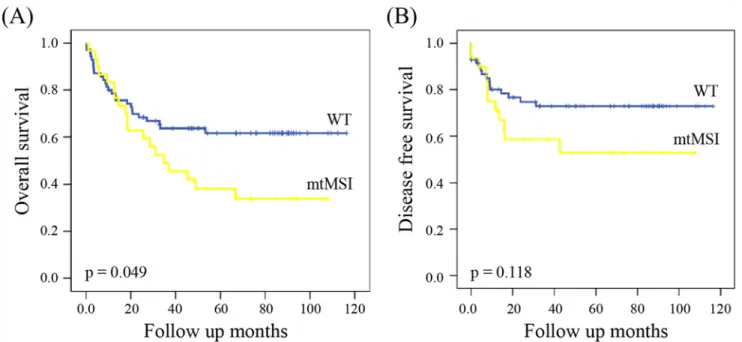
The median follow-up of patients for survival analysis was 47.5 months (1 –113). The results of the univariate analysis in CRCs are presented in S3 Table. Kaplan-Meier curve revealed mtMSI

Table 2. Mitochondrial copy number (mtCN) in colorectal precancerous legions.

LTAs (n = 49) HTAs (n = 29) SPs (n = 34)

Low (n, %) High (n, %) Low (n, %) High (n, %) Low (n, %) High (n, %)

Total * 32 (65.3) 17 (34.7) 19 (65.5) 10 (34.5) 12 (35.3) 22 (64.7)

Gender

Male 22 (62.9) 13 (37.1) 14 (12.7) 4 (19.4) 8 (34.8) 15 (65.2)

Female 10 (71.4) 4 (28.6) 5 (45.5) 6 (54.5) 4 (36.4) 7 (63.6)

Location

Colon 10 (71.4) 4 (28.6) 5 (62.5) 3 (37.5) 3 (37.5) 5 (62.5)

Rectal 22 (62.9) 13 (37.1) 14 (66.7) 7 (33.3) 9 (34.6) 17 (65.4)

mtMSI

(-) 28 (71.8) 11 (28.2) 13 (59.1) 9 (40.9) 11 (40.7) 16 (59.3)

(+) 4 (40.0) 6 (60.0) 6 (85.7) 1 (14.3) 1 (14.3) 6 (85.7)

nMSI

(-) 31 (68.9) 14 (31.8) 18 (64.3) 8 (30.8) 11 (36.7) 19 (63.3)

(+) 1 (25.0) 3 (75.0) 1 (33.3) 2 (66.7) 1 (25.0) 3 (75.0)

KRAS

(-) 28 (70.0) 12 (30.0) 13 (65.0) 7 (35.0) 12 (36.4) 21 (63.6)

(+) 4 (44.4) 5 (55.6) 6 (6637) 3 (33.3) 0 (0) 1 (100)

BRAF

(-) 31 (65.3) 17 (34.7) 19 (65.5) 10 (34.5) 10 (37.0) 17 (63.0)

(+) - - - - 2 (28.6) 5 (71.4)

LTA, low grade of tubular adenoma; HTA, high grade of tubular adenoma; SP, serrated polyp.

* LTA and HTA versus SP, p = 0.003

doi:10.1371/journal.pone.0118612.t002

(6)

Table 3. Clinicopathological Characteristics of Colorectal Cancers According to Genetic Status.

Total (n) mtMSI (n, %) nMSI (n, %) KRAS (n, %) BRAF (n, %)

Total 100 30 (30) 16 (16) 26 (26) 7 (7)

Gender P = 0.022 *

Male 65 18 (29.0) 14 (22.6) 14 (22.6) 2 (3.2)

Female 38 12 (31.6) 2 (5.3) 12 (31.6) 5 (13.2)

Location

Colon 41 14 (34.1) 5 (12.2) 8 (19.5) 5 (12.2)

Rectal 59 16 (27.1) 11 (18.6) 18 (30.5) 2 (3.4)

Stage

I,II (early) 44 11 (25.0) 5 (11.4) 8 (18.2) 4 (9.1)

III,IV (advanced) 56 19 (33.9) 11 (19.6) 18 (32.1) 3 (5.4)

T stage

T1 1 0 (0) 0 (0) 1 (100) 0 (0)

T2 16 6(37.5) 3 (18.7) 2 (12.5) 1 (6.2)

T3 74 20 (27.0) 11 (14.9) 21 (28.4) 6 (8.1)

T4 9 4 (44.4) 2 (22.2) 2 (22.2) 0 (0)

N stage

N0 47 13 (27.7) 5 (9.4) 8 (17.0) 5 (10.6)

N1 30 11 (36.7) 4 (13.3) 11 (36.7) 2 (6.7)

N2 23 6 (26.1) 7 (30.4) 7 (30.4) 0 (0)

M stage

M0 88 27 (30.7) 14 (15.7) 23 (25.8) 6 (6.7)

M1 10 2 (20.0) 2 (18.2) 3 (27.3) 1 (9.1)

Differentiation

Well/Moderate 91 27 (29.7) 13 (14.3) 26 (28.6) 6 (6.6)

Poor/Undifferentiated 9 3 (33.3) 3 (33.3) 0 (0) 1 (11.1)

Vascular invasion

(+) 66 22 (33.3) 14 (21.2) 19 (30.3) 5 (7.6)

(-) 34 8 (23.5) 2 (5.9) 7 (17.6) 2 (5.9)

mtMSI

(-) 70 - 14 (20.0) 22 (31.4) 5 (7.1)

(+) 30 - 2 (6.7) 4 (13.3) 2 (6.7)

nMSI

(-) 84 28 (33.3) - 20 (23.8) 7 (8.3)

(+) 16 2 (12.5) - 6 (37.5) 0 (0)

KRAS

(-) 74 26 (35.1) 10 (13.5) - 4 (5.4)

(+) 26 4 (15.4) 6 (23.1) - 3 (11.5)

BRAF

(-) 93 28 (30.1) 16 (17.2) 23 (24.7) -

(+) 7 2 (28.6) 0 (0) 3 (42.9) -

*p < 0.05

doi:10.1371/journal.pone.0118612.t003

(7)

to be a significant prognostic marker for OS but not DFS (Fig. 1). OS in patients with mtMSI was shorter than in those without mtMSI (HR 3.88; 95% CI, 0.25 –0.89; p = 0.049). When strati- fying for variables, mtMSI was shown to be a statistically significant prognostic marker for OS (S1 Fig.): mtMSI conferred a poor prognosis in colon cancer (HR 4.86; 95% CI, 0.15 –0.87;

p = 0.027). In CRCs without nMSI or BRAF mutation, mtMSI was also an accurate marker for predicting OS (HR 4.75; 95% CI, 0.24 –0.87; p = 0.029 without nMSI, HR 4.38; 95% CI, 0.23–

0.88; p = 0.036 without BRAF mutation).

Discussion

Colorectal cancer is considered a heterogeneous disease, as CRCs and their precursors exhibit distinct pathological features and molecular signatures. To identify colorectal carcinogenesis and potential markers for the prognosis thereof, multitier genetic approaches have been per- formed to investigate CRCs and their precursors [22–26]. Nevertheless, although mitochondri- al microsatellite instability has garnered focus as a cancer marker, no multitier study including mtMSI has been conducted in colorectal tumors [29–37]. Therefore, the present study at- tempted to investigate the clinicopathological characteristics and prognosis of CRCs with mtMSI for the first time.

In the present study, mtMSI was found in 30% of CRCs, which was similar to that reported in previous studies [37–40]. A recent study of a Korean population showed that mtMSI was found in over half (59%) of the patients [49]. This difference might be caused by different ex- perimental methods and sample selection. Notwithstanding, none of the previous studies, nor the present study, confirmed an association between mtMSI and the clinicopathological char- acteristics of CRCs. Instead, CRCs with mtMSI in the present study were shown to be associat- ed with shorter overall survival (OS), and this result was in agreement with the results reported by Lievre et al. [38]. The study about the prognosis of CRCs with mtMSI was rare and their re- sults were considered controversial until now. Previously, Chang et al. [41] reported that mtMSI had no significant impact on prognosis. Inversely, Tsai et al. [40] showed a better

Fig 1. Kaplan —Meier curves for overall survival (A) and disease free survival (B) of colorectal cancer patients according to mitochondrial microsatellite instability status.

doi:10.1371/journal.pone.0118612.g001

(8)

prognosis in Dukes stage C CRCs with mtMSI. Recent research in a Korean population re- vealed a higher frequency of mtMSI in tumors of larger size and of more advanced TNM stage, suggesting mtMSI as a risk factor for poor outcomes [49]. Additionally, other studies suggested that only nMSI and KRAS and BRAF mutations may be significant markers for prognosis in CRCs [19,20,47]. However, our results were not in agreement with these studies. Instead, we showed that combined analysis of mtMSI, nMSI, and BRAF mutation could accurately predict the prognosis of CRCs. Therefore, we cannot rule out the prognostic value of nMSI and BRAF mutations.

To clarify how mtMSI contributes to colorectal development and prognosis, we investigated genetic markers thereof in colorectal precancerous legions. A similar frequency of mtMSI was found in TAs and SPs, and mtMSI was not shown to be associated with their characteristics or with KRAS or BRAF mutations. Most mutations were found in the D-loop, a noncoding sequence of the mitochondrial genome important in mtDNA replication and transcription [30–34]. Within this region, (C)n repeat (D310 sequence) is an essential element for mtDNA replication because it contains the H-strand replication origin [50]. Accordingly, we investigat- ed mtCN in these legions, as well as the relationship between mtCN and mtMSI. The results re- vealed a higher mtCN in SPs than TAs; mtCN was not associated with other markers.

Interestingly, increased mtCN tended to be associated with mtMSI in LTAs, although this did not reach statistical significance (p = 0.059). These results indicated that mtMSI in LTA may

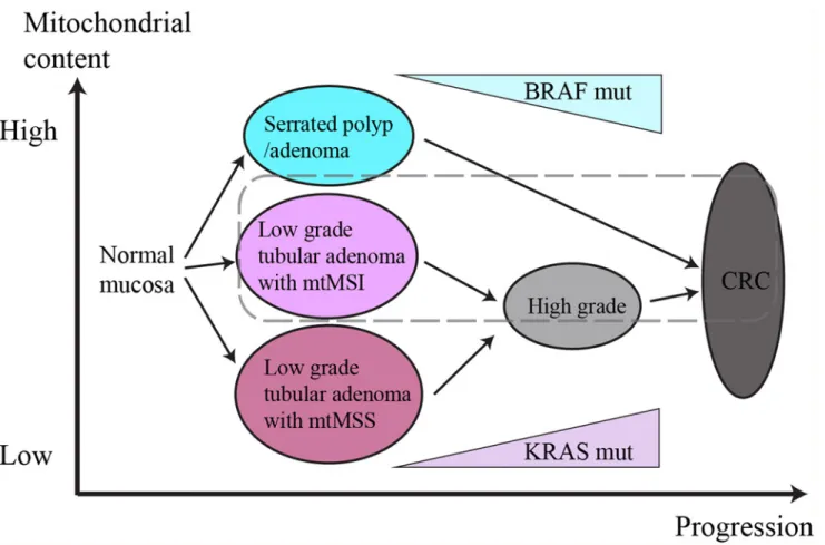
Fig 2. Schematic diagram of the role of mitochondrial DNA in colorectal carcinogenesis. MtMSI is involved in the progression of tubular adenomas as an early event. According to mtMSS and mtMSI, low grade tubular adenomas have different mitochondrial content. And tubular adenomas with mtMSI drive independent carcinogenesis pathway, resulting poor prognosis (dashed line).

doi:10.1371/journal.pone.0118612.g002

(9)

drive higher mitochondrial content, suggesting that mitochondrial DNA status may contribute to the tubular adenoma-carcinoma sequence more than the serrated pathway. In the present study, mtMSI was more significantly associated with the prognosis of CRCs without nMSI or BRAF mutation. This result supports our assertion about its involvement with tubular adenoma-carcinoma sequence, as nMSI and BRAF mutation are representative genetic mark- ers of the serrated pathway.

Lee et al. [32] suggested that mtMSI was associated with decreased mtCN in hepatocellular carcinoma. However, Guo et al. [51] reported recently that mtCN in specimens with mtMSI was significantly higher than in those without mtMSI in laryngeal squamous cell carcinoma.

Our study demonstrated increased mtCN in LTAs with mtMSI, and these data indicated that changes in mtCN as a result of mtMSI may differ from tumor to tumor. Based on these data, we deduced the role of mitochondrial DNA in colorectal carcinogenesis as presented in Fig. 2.

Accordingly, changes in mitochondrial DNA status may drive the progression course of colo- rectal mucosa to TA or SP and may also cause differences in prognosis. These data suggested a new paradigm for colorectal carcinogenesis; nevertheless, further study with a larger number of samples is needed.

In conclusion, this is the first study to suggest the role of mitochondrial DNA in colorectal carcinogenesis to the best of our knowledge. The data herein indicated that mtMSI is an early event in the tubular adenoma-carcinoma sequence, resulting in poor prognosis. In the era of personalized cancer therapy, further study of the characteristics of mitochondrial DNA in pre- cancerous legions may lead to better prognosis and treatment thereof, considering the impor- tance of mitochondria in anticancer drug development.

Supporting Information

S1 Table. Clinicopathological Characteristics of the Patients with Tubular Adenomas and Serrated Polyps.

(DOC)

S2 Table. Clinicopathological Characteristics of the Patients with Colorectal Cancers Ac- cording to Stage.

(DOCX)

S3 Table. Univariate Analysis for Overall Survival and Disease Free Survival of Patients with colorectal Cancer.

(DOCX)

S1 Fig. Kaplan —Meier curves for overall survival according to mitochondrial microsatellite instability status.

(DOC)

Acknowledgments

This study was supported by the research promoting grant from the Keimyung University Medical Research Promoting Grant.

Author Contributions

Conceived and designed the experiments: JHL DKK. Performed the experiments: JHL IH

YNK. Analyzed the data: JHL IH DKK. Contributed reagents/materials/analysis tools: YNK

IJC. Wrote the paper: JHL IJC DKK.

(10)

References

1. Parkin DM, Bray F, Ferlay J, Pisani P (2005) Global cancer statistics, 2002. CA Cancer J Clin 55:

74 –108. PMID: 15761078

2. Shim JI, Kim Y, Han MA, Lee HY, Choi KS, Jun JK, et al. (2010) Results of colorectal cancer screening of the national cancer screening program in Korea, 2008. Cancer Res Treat 42: 191 –198. doi: 10.

4143/crt.2010.42.4.191 PMID: 21253320

3. Fearon ER, Vogelstein B (1990) A genetic model for colorectal tumorigenesis. Cell 61: 759 –767.

PMID: 2188735

4. Peinado MA, Malkhosyan S, Velazquez A, Perucho M (1992) Isolation and characterization of allelic losses and gains in colorectal tumors by arbitrarily primed polymerase chain reaction. Proc Natl Acad Sci U S A 89: 10065 –10069. PMID: 1359533

5. Kern SE, Fearon ER, Tersmette KW, Enterline JP, Leppert M, Nakamura Y, et al. (1989) Clinical and pathological associations with allelic loss in colorectal carcinoma [corrected]. JAMA 261: 3099 –3103.

PMID: 2654431

6. Powell SM, Zilz N, Beazer-Barclay Y, Bryan TM, Hamilton SR, Thibodeau SN, et al. (1992) APC muta- tions occur early during colorectal tumorigenesis. Nature 359: 235 –237. PMID: 1528264

7. Forrester K, Almoguera C, Hank K, Grizzle WE, Perucho M (1987) Detection of high incidence of K-ras oncogenes during human colon tumorigenesis. Nature 327: 298 –303. PMID: 2438556

8. Bos JL, Fearon ER, Hamilton SR, Verlaan-de Vries M, van Boom JH, van der Eb AJ, et al. (1987) Prev- alence of ras gene mutations in human colorectal cancers. Nature 327: 293 –297. PMID: 3587348 9. Thiagalingam S, Lengauer C, Leach FS, Schutte M, Hahn SA, Overhauser J, et al. (1996) Evaluation of

candidate tumor suppressor genes on chromosome 18 in colorectal cancers. Nat Genet 13: 343 –346.

PMID: 8673134

10. Baker SJ, Fearon ER, Nigro JM, Hamilton SR, Preisinger AC, Jessup JM, et al. (1989) Chromosome 17 deletions and p53 gene mutations in colorectal carcinomas. Science 244: 217 –221. PMID: 2649981 11. Greenblatt MS, Bennett WP, Hollstein M, Harris CC (1994) Mutations in the p53 tumor suppressor

gene: clues to cancer etiology and molecular pathogenesis. Cancer Res 54: 4855 –4878. PMID:

8069852

12. Fishel R, Lescoe MK, Rao MR, Copeland NG, Jenkins NA, Garber J, et al. (1993) The human mutator gene homolog MSH2 and its association with hereditary nonpolyposis colon cancer. Cell 75: 1027 –1038.

PMID: 8252616

13. Nicolaides NC, Papadopoulos N, Liu B, Wei YF, Carter KC, Ruben SM, et al. (1994) Mutation of two PMS homologues in hereditary nonpolyposis colon cancer. Nature 371: 75 –80. PMID: 8072530 14. Bronner CE, Baker SM, Morrison PT, Warren G, Smith LG, Lescoe MK, et al. Mutation in the DNA mis-

match repair gene homologue hMLH1 is associated with hereditary non-polyposis colon cancer. Nature 1994; 368:258 –61. PMID: 8145827

15. Cunningham JM, Christensen ER, Tester DJ, Kim CY, Roche PC, Burgart LJ, et al. (1998) Hypermethy- lation of the hMLH1 promoter in colon cancer with microsatellite instability. Cancer Res 58: 3455 –3460.

PMID: 9699680

16. Boland CR, Thibodeau SN, Hamilton SR, Sidransky D, Eshleman JR, Burt RW, et al. (1998) A National Cancer Institute Workshop on Microsatellite Instability for cancer detection and familial predisposition:

Development of international criteria for the determination of microsatellite instability in colorectal can- cer. Cancer Res 58: 5248 –5257. PMID: 9823339

17. Popat S, Hubner R, Houlston RS (2005) Systematic review of microsatellite instability and colorectal cancer prognosis. J Clin Oncol 23: 609 –618. PMID: 15659508

18. Söreide K, Janssen EA, Söiland H, Körner H, Baak JP (2006) Microsatellite instability in colorectal can- cer. Br J Surg 93: 395 –406. PMID: 16555243

19. Samowitz WS, Sweeney C, Herrick J, Albertsen H, Levin TR, Murtaugh MA, et al. (2005) Poor survival associated with the BRAF V600E mutation in microsatellite-stable colon cancers. Cancer Res 65:

6063 –6069. PMID: 16024606

20. Walther A, Johnstone E, Swanton C, Midgley R, Tomlinson I, Kerr D (2009) Genetic prognostic and pre- dictive markers in colorectal cancer. Nat Rev Cancer 9: 489 –499. doi: 10.1038/nrc2645 PMID:

19536109

21. Jass JR (1999) Serrated adenoma and colorectal cancer. J Pathol 187: 499 –502. PMID: 10398112 22. Jass JR, Whitehall VL, Young J, Leggett BA (2002) Emerging concepts in colorectal neoplasia. Gastro-

enterology 123: 862 –876. PMID: 12198712

23. Hawkins NJ, Bariol C, Ward RL (2002) The serrated neoplasia pathway. Pathology 34: 548 –555.

PMID: 12555993

(11)

24. Goldstein NS, Bhanot P, Odish E, Hunter S (2003) Hyperplastic-like colon polyps that preceded micro- satellite-unstable adenocarcinomas. Am J Clin Pathol 119: 778 –796. PMID: 12817424

25. Spring KJ, Zhao ZZ, Karamatic R, Walsh MD, Whitehall VL, Pike T, et al. (2006) High prevalence of sessile serrated adenomas with BRAF mutations: a prospective study of patients undergoing colonos- copy. Gastroenterology 131: 1400 –1407. PMID: 17101316

26. Jass JR, Baker K, Zlobec I, Higuchi T, Barker M, Buchanan D, et al. (2006) Advanced colorectal polyps with the molecular and morphological features of serrated polyps and adenomas: concept of a ‘ fusion’

pathway to colorectal cancer. Histopathology 49: 121 –131. PMID: 16879389

27. Howell N, Kubacka I, Mackey DA (1996) How rapidly does the human mitochondrial genome evolve?

Am J Hum Genet 59: 501 –509. PMID: 8751850

28. Pääbo S (1996) Mutational hot spots in the mitochondrial microsome. Am J Hum Genet 59: 493 –496.

PMID: 8751848

29. Richard SM, Bailliet G, Páez GL, Bianchi MS, Peltomäki P, et al. (2000) Nuclear and mitochondrial ge- nome instability in human breast cancer. Cancer Res 60: 4231 –4237. PMID: 10945635

30. Bianchi NO, Bianchi MS, Richard SM (2001) Mitochondrial genome instability in human cancers. Mutat Res 488: 9 –23. PMID: 11223402

31. Wang Y, Liu VW, Ngan HY, Nagley P (2005) Frequent occurrence of mitochondrial microsatellite insta- bility in the D-loop region of human cancers. Ann N Y Acad Sci 1042: 123 –129. PMID: 15965053 32. Lee HC, Li SH, Lin JC, Wu CC, Yeh DC, Wei YH (2004) Somatic mutations in the D-loop and decrease

in the copy number of mitochondrial DNA in human hepatocellular carcinoma. Mutat Res 547: 71 –78.

PMID: 15013701

33. Yin PH, Lee HC, Chau GY, Wu YT, Li SH, Lui WY, et al. (2004) Alteration of the copy number and dele- tion of mitochondrial DNA in human hepatocellular carcinoma. Br J Cancer 90: 2390 –2396. PMID:

15150555

34. Wu CW, Yin PH, Hung WY, Li AF, Li SH, Chi CW, et al. (2005) Mitochondrial DNA mutations and mito- chondrial DNA depletion in gastric cancer. Genes Chromosomes Cancer 44: 19 –28. PMID: 15892105 35. Tseng LM, Yin PH, Chi CW, Hsu CY, Wu CW, Lee LM, et al. (2006) Mitochondrial DNA mutations and

mitochondrial DNA depletion in breast cancer. Genes Chromosomes Cancer 45: 629 –638. PMID:

16568452

36. Habano W, Nakamura S, Sugai T (1998) Microsatellite instability in the mitochondrial DNA of colorectal carcinomas: evidence for mismatch repair systems in mitochondrial genome. Oncogene 17: 1931 – 1937. PMID: 9788436

37. Polyak K, Li Y, Zhu H, Legnauer C, Wilson JK, Markowitz SD, et al. (1998) Somatic mutations of the mi- tochondrial genome in human colorectal tumours. Nat Genet 20: 291 –293. PMID: 9806551

38. Li èvre A, Chapusot C, Bouvier AM, Zinzindohoue F, Piard F, Roignot P, et al. (2005) Clinical value of mitochondrial mutations in colorectal cancer. J Clin Oncol 23: 3517 –3525. PMID: 15908662 39. Pinheiro M, Veiga I, Pinto C, Afonso L, Sousa O, Fragoso M, et al. (2009) Mitochondrial genome alter-

ations in rectal and sigmoid carcinomas. Cancer Lett 280: 38 –43. doi: 10.1016/j.canlet.2009.02.009 PMID: 19269084

40. Tsai MH, Fang WH, Lin SW, Yen SJ, Chou SJ, Yang YC (2009) Mitochondrial Genomic Instability in Colorectal Cancer: No Correlation to Nuclear Microsatellite Instability and Allelic Deletion of hMSH2, hMLH1, and p53 Genes, but Prediction of Better Survival for Dukes ’ Stage C Disease. Ann Surg Oncol 16: 2918 –2925. doi: 10.1245/s10434-009-0581-7 PMID: 19582509

41. Chang SC, Lin PC, Yang SH, Wang HS, Liang WY, Lin JK (2009) Mitochondrial D-loop mutation is a common event in colorectal cancers with p53 mutations. Int J Colorectal Dis 24: 623 –628. doi: 10.

1007/s00384-009-0663-9 PMID: 19198855

42. Feng S, Xiong L, Ji Z, Cheng W, Yang H (2011) Correlation between increased copy number of mito- chondrial DNA and clinicopathological stage in colorectal cancer. Oncol Lett 2: 899 –903. PMID:

22866147

43. Jeong CW, Lee JH, Sohn SS, Ryu SW, Kim DK (2010) Mitochondrial microsatellite instability in gastric cancer and gastric epithelial dysplasia as a precancerous lesion. Cancer Epidemiol 34: 323 –327. doi:

10.1016/j.canep.2010.03.015 PMID: 20409774

44. Lee JH, Ryu TY, Cho CH, Kim DK (2011) Different characteristics of mitochondrial microsatellite insta- bility between uterine leiomyomas and leiomyosarcomas. Pathol Oncol Res 17: 201 –205. doi: 10.

1007/s12253-010-9297-z PMID: 20853077

45. Brennetot C, Buhard O, Jourdan F, Flejou JF, Duval A, Hamelin R (2005) Mononucleotide repeats

BAT-26 and BAT-25 accurately detect MSI-H tumors and predict tumor content: implications for popula-

tion screening. Int J Cancer 113: 446 –450. PMID: 15455342

(12)

46. O ’Brien MJ, Yang S, Mack C, Xu H, Huang CS, Mulcahy E, et al. (2006) Comparison of microsatellite in- stability, CpG island methylation phenotype, BRAF and KRAS status in serrated polyps and traditional adenomas indicates separate pathways to distinct colorectal carcinoma end points. Am J Surg Pathol 30: 1491 –1501. PMID: 17122504

47. Baldus SE, Schaefer KL, Engers R, Hartleb D, Stoecklein NH, Gabbert HE (2010) Prevalence and het- erogeneity of KRAS, BRAF, and PIK3CA mutations in primary colorectal adenocarcinomas and their corresponding metastases. Clin Cancer Res 16: 790 –799. doi: 10.1158/1078-0432.CCR-09-2446 PMID: 20103678

48. Chen T, He J, Shen L, Fang H, Nie H, Jin T, et al. (2011) The mitochondrial DNA 4,977-bp deletion and its implication in copy number alteration in colorectal cancer. BMC Med Genet 12: 8. doi: 10.1186/

1471-2350-12-8 PMID: 21232124

49. Lim SW, Kim HR, Kim HY, Huh JW, Kim YJ, Shin JH,et al. (2012) High-frequency minisatellite instability of the mitochondrial genome in colorectal cancer tissue associated with clinicopathological values. Int J Cancer 131: 1332 –1341. doi: 10.1002/ijc.27375 PMID: 22120612

50. Lee HC, Wei YH (2009) Mitochondrial DNA instability and metabolic shift in human cancers. Int J Mol Sci 10: 674 –701. doi: 10.3390/ijms10020674 PMID: 19333428

51. Guo W, Yang D, Xu H, Zhang Y, Huang J, Yang Z, et al. (2013) Mutations in the D-loop region and in-

creased copy number of mitochondrial DNA in human laryngeal squamous cell carcinoma. Mol Biol

Rep 40: 13 –20. doi: 10.1007/s11033-012-1939-7 PMID: 23114912

수치

Table 1. Mitochondrial microsatellite instability (mtMSI) in colorectal precancerous legions.
Table 2. Mitochondrial copy number (mtCN) in colorectal precancerous legions.
Table 3. Clinicopathological Characteristics of Colorectal Cancers According to Genetic Status.
Fig 1. Kaplan —Meier curves for overall survival (A) and disease free survival (B) of colorectal cancer patients according to mitochondrial microsatellite instability status.
+2

참조

관련 문서

To examine the characteristics of its cutting edge, the material was trimmed to make a rectilinear cutting edge, and in addition to the cutting edge of the

In this study, water flow system of Danghang Bay, which has the narrow and long topographical characteristics with the narrow bay mouth and its flow

GDP impact of COVID-19 spread, public health response, and economic policies. Virus spread and public

Chapter 3 proposes the educational values and significance of chalk art through viewing its characteristics in public interest and comparing the example

We determined the nucleotide sequences of the mitochondrial DNA (mtDNA) control region using cloning and sequencing, and obtained the complete sequence from the cattle bones

Seed germination, cutting propagation and early growth characteristics were investigated in order to develop the method of cutting propagation and to find

In this study, we found that Pin1 plays a pivotal role in insulin- induced AP-1 activation through its interaction with p70S6K and prolongs activity of

The aim of this study was to determine the serum levels of the prohormone form of hepcidin, pro-hepcidin, and its relations with iron status and its