약학회 지 제 49 권 제 2 호 128~133 (2005)
Yakhak HoejiVol. 49,No. 2 稱學合職
Diclofenac에 의해 유발된 장내세균전위오ᅡ 지질과산화에 대한 글루타민의 효과
김은정 • 김정욱*’#
숙명여자대학교 약학대학, *중앙대학교 의과대학내과학교실 (Received October 23,2004; Revised December 9,2004)
Effect of Glutamine on the Diclofenac Induced Bacterial Translocation and Lipid Peroxidation
Eun Jeong K im and Jeong Wook K im *,#
College of Pharmacy, Sookmyung Women's University, Seoul 140-742,Korea
^Department of Internal Medicine, Chungang University of College of Medicine, Seoul 140-757,Korea
Abstract — The aim of this study was to examine whether administration of glutamine are able to prevent the NSAID induced bacterial translocation and lipid peroxidation in the rats. The animals with glutamine were fed with L-glutamine for 5 days before diclofenac administration (100 mg/kg orally). 48 hour after diclofenac administration, intestinal permeability, serum biochemical profiles, and malondialdehyde levels of ileum were measured for evaluation of gut damage. Also, enteric aerobic bacterial counts, number of gram-negatives in mesenteric lymph nodes, liver, spleen and kidney and malond
ialdehyde levels in liver, spleen, kidney and plasma were measured. Diclofenac caused the gut damage, enteric bacterial overgrowth, increased bacterial translocation and increased lipid peroxidation. Co-administration of glutamine reduced the gut damage, enteric bacterial overgrowth, bacterial translocation and lipid peroxidation induced by diclofenac. This study suggested that glutamine might effectively prevent non-steroidal anti-inflammatory drug induced bacterial translocation and lipid peroxidation in the rat.
Keywords □ anti-inflammatory agents, non-steroidal, bacterial translocation, lipid peroxidation, glutamine
비스테로이드성 힝■염제 (nonsteroidal anti-inflammatory drugs) 는 전세계적으로흔히 시용되고있는약물중의 하나로골관절 질환및통중의 치료, 허혈성심질환과뇌질환의 예방, 해열등의
목적으로사용되고있는약제이다.1> 그러나이와 같은효과에도
불구하고비스테로이드성 항염제에 의한부작용으로약물시용 에제한을받는경우가흔히 있다. 이중소화기계에서 발생하는 부작용이 가장흔하며2,3) 장관장벽의손상과장내세균과중식에 의한 장내세균전위를유발하기도한다. 비스테로이드성 항염제 에의한장내세균전위는비대상성 심부전환자에서 그중요성 이강조되고있는데면역반응을촉진하여심부전악화께의한입
원기간의 증대를유발하는 것으로예상되고있다.4’5) 또한복부
수술을받는 환자에서수술전비스테로이드성 항염제의투여는 수술중및수술후장내세균전위를유발한다.6) 이와같은장내 세균전위는유발원인과상관없이 병원내감염과패혈증이나전
#본 논문에 관한 문의는 저자에게로 (전화) 02-748-9941 (팩스) 02-3785-0160 (E-mail) [email protected]
신성염증중후군과동반되는다발성 장기부전의 주요발생 기전 중의하나로 제시되고있다.71
비스테로이드성 항염제는장관손상이외에도 간손상및신장 손상을유발하기도하는데이와같은상기약제에의한장관및간,
신장과같은장기의 손상에는산화적스트레스가관여한다.8 또한산화적 스트레스는장내세균전위와더불어 패혈중이나전 신성염중증후군과동반되는다발성장기부전의유발에관여하므 로 1U 상기질환과같은중증의 질환자에서사용되는비스테로이 드성 항염제는 장내세균전위와산화적스트레스를중가시켜환 자의상태를악화시킬수있다. 또한다른병태를이용한연구에 서산화적 스트레스가장관손상및장내세균전위가산화적스트
레스, 특히 지질과산화와연관성이 있다고보고하였다.12~14) 글루타민은소장및대장의상피세포와면역기능과연관된세 포들에서시용되는주요물질중에하나로장관점막세포의 중식 을유발하며 여러 병태모델에서장관손상을 방지하고장내세균
전위의 억제및예방효과가보고되었다;15’16) 또한글루타민은
패혈증에 의하여유발된 장관에서의산화적 스트레스를감소시 키고17) 화상에서의 산화적스트레스를감소시켜 생존율을중가
128
시킨다.18) 그러나 비스테로이드성 항염제에 의한 장내세균전위 와 산화적 스트레스에 대한 글루타민의 효과는 아직 입증된 바 가 없으며 다만 실험동물에서 장관의 손상을 감소시키며,19) 임 상연구에서 indomethadn 투여에 의한 장관장벽손상에 의한 장 투과성의 증가를 글루타민이 억제할 수 있는 것으로 보고 되고 있다.20)
이에 본 연구에서는 비스테로이드성 항염제에 의한 장내세균 전위 및 지질과산화 유발 백서에서 글루타민의 효과를 알아보았다.
실험 방법
대상
7주령의 체중 180-210 gm 정도의 수컷 Sprague-Dawley 백 서 32마리를 오리엔트(Orient Co., Ltd., Seoul, Korea)에서 공급 받아 실험 전 7일간 적응시켰다. 백서의 사육 시 12시간 간격으 로 낮과 밤을 구별하고 사육 온도는 22±10C로 유지하였으며 고 형사료인 Basal diet 5755(PMI Nutrition International, Inc., Richimond, California, USA)와 물을 자유롭게 섭취하게 하였다.
백서들은 실험 시작 전 감염이나 기타 이상소견이 관찰되지 않 았다. 백서들은 모두 4군으로 나누었으며 대조군, diclofenac 투 여군,diclofenac 및 글루타민 0.5 g/kg/day 투여군, diclofenac 및 글루타민 1.0g/kg/day 투여군으로 하였으며 각 군 당 백서 8마 리를 배정하였다.
Diclofenac에 의한 장손상 유발 및 글루타민의 투여
매일 백서의 물과 사료의 섭취량과 몸무게의 변화를 측정하였 다. 밑망을 설치하여 배설물이나 깔집 등 음식물이 아닌 것을 섭 취하는 것을 방지하였다. 투여용량 증가에 따른 글루타민의 효 과를 관찰하기 위해 글루타민 투여군은 diclofeanc 투여 시작 5 일 전부터 L-glutamine(Daesang, Co., Ltd., Seoul, Korea)을 각 각 0.5 및 1.0g/kg/day의 용량으로 일일 2차례 금속 경구 투여 관을 이용하여 경구투여 하였으며 대조군과 diclofenac 투여군은 대조약물로 생리식염수를 같은 방법으로 경구투여 하였다. 장관 손상과 장내세균전위에 대한 동물실험들에서 대부분 글루타민을 병태 유발 3일 이전부터 f 며해야 효과가 있었으며 1.0g/kg/day 이하의 용량으로 충분한 효과가 보고 되었다. 비스테로이드성 항 염 제에 의한 장관손상은 diclofenac sodium(Sigma Chemical Co., St. Louis, MO, USA)을 100 mg/kg 용량으로 일회 경구투 여 하여 유발하였다.
장투과성의 측정
Diclofenac 투여 48시간 후에 10 mg의 phenolsulfonphthalein (PSR Sigma Chemical Co., St. Louis, MO, USA)을 백서에 경 구 투여 후 대사케이지에서 PSP 투여 2시간 후부터 24시간 동
안 소변을 수집하였다. 채집된 소변에서의 PSP 농도를 구하기 위하여 중류수로 부피를 일정하게 한 후 2,500 rpm, 10분간 원 심분리 하였다. 상등액 lm /에 10% NaOH 5 m/로 알칼리화 시 킨 후 560 nm의 파장에서 분광광도계 (Smartspect 300,Biorad, Hercules, CA, USA)로 측정하여21) 회수된 PSP양을 백분율•로 표 시하였다.
장투과성 검사를 위한 24시간 소변 채취 후 백서는 ketamine hydrochloride 80 mg/kg 및 xylazine hydrochloride 8 mg/kg를 근주하여 마취한 후에 무균조작으로 복부를 정중앙에서 절개하 였다. 절개 직후 맹장으로부터 상방 40 cm의 회장에서 장 내용 물을 100mg을 채취하였다. 이후 횡경막을 절개하고 심장에서 혈액을 채취하여 백서를 사망시켰다. 채취한 혈액에서 혈액검•사 와 지질과산화 측정을 위하여 혈청과 혈장을 분리하였다. 장내 세균전위 유무를 알아보기 위하여 무균조작으로 장간막 림프절, 간, 비장 및 신장 100 mg을 채취하고 지질과산화 측정을 위하여 간, 비장 및 신장을 500 mg 각각 이상 채취하였다. 맹장의 장내 세균수를 측정하기 위하여 맹장을 1 cm 정도 절개한 후 대변 100 mg을 채취하였다.
장내 호기성 세균수 및 장내세균전위에 대한 검사
회장 및 맹장에서 획득한 장내용물 100mg을 연속적으로 희 석한 후 장내 호기성균수를 측정하기 위하여 혈액한천배지와 장 내 그람 음성균수를 측정하기 위하여 MacConkey 한천배지에 접 종하여 배양기에서 37°C로 24시간 동안 배양하였다. 배양 후 각 배지에서 자란 집락 수를 관찰하여 CFU(colony-forming unit)/g 으로 표시하였다. 장내세균전위를 알아보기 위하여 장간막림프 절, 간,비장 및 신장 100 mg을 소독된 Ten Broeck 조직파쇄기 를 이용하여 균질화한 후 연속적으로 희석하고 그람 음성균수를 측정하기 위하며 MacConkey 한천배지에 접종하여 배양기에서 37°C로 24시간 동안 배양하였다. 배양 후 각 배지에서 자란 집 락 수를 관찰하여 CFU(colony-forming unit)/g로 표시하였다.
지질과산화측정
간, 비장,신장 및 소장의 각 조직을 절단하여 무게를 잰 후 10배의 0.1 M phosphate buffer를 가한 후 Ten Broeck 조직 파 쇄기를 이용하여 분쇄하여 조직 균질액을 제조하였다. 조직 및 혈장 중 과산화지질은 TBA를 이용한 비색법으로 측정하였다.22) 각 조직 균질액 및 혈장 lm /에 TBA 시액 2m/를 혼합한 후 100oC의 항온조에서 15분간 가열하여 발색시켰다. 반응액을 3000 rpm, 15분간 원심분리한 후 상등액을 분광광도계로 535nm에서 흡광도를 측정히여 MDA(malondialdehyde) 양올 구하였다. MDA 표준물질로 1,1,3,3,-tetramethoxy propane을: 시용하였으며 MDA
Vol. 49, No. 2’ 2005
130 김은정 • 김정욱
양은 nmol/mg protein으로 표현하고 간, 비장, 신장 및 소장의 단백질양은 bovine serum albumin을 표준으로 하여 Bradford 법23>을 이용하여 측정하였다.
혈액 검사
장손상에 따른 장관에서의 단백소실을 측정하기 위하여 백서 에서 채취한 혈액으로 총단백, 알부민 수치를 측정하고 총단백 및 알부민 수치 변화가 diclofenac의 간독성에 의한 것이 아님을 중명하기 위하여 ALT, AST 및 총 빌리루빈을 동시에 측정하며 비교하였다.
Table I - Body weight changes and amounts of food Intakes in the animals
Trial population Body weight change (%/7 days)
Chow intakes (g/100 g BW/7 days)
Protein intakes (g/100 g BW/7 days)
Calorie intakes (cal/100 g BW/7 days) Control
NSAID GLN 0.5 GLN 1.0
27.19±5.99 6.09±6.62*
5.28±5.13*
5.28 ±7.92*
69.83 ±7.01 50.53±4.21*
51.43±6.49*
50.59±7.98*
13.62 ±1.37 10.04±1.02*
10.47±1.19*
10.62 ±1.50*
284.94±28.60 209.42 ±20.46*
216.45 ±25.48*
216.48±32.90*
Body weight change was expressed in the percent weight change for 7 days based on the body weight at operation.
BW, body weight; Control, control group; NSAID, group with diclofenac; GLN 0.5, group with diclofenac and glutamine 0.5 g/kg/day;
GLN 1.0,group with diclofenac and glutamine 1.0 g/kg/day.
*/>< 0.001 compared with control.
통계 처리
모든 수치는 평균±표준편차로 표시하고 각 군 간의 차이는 SPSS 11.0 프로그램 (SPSS Inc, Chicago, IL, USA)을 이용하여 Mann-Whitney 검정법..0■로 p value가 0.05 이하인 경우를 유의 성이 있는 것으로 판정하였다.
결과 및 고찰
장관 손상
본 연구에서는 장관손상을 알아보기 위하여 장투과성 검사와 장관에서의 지질과산화 측정 뿐 만 아니라 백서의 식이량과 체 중변화와 더불어 혈청 내 단백과 알부민 수치를 측정하였다. 실 험동물에서 비스테로이드성 항염제에 의한 장관손상 시 체중변 화가 없으며 식이량이 일정하다면 간기능 수치의 변화 없이 변 화하는 혈청 내의 단백과 알부민 수치는 비스테로이드성 항염제 에 의한 장관에서의 단백소실을 대변한다.24)
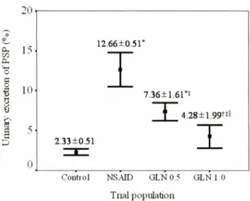
장관장벽의 손상을 측정하기 위한 PSP를 이용한 장투과성 검 사에서는 대조군보다 diclofeanc을 단독 투여한 군에서 4배 이상 중가소견을 보였으며여<0.001), 글루타민의 투여에 의해 diclofenac에 의한 장투과성의 중가가 감소하였다(^<0.001)(Fig.
1). Diclofenac 투여에 의한 소장에서의 지질과산화의 중가도 같 은 잉상을 보였다여<0.05)(Table V).
백서의 7일간 체중변화와 섭취한 식이,열량 및 단백질의 양
0
Control NSAID G LN 0.5 GLN 1 0
Tnal population
Fig. 1 - The Changes of intestinal permeability measured by 24 hour urinary excretion of PSP (phenolsulfonphthalein).
Values are means ±SD. Control, control group; NSAID, group with diclofenac; GLN 0.5, group with diclofenac and glutamine 0.5 g/kg/day; GLN 1.0, group with diclofenac and glutamine 1.0 g/kg/day. */><0.001 and V<0.01 compared with control. $<0.001 compared with NSAID. s/><0.01 compared with GLN 0.5.
은 대조군보다 diclofeanc을' 단독 투여한 군에서 적었으며 글루 타민을 병합투여한 군에서도 대조군보다 적었으나여<0.001) diclofeanc을 단독 투여한 군과 차이를 보이지 않았다(Table I).
혈액검사세서는 diclofenac 투여군에서 AST, ALT 및 총빌리루빈 수치의 중가소견 없이 대조군보다 혈청의 총단백 및 알부민 수 치가 감소하였고여<0.001),글루타민을 투여한 군에서는 diclofenac을 단독 투여한 군에 비해 저단백혈증 개선여<0.05)과 저알부민혈중 개선여<0.01)이 관찰되었다(Table II). 이는 diclofenac에 의한 장관손상으로 유발된 장관에서의 단백소실이 글루타민의 투여에 의하며 개선됨을 의미한다.
이와 같이 diclofenac에 의한 장관손상으로 발생한 장투과성 및 장관에서의 지질과산화의 중가와 장관에서의 단백소실은 글 루타민의 투여에 의해 감소되었으며 글루타민의 투여는 비스테 로이드성 항염제에 의한 장관손상을 감소시킴을 알 수 있다.
12.66 + 0.51*
7.36土1.61*1
4.28土1.99벼 2.33+0.51
- T - I
0505그
1
1
1%
) d
ds G,
UOTP.IOB
.
^ 3 5
/. Pharm. Soc. Korea
Table II - Serum biochemical findings 48 hours after diclofenac administration
Trial population Total protein (g/d/) Albumin (g/d/) ALT (IU//) AST (IU//) Total bilirubin (mg/d/)
Control 5.82 ±0.30 3.67±0.34 26.7±2.9 108.4±14.5 0.25±0.11
NSAID 4.57±0.32* 2.55±0.25* 25.1 ±4.5 101.6±13.2 0.23 ±0.07
GLN 0.5 5.03±0.42*§ 2.98±0.23n 22.7±7.8 99.8±36.4 0.28±0.07
GLN 1.0 5.17±0.53^ 3.05±0.38n 26.8±7.1 103.0 ±34.8 0.28±0.04
ALT, alanine aminotransferase; AST, aspartage aminotransferase; Control, control group; NSAID, group with diclofenac; GLN 0.5,group with diclofenac and glutamine 0.5 g/kg/day; GLN 1.0, group with diclofenac and glutamine 1.0 g/kg/day
> < 0.001 and 〈ᄋ.아 compared with control.
+/><0.01 and §^< 0.05 compared with NSAID.
장내세균 고®식 및 장내세균전위
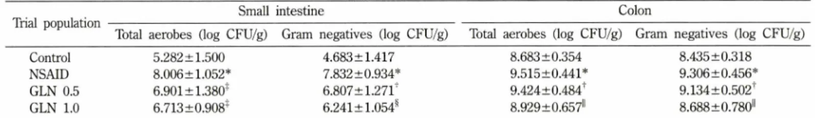
비스테로이드성 항염제는 장관손상에 의한 장투과성의 중가뿐 만 아니라 장내세균의 과증식도 유발하는데25) 본 연구에서도 diclofenac 단독 투여군에서 대조군보다 소장 및 대장에서 호기 성균 및 그람음성균의 과증식이 관찰되었다여<0.001). 글루타민 을 투여한 군에서는 소장의 그람음성균의 과증식이 감소되었고 여<0.01), 대장의 호기성 균 및 그람음성균의 과증식이 개선되는 양상을 보였다여<0.05)(Table III). 글루타민의 비스테로이드성 항염제에 의한 장내세균 과증식에 대한 효과는 현재까지의 비스 테로이드성 항염제에 의한 장손상 연구에서는 보고 되지 않았으 며 장관손상 및 장내세균전위가 유발되는 다른 병태모델의 일부 에서 글루타민이 장내세균의 과증식을 억제하였다.26) 하지만 글 루타민은 장관의 점막세포 뿐만 아니라 면역과 연관된 세포의 증 식을 촉진하여 GALT(gut associated lymphoid tissue)의 훨성화 로 장관의 면역력을 강화시키므로 장내세균의 과증식을 억제할 수 있다.275
장내세균전위를 관찰하기 위한 간,비장 및 신장에서의 그람 음성 세균수의 측정결과 diclofenac 투여군에서 대조군보다 장간 막 림프절, 간,비장 및 신장에서 그람음성 세균수의 증기를 관 찰 할 수 있었다^)<0.001). 글루타민 투여군에서는 diclofenac 단 독 투여군보다 장간막 림프절,간, 비장 및 신장에서 그람음성 세 균수가 감소하였는데, 간에서는 글루타민 투여 용량의 증가에 따 라 그람음성 세균수가 감소하였으며여<0.05) 장간막 림프절에서 는 0.5g/kg의 투여량에도 MDA가 감소하였으나여<0.01) 1.0 g/
kg 투여군 과는 차이가 없었다. 이에 비해 비장에서는 0.5g/kg 의 투여량에서는 효과가 없었으나 1.0g/kg 투여시 diclofenac 단 독 투여군보다 그람음성 세균수가 감소하였으며여<0.01),신장 에서도 같은 양상이 관찰되었다여<0.05)(Table IV). 이와 같이 비스테로이드성 항염제에 의한 장관손상과 장내세균의 과증식을 억제하는 글루타민은 상기 약제에 의한 장내세균전위를 억제하 였다. 하지만 투여 용량에 따른 글루타민의 장내세균전위에 대 한 효과는 각 장기에서 다양하게 관찰되며 이에 대한 추가적인
Table III - Changes of the number of enteric bacterial numbers in the small intestine and colon
Trial population Small intestine Colon
Total aerobes (log CFU/g) Gram negatives (log CFU/g) Total aerobes (log CFU/g) Gram negatives (log CFU/g)
Control 5.282 ±1.500 4.683 ±1.417 8.683 ±0.354 8.435±0.318
NSAID 8.006± 1.052* 7.832±0.934* 9.515±0.441* 9.306±0.456*
GLN 0.5 6.901 ±1.380* 6.807±1.271t 9.424±0.484' 9.134±0.502f
GLN 1.0 6.713±0.908+ 6.241 ±1.054§ 8.929±0.657y 8.688 ±0.780°
Control, control group; NSAID, group with diclofenac; GLN 0.5, group with diclofenac and glutamine 0.5 g/kg/day; GLN 1.0,group w ith diclofenac and glutamine 1.0 g/kg/day.
<0.001, ^ c O .O l and 4多 <0.05 compared with control.
§^<0.01 and ^< 0 .0 5 and compared with NSAID.
Table IV - Bacterial colony counts obtained from culture of the mesenteric lymph node, liver, spleen and kidney
Trial population M LN (log CFU/g) Liver (log CFU/g) Spleen (log CFU/g) Kidney (log CFU/g)
Control 0.742±0.391 1.542 ±1.651 1.658± 1.830 1.915± 1.365
NSAID 6.060 ±1.037* 5.946±0.448* 5.514±0.755* 4.745 ±0.599*
GLN 0.5 5.003 ±0.764*" 5.188±0.786*11 5.050±0.810* 4.448 ±0.797*
GLN 1.0 4.369± 1.045*" 4.457±0.875*§** 3.974±0.870n
MLN, mesenteric lymph node; Control, control group; NSAID, group with diclofenac; GLN 0.5, group with diclofenac and glutamine 0.5 g/kg/day; GLN 1.0,group with diclofenac and glutamine 1.0 g/kg/day.
*/)< 0.001, T/)<0.01 and ^ < 0 .0 5 compared with control.
<0.001, ᅵᅵp<0.01 and V 〈ᄋ.ᄋ5 compared with NSAID.
**/><0.05 compared with GLN 0.5.
Vol. 49, No. 2, 2005
132 김은정 • 김정욱
Table V - Malondialdehyde levels of the small intestine, liver, spleen, kidney and plasma Trial population Small intestine
(nmol/mg protein)
Liver (nmol/mg protein)
Spleen (nmol/mg protein)
Kidney (nmol/mg protein)
Plasma (nmol/ml) Control
NSAID GLN 0.5
GLN 1.0 OIOO OIto «£>CT5^ h-* o ^ CD CO 1+ 1+ 1+ 1+ oioi• tss o bot-* Id oo cd o 4^- * cnco=a on
0.559±0.280 2.783 ±0.540*
1.714±0.809*"
l.030±0.4831:§tt
1.948±0.559 6.502 ±1.155*
4.066±1.394+§
3.546±2.805ffi
2.146±0.849 6.482 ±1.800*
5.981 ±3.344*
6.066±2.055 40.501 ±8.291*
35.416±9.336*
20.430±6.527*§**
MLN, mesenteric lymph node; Control, control group; NSAID, group with diclofenac; GLN 0.5, group with diclofenac and glutamine 0.5 g/kg/day; GLN 1.0, group with diclofenac and glutamine 1.0 g/kg/day.
*^<0.001,t/)<0.01 and ><0.05 compared with control.
§/)<0.001, 1 j/)<0.01 and ^cO.OS compared with NSAID.
**/)<0.01 and ft/)<0.05 compared with GLN 0.5.
연구가 필요하다.
간,비장 및 신장에서의 지질 고[산화
Diclofenac에 의한 간, 비장, 신장 및 혈장에서 산화적 스트레 스인 지질과산화를 보기 위해 측정한 MDA 수치는 diclofenac투 여 군에서 대조군보다 상승하였다여<0.001). 글루타민을 투여한 경우 간, 비장, 신장 및 혈장에서 비스테로이드성 항염제에 의하 여 증가한 MDA가 감소하였다. 간에서는 글루타민의 투여량이 중가 할수록 중가된 MDA가 감소하였으며 비장에서는 0.5g/kg 의 투여량에도 MDA가 감소하였으나여<0.001) 1.0g/kg 투여군 과는 차이가 없었다. 이에 비해 신장에서는 0.5g/kg의 투여량에 서는 유의한 효과를 보이지 않았으나 1.0g/kg 투여시 diclofenac 단독 투여군보다 MDA가 감소하였으며여<0.05) 혈장에서도 같 은 양상이 관찰되었다여<0.001)(Table V). 이와 같이 각 장기에 서의 MDA 변화의 차이는 각 장기의 비스테로이드성 항염제에 의한 지질과산화 및 글루타민 효과의 차이를 그 원인을 생각할 수도 있으나 아직 이에 대한 구체적인 보고가 없으므로 추가적 인 연구가 필요하다.
결 론
비스테로이드성 항염제 사용 전의 글루타민의 투여는 상기약 제에 의한 장관손상", 장내세균 과중식 및 장내세균전위와 지질 과산화를 억제하며, 비스테로이드성 항염제에 의한 감염성 합병 중 및 다발성 장기부전의 발생과 악화의 예방에 효과적일 것으 로 생각된다. 그러나 비스테로이드성 항염제에 의한 장내세균전 위와 각 장기에서의 산화적 스트레스와의 연관성에 대한 추가적 인 연구가 필요하며 동물실험 결과를 기초로 한 임상연구가 병 행 되어야 할 것으로 생각된다.
참 고 문 헌
1) Davies, N. M .: Review article: non-steroidal anti-inflammatory drug-induced gastrointestinal permeability. Aliment. Pharmacol
Ther. 12, 303 (1998).
2) Allison, M. C.,Howatson, A. G., Torrance, C., Lee E D. and Russell, R. I . : Gastrointestinal damage associated with the use of non-steroidal anti-inflammatory drugs. N. Engl. J. Med. 327, 749 (1992).
3) Bjamason, I., Zanelli, G., Smith, T, Prouse, E, Williams, R, Smethurst, R, Delacey, G., Gumpel, M. J. and Levi, A. J. : Nonsteroidal anti-inflammatory drug-induced intestinal inflam - mation in humans. Gastroenterology 93,480 (1987).
4) Page, J. and Henry, D. : Consumption of NSAIDs and the development of congestive heart failure in elderly patients: an underrecognized public health problem. Arch. Intern. Med.
160,777 (2000).
5) Rauchhaus, M., Sharma, R. and Bolger, A .: NSAIDs, intestinal cell integrity, and bacterial translocation in chronic heart failure. Arch. Intern. Med. 160, 3004 (2000).
6) Brinkmann, A., Wolf, C. E, Berger, D.,Kneitinger, E.,
Neumeister, B., Buchler, M.,Radermacher, R, Seeling, W. and Georgieff, M. : Perioperative endotoxemia and bacterial translocation during major abdominal surgery: evidence for the protective effect of endogenous prostacyclin? Crit. Care. Med.
24,1293 (1996).
7) Wiest, R. and Rath, H. C. : Gastrointestinal disorders of the critically ill. Bacterial translocation in the gut. Best. Pract. Res.
Clin. Gastroenterol. 17, 397 (2003).
8) Basivireddy, J., Vasudevan, A., Jacob, M. and Balasubramanian, K. A. : Indomethacin-induced mitochondrial dysfunction and oxidative stress in villus enterocytes. Biochem. Pharmacol. 64, 339 (2002).
9) Basivireddy, J., Jacob, M., Pulimood, A. B. and Balasubramanian, K. A. : Indomethacin-induced renal damage: role of oxygen free radicals. Biochem. Pharmacol 67, 587 (2004).
10) Galati, G., Tafazoli, S., Sabzevari, 0.,Chan, T. S. and O'Brien, R J. : Idiosyncratic NSAID drug induced oxidative stress.
Chem. Biol. Interact. 142,25 (2002).
11) Alonso de Vega, J. M., Diaz, J., Serrano, E. and CarboneU, L. E : Oxidative stress in critically ill patients with systemic inflam - matory response syndrome. Crit. Care. Med. 30, 1782 (2002).
J. Pharm. Soc. Korea
12) Eleftheriadis, E., Kotzampassi, K., Papanotas, K., Heliadis, N.
and Sams, K. : Gut ischemia, oxidative stress, and bacterial translocation in elevated abdominal pressure in rats. World. J.
Surg. 20,11 (1996).
13) Chiva, M., Soriano, G., Rochat, I., Peralta, C., Rochat, E, Llovet, T., Mirelis, B.,Schiffrin, E. J., Guamer’ C. and Balanzo, J. : Effect of Lactobacillus johnsonii L ai and antioxidants on intestinal flora and bacterial translocation in rats with experimental cirrhosis. J. Hepatol. 37, 456 (2002).
14) Ocal, K.,Avian, D., Cinel, I., Unlu, A., Ozturk, C., Yaylak, E, Dirlik, M.,Camdeviren, H. and Aydin, S. : The effect of N- acetylcysteine on oxidative stress in intestine and bacterial translocation after thermal injury. Bum s 30,778 (2004).
15) Buchman, A. L. : Glutamine: commercially essential or con
ditionally essential? A critical appraisal of the human data. Am.
J. Clin. Nutr. 74,25 (2001).
16) Kudsk, K. A., Wu, Y, Fukatsu, K.’ Zarzaur, B. L., Johnson, C.
D., Wang, R. and Hanna, M. K. : Glutamine-enriched total parenteral nutrition maintains intestinal interleukin-4 and mucosal immunoglobulin A levels. J. Parenter. Enteral. Nutr.
24, 270 (2000).
17) Jung, S. E., Youn, Y. K.,Lim, Y. S., Song, H. G., Rhee, J. E. and Suh, G. J. : Combined administration of glutamine and growth hormone synergistically reduces bacterial translocation in sepsis. J. Korean. Med. Sci. 18,17 (2003).
18) Yeh, S. L.,Shang, H. E, Lin, M. T, Yeh, C. L. and Chen, W J. : Effects of dietary glutamine on antioxidant enzyme activity and immune response in burned mice. Nutrition 19,880 (2003).
19) Arndt, H., Kullmann, E, Reuss, F, Scholmerich, J. and Palitzsch, K. D. : Glutamine attenuates leukocyte-endothelial
cell adhesion in indomethacin-induced intestinal inflammation in the rat. JPEN. J. Parenter. Enteral Nutr. 23,12 (1999).
20) Hond, E. D., Peeters, M .,Hiele, M., Bulteel, V, Ghoos, Y. and Rutgeerts, E : Effect of glutamine on the intestinal permeability changes induced by indomethacin in humans. Aliment.
Pharmacol Ther. 13,679 (1999).
21) Nakamura, J., Takada, S., Ohtsuka, N.’ Heya, T., Yamamoto, A., Kimura, T. and Sezaki, H. : An assessment of indomethacin- induced gastrointestinal mucosal damage in-vivo: enhancement of urinary recovery after oral administration of phenolsulfon- phthalein in rats. J. Pharm. Pharmacol. 35,369 (1983).
22) Buege, J. A. and Aust, S. D. : Microsomal lipid peroxidation.
Method Enzymol. 52,302 (1978).
23) Bradford, M. M. : A rapid and sensitive method for the quantitation of microgram quantities of protein utilizing the principle of protein-dye binding. Anal. Biochem. 72, 248 (1976).
24) Atchison, C. R., West, A. B., Balakumaran, A., Hargus, S. J., Pohl, L. R., Daiker,D. H.,Aronson, J. E, Hoffinann, W. E., Shipp, B. K. and Treinen-Moslen, M. : Drug enterocyte adducts: possible causal factor for diclofenac enteropathy in rats. Gastroenterology 119, 1537 (2000).
25) Reuter, B. K.,Davies, N. M. and Wallace, J. L. : Nonsteroidal anti-inflammatory drug enteropathy in rats: role of permeability, bacteria, and enterohepatic circulation. Gastroenterology 112,
109 (1997).
26) Choi, S. H.,Lee, S. and Lee, M. D .: Glutamine on the luminal microbial environment after massive small bowel resection. J.
Korean. Med. Sci. 17, 778 (2002).
27) Miller, A. L. : Therapeutic considerations of L-glutamine: a review of the literature. Altem. Med Rev. 4,239 (1999).
Vol. 49,No. 2’ 2005