J Korean Neurosurg Soc/Volume 30/September, 2001 1127 KISEP Case Reports J Korean Neurosurg Soc 30::::1127-1129, 2001
요근에 단독으로 발생한 신경초종
- 증 례 보 고 -
대구가톨릭대학교 의과대학 신경외과학교실
석상훈・김문철・정 훈・이상평・최기환・여형태
=
=
=
= Abstract ====
Solitary Schwannoma in the Psoas Muscle
- A Case Report -
Sang Hun Seok, M.D., Mun Chul Kim, M.D., Hoon Chung, M.D., Sang Pyung Lee, M.D., Gi Hwan Choi, M.D., Hyung Tae Yeo, M.D.
Department of Neurosurgery, School of Medicine, Catholic University of Taegu, Taegu, Korea
he vast majority of schwannomas occur on cranial nerves, and rarely in the retroperitoneum. Solitary schwan- noma in the psoas muscle is extremely rare. The authors present a case of retroperitoneal neoplasm in the psoas muscle identified as schwannoma which is not associated with von Recklinghausen’s disease. A 68 years old female patient was admitted because of low back pain and weakness at the left leg. CT and MRI revealed a large cystic mass with well-defined margin and multiple internal septation within the left psoas muscle. The tumor was totally extirpated and histologically confirmed as a schwannoma.
KEY WORDS:Solitary schwannoma・Retroperitoneum・Psoas muscle.
서 론
신경초종은 대부분 두경부와 사지에 호발하며 후복강내에 서 일차적으로 발견되는 경우는 전체 신경초종 중에 약 3%
정도로 드물며 후복강내에 생기는 종양의 약 4%정도를 차 지하는 것으로 보고되고 있다2). 특히 요근내에 발생한 신경 초종은 극히 드물어 지금까지 6증례의 보고를 확인할 수 있
었다1)3)4)6). 본 교실에서는 후복강내의 요근에 단독으로 발
생한 양성 신경초종 1예를 치험 하였기에 문헌고찰과 함께 보고하고자 한다.
증 례
환자는 67세 여자로서 하부 요추에 동통 및 좌하지의 쇠 약을 주소로 입원하였다. 환자는 내원 약 1년 전부터 요통 및 좌하지에 힘이 약해져 지팡이를 짚고 다녀야 할 정도였으
나 증상발현 수주전에 있은 교통사고로 인한 좌하지의 비골 골절 때문으로 생각하고 그냥 지내오다가 증상이 차츰 악화 되어 내원하였다. 과거력 및 가족력 상에는 1년전 교통사 고 외에는 특이소견이 없었으며 이학적 소견상 활력징후는 정상 범위였고 전신상태는 특기할 만한 사항이 없었다. 피 부에 담갈색반점(cafe-au-lait spots)등 폰레크링하우젠 병(von Recklinghausen’s disease)을 의심할 만한 소견 은 관찰되지 않았으며 혈액검사 상에도 이상소견은 없었고 신경학적 검사 상 좌측 족관절의 배굴 및 저굴력 감소소견 이 관찰되었다. 요추부 자기공명영상 상 좌측 요근 내에 약 9×6×5cm 크기의 낭성 종괴가 제 3~4 요추간의 높이에서 부터 제 5 요추-천추 간의 높이까지 발견되었다. 이 종괴는 경계가 분명하였고 내부에 여러 개의 격막이 형성되어 있었 으며 이 격막은 조영제에 증강된 신호영상으로 관찰되었고 종괴 내에는 출혈이나 괴사조직으로 추정되는 내부체액수위 (fluid-fluid level)가 관찰되었다(Fig. 1). 수술은 후복막 전외측접근법(retroperitoneal anterolateral approach)으
TTTT
요근에 단독으로 발생한 신경초종
J Korean Neurosurg Soc/Volume 30/September, 2001 1128
로 완전제거 하였으며 수술 중 종양과 신경과의 연결은 찾 을 수 없었다(Fig. 2). 수술적 소견으로는 비교적 연성의 황 갈색의 종양이 방사선학적 소견과 같은 크기로 요근 내에 관 찰되었고 그 종괴를 가로지르는 혈관이 관찰되었으며 윤기 나는 피막에 잘 둘러싸여 있었다. 조직학적으로 섬유막으 로 둘러싸인 전형적인 신경초종으로 확인되었다(Fig. 3).
고 찰
단발성 신경초종은 약 18%에서 폰레크링하우젠병을 동 반하는 것으로 보고되고 있다2)7). 그러나 문헌고찰에 의하면 신경초종이 폰레크링하우젠병을 동반하지 않고 요근 내에 단독으로 발생한 경우는 지금까지 총 6례에서 보고되고 있
다. 4례는 비뇨기과 의사가, 1례는 방사선과 의사가, 나머지 1례는 신경외과 의사가 보고하였다. 환자의 나이는 29세에 서 56세까지 다양하게 보고되고 있었으며, 이 중에서 1례는 증상없이 우연히 발견되었고 나머지 5례는 요통, 하지 방사 통 또는 하지 쇠약 등의 임상증상을 가지고 있었다. 진단을 위한 술전검사에서 전산화단층촬영이나 자기공명영상을 이 용한 경우가 각각 1례씩 있었다. 종양의 크기는 3×5cm에 서 12×8×7.5cm으로 대체로 다양한 상태였고 위치는 우 측 요근에 발생된 경우가 3례, 좌측이 3례로 보고되었다. 수 술 소견상 전 예에서 잘 형성된 피막에 의해 덮혀 있었고 병 리학적으로 악성의 증거는 찾을 수 없었다. 수술 중 주위 신 경과의 연결에 대해 언급한 경우는 2례에서 있었고 그 중 1 례에서는 연결을 확인할 수 없었고 1례에서는 우측 제 3 요추 신경과의 연결을 보고하고 있다1)3)4)6). 본 예에서의 수술 소 견으로 잘 형성된 피막이 역시 관찰되었고 수술 중 종양과 신경과의 연결은 찾을 수 없었으며 좌측 요천골신경총이 종 양에 의해 압박되어 있었다. 이로 인해 좌하지의 쇠약이 생 긴 것으로 판단되었으며 종양제거 후 좌하지의 쇠약은 완전 히 회복되었다. 병리학적 검사상 전형적인 신경초종으로 확 인되었으며 역시 악성의 증거는 찾을 수 없었다.
후복강내에 생기는 신경초종은 방사선학적으로 독특한 소 견을 가지는 것으로 보고되고 있다. Takatera 등8)은 133례 를 분석하여 양성 신경초종의 약 63%에서, 악성 신경초종의 약 75%에서 낭종성 변화를 보이며 이는 후복강내 신경초종 의 특징적인 소견으로 보고하였으며, Kim 등5)도 다발성의
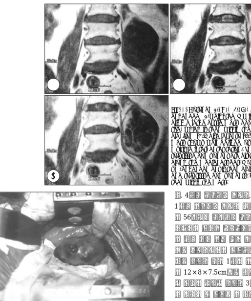
Fig. 1. Coronal T1-(A), T2-(B), and gadolinium- enhanced T1-weighted MR images(C) reve- aling a large cystic mass, about 9×6×5cm in size, which is seen within the left psoas mus- cle, from L3-4 disc level to L5-S1 disc level. This mass shows well defined outer margin and multiple internal septation. Small areas of the soft tissue component are also seen, especially along the medial aspect. Moderate gadolini- um-enhancement is seen along the septation and soft tissue components. Fluid-fluid level is seen within the mass.
Fig. 2. Intraoperative photograph:A relative soft well-enca- psulated mass within the psoas muscle.
A A A
A BB BB
CC CC
석상훈·김문철·정 훈·이상평·최기환·여형태
J Korean Neurosurg Soc/Volume 30/September, 2001 1129
잘 분획 되어진 낭종성의 괴사영역을 가진 경계가 분명한 원 형형태를 그 특징으로 보고하고 있다. 또한 자기공명영상상 T1 강조영상에서는 저신호강도를, T2 강조영상에서는 고 신호강도를 보이며 비괴사된 종양부위 중에 세포가 많은 Antoni A형 부위는 상대적으로 저신호강도를, 세포가 적은 Antoni B형 부위는 상대적으로 고신호강도를 보이는 것으로 보고하고 있다. 조영증강된 T1 강조영상에서는 잘 분획되어 진 낭종성의 괴사영역이 관찰되고 비괴사된 종양부위에 잘 조영증강된 소견이 관찰된다고 보고하고 있다5). 본 증례에 서도 방사선학적으로 특징적인 신경초종의 소견을 보였으며 수술 전 병을 진단하는 데 많은 도움이 되었다.
수술은 후복막 접근법이 요척추 주변부에 직접 접근 할 수 있는 방법으로 알려져 있다. 그외 다른 수술 접근법으로 는 복막을 경유하는 접근법과 천골가시근육(sacrospinal mu-scle)을 통한 후외측접근법이 있으나, 전자의 경우는 하 대정맥과 요관의 손상 위험이 있으며, 후자는 수술 후에도 장기간 지속되는 근육통의 합병증이 있다고 알려져 있다3).
결 론
요근에 단독으로 발생한 신경초종은 지금까지 6례에서 만 보고될 만큼 드물게 발생되는 것으로 알려져 있다. 이에 저자들은 67세 여자에서 요근내에 발생한 단발성 신경초종 을 1례 치험 하였기에 문헌 고찰과 함께 보고 하는 바이다.
•논문접수일:2000년 6월 21일
• 심사완료일:2001년 7월 20일
•책임저자:김 문 철
705-718 대구광역시 남구 대명4동 3056-6 대구가톨릭대학교 의과대학 신경외과학교실 전화:053)650-4253, 전송:053)650-4932 E-mail:mckim@cuth.cataegu.ac.kr
References
1) Claes H, Oyen R, Stessens R, Vereecken R:Solitary benign schwannoma in the psoas muscle. J Urol 137:753-756, 1987 2) Donnal JF, Baker ME, Mahony BS, Leight GS:Benign retro-
peritoneal schwannoma. Urology 31:332-334, 1988 3) Hida K, Iwasaki Y, Abe H, Itamoto K, Kaneda K:Schwan-
noma in the psoas muscle removed by the retroperitoneal ap- proach. Br J Neurosurg 7:213-215, 1993
4) Johenning PW, D’Angelo J:Neurilemoma and the urologist.
J Urol 109:377-380, 1973
5) Kim SH, Choi BI, Han MC, Kim YI:Retroperitoneal Neuril- emoma:CT and MR findings. AJR 159:1023-1026, 1992 6) Kuyumcuoglu U, Germiyanoglu C:Solitary benign schwan-
noma in the psoas muscle. Int Urol Nephrol 22:107-111, 1990 7) Stout AP:The peripheral manifestations of the specific nerve
sheath tumor(neurilemmoma). Am J Cancer 24:751-796, 1935 8) Takatera H, Takiuchi H, Namiki M, Takaha M, Ohnishi S Sonoda
T:Retroperitoneal schwannoma. Urology 28:529-531, 1986 Fig. 3. A:The tumor is composed of spindle cells arranged in a pali- sading or streaming pattern. Blood vessels are prominent(H & E ×200).
B:The tumor cells showing diffuse immunoreactivity for S-100 protein.
AAA
A BBBB