고추의 엽록소 형광 이미지 분석법에 의한 한발스트레스 지표화 가능성
유성녕·엄기철·박소현·김태완1*
세종데이터해석연구원, 1국립한경대학교
Possibility of Drought stress Indexing by Chlorophyll Fluorescence Imaging Technique in Red Pepper (Capsicum annuum L.)
Sung-Yung Yoo, Ki-Cheol Eom, So-Hyun Park, and Tae-Wan Kim1* SEjong Institute of Data Analysis (SEIDA), Suwon 443-766, Korea
1HanKyong National University, Ansung, 456-749, Korea
The objectives of this study focused on measuring chlorophyll fluorescence related to drought stress comparing some parameters. Almost parameters were declined although they were not significant on the basis of mean values of fluorescence of total leaf area. While the ratio of fluorescence intensity variable chlorophyll (FV) to fluorescence intensity maximal chlorophyll (FM) was not changed, the effective quantum yield of photochemical energy conversion in photosystemII (ΦPSII) and chlorophyll fluorescence decrease ratio (Rfd) were slightly reduced, indicating inhibition of the electron transport from quinone bind protein A (QA) to quinone bind protein B (QB). Some parameters such as non-photochemical quenching rate (NPQ_LSS) and coefficients of non-photochemical quenching of variable fluorescence (qN) in mid-zone of leaf and near petiole zone leaf were significantly enhanced within 4 days after drought stress, which can be used as physiological stress parameters. Decrease in ΦPSII could was significantly measured in all leaf zones. In conclusion, three parametric evidences for chlorophyll fluorescence responses such as ΦPSII, NPQ, and qN insinuated the possibility of photophysiological indices under drought stress.
Key words: Electron transport, Nonphotochemical quenching (NPQ), Photosystem II
접수 : 2012. 7. 27 수리 : 2012. 8. 30
*연락저자 : Phone: +82316705081
E-mail: [email protected]
서 언
고추 (Capsicum annuum. L)는 중부아메리카가 원산지로 국내에서는 17세기 초 전래되어 현재까지 재배하고 있는 주 요 채소작물이다. 고추는 국내 생산액 규모가 연간 1조 1,096 억원 (통계청, 2010)으로 채소작물 생산액의 약 13% (채소작 물 중 1위)를 차지하고 있다.
고추는 토양수분 등 재배환경에 따라 매운맛이 다르다 (Jeong 등, 1996). 따라서 고품질 고추를 생산하기 위해서는 적절한 물관리 및 양분관리를 통한 친환경 고추 생산기술 개발이 요구되고 있다. 우리나라의 최근 30년간 (통계청, 1981~2010) 연평균 강수량은 1,308 mm로 세계평균인 973 mm보다 약 1.3배가량 많으나 그 강수량의 대부분이 여름철 에 집중되고 있어 효율적인 물관리가 어려워 작물이 수분스 트레스를 받게 되는 경우가 있다.
지금까지 비파괴적 분석을 통한 식물체의 비생물학적 스 트레스 (abiotic stress) 지표 연구는 오랫동안 수행되어왔
으며, 엽록소형광분석법에 의한 식물체의 건전생장에 관한 연구가 대표적이다. 엽록소 형광분석에 관한 최초의 연구는 Kautsky and Hirsch (1931)의 광합성과정 중의 빛의 섬광에 의한 CO2 흡수동화의 변화원인에 대한 연구에서 비롯되었다.
또한, chlorophyll a (Chla)의 분자구조가 밝혀진 이래 Chla의 형광현상을 응용하여 광합성의 일련반응을 해석하려 는 연구가 1966년 Butler의 선도적 실험 이후 많이 시도되 어 왔다 (Butler et al., 1966; Goedheer, 1972; Govindjee et al., 1973, 1979, 1995, 2004; Butler, 1978; Briantais et al., 1986; Renger et al., 1986; Horton et al., 1990; Krause et al., 1991; Lazár, 1999; Strasser et al., 2000, 2004; Maxwell et al., 2000; Mosenqvist et al., 2003; Schreiber, 2004; Papage- orgiou et al., 2007; Baker, 2008; Lazár et al., 2009).
이와 같은 엽록소 형광현상의 측정방법은 근본적으로 빛 에 노출된 식물체의 형광유발현상을 응용한 Kautsky effect 를 측정하는 방식에서 발전되어 왔다. 빛에 노출된 엽록소 는 흡수된 빛의 파장과 강도에 따라 형광 양상이 변화하게 되며 일반적으로 15분에서 1시간의 암 (어두운) 기간을 거치 면 다시 원상태의 형광현상이 나타날 수 있는 상태로 복귀 하는 것으로 알려져 있다 (Starsser, 1985 ; Srivastava et
Optimum watering Drought condition
Fig. 1. Comparison of red pepper grown under different water conditions. Photos were taken 4 days after drought stress.
al., 1995). 식물의 스트레스와 관련된 초기의 연구는 주로 암상태에 식물체를 방치한 다음 빛을 조사하여 최대형광값/
최소형광값 비 (Fm/FO ratio)를 이용하여 식물의 건전성을 추정하였으며, 정상적인 식물체에서는 대부분 5~6 사이의 값을 갖는 것으로 알려져 있다 (Bjorkman and Demmig, 1987). 최근에는 Fm/FO ratio 보다는 0.78~0.84 범위의 값 을 나타내는 변동형광값/최대형광값 비 (Fv/Fm Ratio) 가 보 편적으로 사용되고 있으며, 이러한 Fv/Fm Ratio는 1차적인 PSII 광화학반응에서의 최대 양자수득율을 나타내는 변수 로 활용되고 있다 (Govindjee, 1995, 2004; Butler and Kitajima, 1975; Paillotin, 1976; Genty et al., 1989).
본 연구에서는 엽록소 형광분석법을 이용하여 생육초기 물 부족에 따른 수분 스트레스 조건이 고추 생육에 미치는 영향을 구명하고, 이를 통해 수분스트레스 지표를 선발하고 자 하였다.
재료 및 방법
공시재료 50구 육묘판에서 재배한 발아 후 5주된 고추 (농그린) 유묘를 구입하여 육묘판에서 3일 동안 1일 간격으 로 고추 한 주당 10 ml씩 관수하여 초기 근권 수분조건을 동일하게 조절하였다. 육묘판 상토의 수분상태는 관수 3일 후 부터 대조구와 무관수구로 구분하여 대조구는 고추 유묘 를 같은 방법으로 1일 간격으로 10 ml씩 관수하여 재배하였 고, 무관수구는 관수를 하지 않고 시험에 사용하였다 (Fig. 1).
엽록소 형광분석 무관수 시점으로부터 4일 (96시간) 동안 24시간 간격으로 고추의 엽록소 형광반응 변화를 다음과 같이 분석하였다. 엽록소 형광분석은 Handy Cam (FlorCam,
CZ)을 이용하여 암적응 (엽록소 형광분석기 챔버에서 20분 간 암처리)상태에서 Quenching kinetics analysis 방법으로 측정하였다 (Barbagallo, et al. 2003; Genty, et al. 1989, 1990). 측정시 엽록소 형광 유도를 위해 화학광선과 연속광 원을 사용하였다. 이때 Handy Cam의 분석조건은 화학 광 선 (actinic light, red LED); 200μmol m-2 s-1, 연속광원 (saturating light, moderate light); 1,250μmol m-2 s-1 이 었다. 측정된 데이터는 Gorbe et al. (2012)이 제시한 방법 에 의해 분석하였다 (Table 1). 또한, 같은 잎에서 공간적인 차이에 의한 엽록소 형광 반응을 분석하기 위해 엽록소 형 광이미지 분석기를 이용해 잎 하나를 세부분으로 구분하였 고, 부위별 구분은 (1)줄기에 붙어 있는 엽병과 가장 가까운 부위, (2)잎 중간부위, (3)엽병에서 가장 멀리 떨어진 잎 끝 부위로 구분해 조사하였다 (Fig. 2).
통계분석 ANOVA 분석 및 t-검정은 SAS program (verion 9.2)를 이용하여 통계분석을 실시하였다.
결과 및 고찰
잎의 평균 엽록소 형광반응 변화 수분 부족은 잎에 서 광합성의 감소를 직접 유도하고 연속적으로 RuBisCO 와 같은 광합성 조절 효소의 불활성화 및 광억제 충격 (Photoinhibition damage)를 초래하기 때문에 궁극적으로 작물의 수량에 치 명적인 작용을 나타내는 생리적 작용을 하는 것으로 알려져 있다 (Medrano et al., 1997). 본 연구 결과 한발스트레스 4 일째 고추시료의 엽록소형광반응의 차이를 보면 대조구에 비해 무관수구에서 증가된 엽록소 형광값은 총 16개 매개변 수 중 최소 형광값 (FO), 연속 광에서의 비광학적 소광에 의
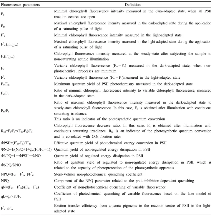
Table 1. Summary of the major Chlorophyll fluorescence parameters (modified from Gorbe et al’s data, 2012).
Fluorescence parameters Definition
Fo Minimal chlorophyll fluorescence intensity measured in the dark-adapted state, when all PSII reaction centres are open
Fm Maximal chlorophyll fluorescence intensity measured in the dark-adapted state during the application of a saturating pulse of light
F′o Minimal chlorophyll fluorescence intensity measured in the light-adapted state
F′m(Fm_Lss) Maximal chlorophyll fluorescence intensity measured in the light-adapted state during the application of a saturating pulse of light
Fs(Ft_Lss) Chlorophyll fluorescence intensity measured at the steady-state after subjecting the sample to non-saturating actinic illumination
Fv Variable chlorophyll fluorescence (Fm−Fo) measured in the dark-adapted state, when non- photochemical processes are minimum
F′v Variable chlorophyll fluorescence (Fm−Fo)measured in the light-adapted state Fv/Fm Maximum quantum yield of PSII photochemistry measured in the dark-adapted state Fo/Fv
Ratio of minimal chlorophyll fluorescence intensity to variable chlorophyll fluorescence, measured in the dark-adapted state
Fm/Fs
Ratio of maximal chlorophyll fluorescence intensity measured in the dark-adapted state to steady-state chlorophyll fluorescence. In this case, Fs is obtained after illumination with continuous saturating irradiance.
This ratio is an indicator of the photosynthetic quantum conversion
Rfd=Fd/Fs=(Fm-Fs)/Fs
Chlorophyll fluorescence decrease ratio. In this case, Fs is obtained after illumination with continuous saturating irradiance. Rfd is an indicator of the photosynthetic quantum conversion and is correlated with CO2 fixation rates
ΦPSII=(F′m-Fs)/F′m Effective quantum yield of photochemical energy conversion in PSII ΦNO=1/(NPQ+1+qL(Fm/Fo−1)) Quantum yield of non-regulated energy dissipation in PSII
ΦNPQ=1−ΦPSII−ΦNO Quantum yield of regulated energy dissipation in PSII
ΦNPQ/ΦNO Ratio of quantum yield of regulated to non-regulated energy dissipation in PSII, which is related to the capacity of photoprotection of the photosynthetic apparatus
NPQ=(Fm−F′m )/F′m Stern-Volmer non-photochemical quenching coefficient
NPQI Component of the NPQ parameter related to the photoinhibition-dependent quenching qN=(Fm−F′m)/(Fm−F′o) Coefficient of non-photochemical quenching of variable fluorescence
qL=qP×Fo/Fs
Coefficient of photochemical quenching of variable fluorescence based on the lake model of PSII
F′v /F′m
Exciton transfer efficiency from antenna pigments to the reaction centre of PSII in the light- adapted state
한 정류상태 형광값 (Ft_LSS), 첨두 형광값 (FP) 및 가변형광 의 비광화학적 소광 계수 (qN) 등 4개였다. 대조구 대비 무 관수구에서 감소한 형광 매개변수는 10개로 대부분 감소하 는 경향을 보였다 (Table 2). 특히 빛이 없는 상태 (암적응) 에서의 매개변수인 최대형광값 (Fm)과 변동형광값 (Fv)에서 기인하는 PSII 최대 양자 수득율을 나타내는 중요한 변수인 Fv/Fm는 거의 변화가 없었으며, 이는 지금까지 여러 연구를 통해 증명되어진 결과 (Calatayud et al., 2006; Massacci et al., 2008)와 일치하였다. 또한, 지속적인 한발스트레스 조건이 아닌 짧거나 가벼운 정도의 한발스트레스 조건에서 는 기공의 닫힘에 의한 광계II 광화학 반응이나 Fv/Fm과 같은 엽록소 형광 매개변수에 영향을 주지 않았다는 결과와도 일
치하였다 (Massacci and Jones, 1990 ; Jeffries, 1994). 그 중 명적응 기간 동안 연속광에 의한 정류상태 PSII 양자 수득율 (QY_LSS = ΦPSII) 또한 대조시료보다 5% 감소하였다. 식물 의 체력을 (Fp – Ft_LSS)/ Fp 의 식에 의해 정량화 할 수 있는 매개변수인 형광 감쇄율 (Rfd) 또한, 관수한 시료에 비해 9% 감소하였다. 이와 같이 한발스트레스에 의해 각 매개변 수가 감소함을 확인 할 수 있었으나, 통계처리 (t-검정)을 한 결과 전체 잎의 총 면적을 기준으로 평균한 광화학 매개 변수는 스트레스 산정의 지표로 활용하기에는 통계적 유의 성이 인정되지 않았다.
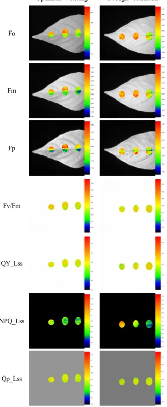
잎의 공간적 엽록소 형광반응 변화 잎의 공간적인
Optimum watering Drought condition
Fo
Fm
Fp
Fv/Fm
QY_Lss
NPQ_Lss
Qp_Lss
Fig. 2. Spatial distribution of Chlorophyll fluorescence parameters in the leaves of red pepper grown under different water conditions.
Table 2. Comparison of Chlorophyll fluorescence parameters in the leaves of red pepper grown under different water conditions
Parameter optimum watering
drought condition
(4 days after treatment) significance
Fo 517±17.37 531±45.8 NS
Fm 2432±182.2 2437±284.4 NS
Fv 1915±185.1 1907±276.1 NS
Fp 1533±210.9 1586±382.4 NS
Ft_LSS 672±40.8 715±117.0 NS
Fv/Fm 0.79±0.02 0.78±0.03 NS
ΦPSII 0.45±0.04 0.42±0.01 NS
NPQ_LSS 1.04±0.43 0.99±0.15 NS
Qp_LSS 0.69±0.04 0.65±0.06 NS
Rfd_LSS 1.37±0.46 1.25±0.21 NS
qN 0.571±0.11 0.582±0.05 NS
qL 0.444±0.11 0.445±0.12 NS
NS : no significance.
엽록소 형광 반응을 Fig. 2와 같이 세부분으로 구분하여 분 석한 결과 관수를 계속하고 있는 고추의 잎에서는 공간적으 로 크게 차이가 없었으나, 한발스트레스 4일 후 한발스트레 스를 받은 고추의 잎에서는 공간적으로 형광 매개 변수들 간의 차이를 보였다.
Table 3의 엽록소 형광 반응 결과에서와 같이 한발 스트
레스를 받은 대부분의 매개변수에서 엽병 부근에서 잎의 끝 으로 갈수록 감소하거나 증가하는 일정한 패턴을 보였다.
PSII 최대 양자수율 (FV/Fm=QY_MAX)는 대조시료와 동일하 게 값의 변화가 없었다. 그리고 FO, Fm, FV와 같은 암적응 형광값과 Fs (Ft_LSS), 정류 상태 최대 형광값 (F’m; Fm_LSS), 정류상태 광화학적 소광 (NPQ_LSS)와 같은 명적응 정류 상 태 형광값, 그리고 PSII에서 조절되지 않는 양자 수득율 ΦNO 는 증가하였다. 따라서 엽병에서 멀어 질수록 NPQ_LSS와 qN의 증가는 결국 ΦPSII (QY_LSS)의 감소로 이어지는 것으 로 확인되었다.
Calatayud et al. (2006)은 점진적으로 한발스트레스를 받게 한 장미에서 PSII의 광합성 능력에서 공간적-시간적 변화 연구를 수행하였는데, 비광화학 과정은 한발스트레스 조건에서 처음 며칠 동안 증가하고 상대수분 함량은 시간이 흐름에 따라 더 심하게 감소하였으며, 이 때 ΦPSII, QP, 및 qL은 급격히 감소하고 ΦNO는 증가하였으나, FV/Fm, QP와 qN은 공간적으로 전체 잎이 동일하게 작용한다고 했다. 이 는 본 연구결과와 일치함을 알 수 있었다. 반대로, Fo및 Fm
은 전체 무관수 기간 (9일) 내내 잎의 중앙맥 옆이 높게 나 타났으며 ΦPSII, NPQ, ΦNPQ과 ΦNO는 무관수 기간 중 (4
∼5일)에 공간적 차이를 보였다. 특히 한발스트레스를 받은 고추의 잎 끝에서 엽병에 가까워질수록 ΦPSII가 감소하는 것으로 나타났다. 한발스트레스에 대한 각 형광매개변수의 유의성 분석을 한 결과 ΦPSII는 잎 끝부위에서 5% 수준 유 의성이 인정되었으며, 잎 중간부위와 엽병 부근에서는 1%
수준의 유의성이 인정되었다. NPQ_LSS 및 qN은 잎 중간 부 위와 엽병부근에서 1% 수준의 유의성이 인정되었으며, Φ NO는 잎 중간 부위에서 1% 수준의 유의성이 인정되었다.
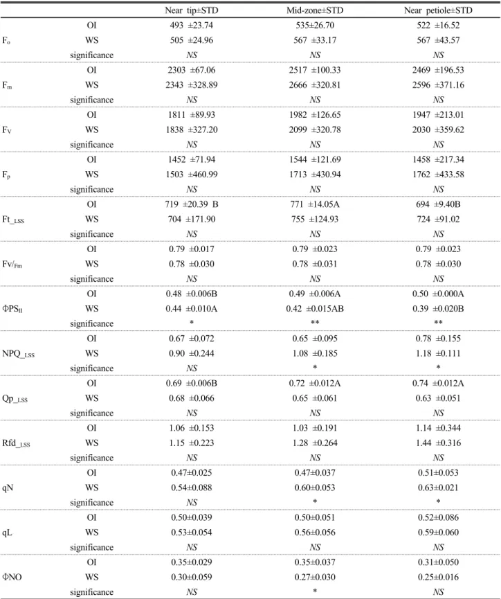
Table 3. The Spatial differences of Chl fluorescence parameter in the leaves of red pepper grown under optimum irrigation (OI) and water stress (WS).
Near tip±STD Mid-zone±STD Near petiole±STD
Fo
OI 493 ±23.74 535±26.70 522 ±16.52
WS 505 ±24.96 567 ±33.17 567 ±43.57
significance NS NS NS
Fm
OI 2303 ±67.06 2517 ±100.33 2469 ±196.53
WS 2343 ±328.89 2666 ±320.81 2596 ±371.16
significance NS NS NS
FV
OI 1811 ±89.93 1982 ±126.65 1947 ±213.01
WS 1838 ±327.20 2099 ±320.78 2030 ±359.62
significance NS NS NS
Fp
OI 1452 ±71.94 1544 ±121.69 1458 ±217.34
WS 1503 ±460.99 1713 ±430.94 1762 ±433.58
significance NS NS NS
Ft_LSS
OI 719 ±20.39 B 771 ±14.05A 694 ±9.40B
WS 704 ±171.90 755 ±124.93 724 ±91.02
significance NS NS NS
Fv/Fm
OI 0.79 ±0.017 0.79 ±0.023 0.79 ±0.023
WS 0.78 ±0.030 0.78 ±0.031 0.78 ±0.030
significance NS NS NS
ΦPSII
OI 0.48 ±0.006B 0.49 ±0.006A 0.50 ±0.000A
WS 0.44 ±0.010A 0.42 ±0.015AB 0.39 ±0.020B
significance * ** **
NPQ_LSS
OI 0.67 ±0.072 0.65 ±0.095 0.78 ±0.155
WS 0.90 ±0.244 1.08 ±0.185 1.18 ±0.111
significance NS * *
Qp_LSS
OI 0.69 ±0.006B 0.72 ±0.012A 0.74 ±0.012A
WS 0.68 ±0.066 0.65 ±0.061 0.63 ±0.051
significance NS NS NS
Rfd_LSS
OI 1.06 ±0.153 1.03 ±0.191 1.14 ±0.344
WS 1.15 ±0.223 1.28 ±0.264 1.44 ±0.316
significance NS NS NS
qN
OI 0.47±0.025 0.47±0.037 0.51±0.053
WS 0.54±0.088 0.60±0.053 0.63±0.021
significance NS * *
qL
OI 0.50±0.039 0.50±0.051 0.52±0.086
WS 0.53±0.054 0.56±0.056 0.59±0.060
significance NS NS NS
ΦNO
OI 0.35±0.029 0.35±0.037 0.31±0.050
WS 0.30±0.059 0.27±0.030 0.25±0.016
significance NS * NS
NS : no significance, * : p<0.05 significance level in student’s t-test, ** : p<0.01 significance level in student’s t-test. Means with same letter are not significantly different.
그밖에 형광매개변수에서는 유의성이 인정되지 않는 것으 로 나타났다.
수분 스트레스에 따른 각각의 형광변수가 공간적인 상관
을 보이는지 확인하기 위해 대조군 고추의 잎을 1로 고정하 여 polar plot 분석을 하였다. 그 결과 엽병 부근에서 NPQ_LSS
와 Rfd_LSS의 현격한 차이를 볼 수 있었다 (Fig. 3).
Fig. 3. Polar plot analysis of fluorescence parameters at different parts of leaves under drought condition. The values of control parameter were radially fixed on 1.0 comparing to others under drought stress. For the distributional analysis of chlorophyll fluorescence parameters the measurements were performed in three parts of leaves such as leaf zone near petiole, mid-zone of leaf and leaf zone near tip.
Massacci 등 (2008)도 한발스트레스 조건에서 면화 잎에 서 FV/Fm 및 ΦNPQ의 공간적변화의 매우 동질적인 분포를 측정하였으며, Calatayud 등 (2006)의 결과와 다른 ΦPSII의 증가와 같은 동질적인 패턴을 측정했다. 이것은, 식물이 한 발스트레스를 일종의 환경변화로 인식하고 환경에 순응하 는 과정에서 광호흡속도의 향상에 기인한 것으로 제시하였 다. 그러나 본 연구에서는 ΦPSII의 증가는 없었으며, 이는 짧은 한발스트레스 기간에 기인하는 것으로 판단되었다.
결론적으로 엽록소 형광반응 분석을 이용하여 물 부족 조건에서 고추의 한발스트레스를 판단할 수 있는 생리적 지 표로 ΦPSII (QY_LSS), NPQ_LSS, qN 등의 형광매개 변수가 활 용 가능한 것으로 사료되었다. ΦNO는 광에너지 전환에서 광계에 의해 형광 조절이 불가능한 비광학적 소광량으로 수 분스트레스 지표를 설정할 경우 잎 중간 부위만 가능할 것 으로 예측되었다.
요 약
본 연구에서는 생육초기 고추의 한발 스트레스 조건에서 작물 생육에 미치는 영향을 구명하기 위해 엽록소 이미지형 광분석을 이용하여 엽록소 형광 매개변수를 비교분석 하였 다. 비록 통계적 유의성이 인정되지는 않았지만 전체 잎의 이미지에 나타나는 형광매개변수들의 평균값은 감소하는
경향이었다. 가변형광값에대한 최대형광값의 비는 변동이 없는 반면, 광계II의 정류상태에서의 양자수득율 및 형광감 쇄율은 다소 감소하였다. 이러한 현상은 퀴논 A 단백질로부 터 퀴논 B 단백질로의 전자전달의 감소를 의미하였다. 잎의 중앙부위와 엽병부근에서의 비광학적 소광 및 가변형광의 비광학적 소광 상수는 한발스트레스 4일 후 유의성있게 증 가하였다. 즉 생리적 스트레스 매개변수로 활용이 가능하였다.
광계II에서의 정류상태 양자수득율의 감소는 고추잎의 모 든 부위에서 공통적으로 유의성있게 측정되었다. 결론적으 로 ΦPSII (QY_LSS), NPQ_LSS, qN 등의 엽록소형광 매개변수 들은 고추의 한발스트레스를 판단할 수 있는 생리적 지표로 활용 가능한 것으로 유의성이 인정되었다.
사 사
본연구는 농촌진흥청 지원과제 (과제명 : 지역 바이오매 스 자원 인벤토리 작성; 과제번호: PJ007391)로 수행되었음.
인 용 문 헌
Baker, N.R. 2008. Chlorophyll fluorescence. a probe of photosynthesis in vivo. Annu. Rev. Plant Biol. 59:659-668.
Barbagallo, R.P., K. Oxborough., K.E. Pallett, and N.R.
Baker. 2003. Rapid, non-invasive screening for pertubations of metabolism and plant growth using chlorophyll fluorescence imaging. Plant Physiol. 132:485-493.
Bjorkman, O. and B. Demmig. 1987. Photon yield of O2 evolution and chlorophyll fluorescence characteristics at 77 K among vascular plants of diverse origins. Planta.
170:489-504.
Briantais, J.M., C. Vernotte., G.H. Krause, and E. Weis.
1986. Chlorophyll a fluorescence of higher plants. : chloroplasts and leaves, in: Govindjee, J. Amesz, D.C.
Fork (Eds.). Light Emission by Plants and Bacteria.
Academic Press. New York. 539-583.
Butler, W.L. 1966. Fluorescence yield in photosynthetic systems and its relation to electron transport. Curr. Top.
Bioenerg. 1:49-73.
Butler, W.L. 1978. Energy distribution in the photochemical apparatus of photosynthesis. Annu. Rev. Plant Physiol.
29:345-378.
Butler, W.L. and M. Kitajima. 1975. Fluorescence quenching in photosystem II of chloroplasts. Biochim. Biophys. Acta.
376:116-125.
Calatayud, A., D. Roca, and P.F. Martinez. 2006. Spatial- temporal variations in rose leaves under water stress conditions studied by chlorophyll fluorescence imaging.
Plant Physiology and Biochemistry. 44(10):564-573.
Horton, P, and J.R. Bowyer. 1990. Chlorophyll fluorescence transients. in: J.L. Harwood, J.R. Bowyer (Eds.). Methods in Plant Biochemistry. vol. 4. Academic Press. New York.
259-296.
Jeong, C.S., Y.R. Yeuong., H.K. Yun., K.C. Yoo, and M.
Nagaoka. 1996. Effect of light intensities and temperatures on capsaicin and sugar contents of each growth stage in Capsicum annuum L. Inst. Agri. Sci. Kangwon Nat.
Univ. J. Agr. Sci. Vol. 7.
Kautsky, A. and Hirsh. A. 1931. Neue Versuche zur Kohlensäureassimilation.” Naturwissenschaften. 19:964.
Krause, G.H, and E. Weis. 1991. Chlorophyll fluorescence and photosynthesis. : the basics, Annu. Rev. Plant Physiol.
Plant Mol. Biol. 42:313-349.
Genty, B., J.M. Briantais, and N.R. Baker. 1989. The relationship between the quantum yield of photosynthetic electron transport and quenching of chlorophyll fluorescence.
Biochim. Biophys. Acta. 990:87-92.
Genty, B., J. Harbinson, and Baker, N. R. 1990. Relative quantum efficiencies of the two photosystems of leaves in photorespiratory and non-photorespiratory conditions.
Plant Physiol. Biochem. 28:1-10.
Goedheer, J.H.C. 1972. Fluorescence in relation to photosynthesis.
Annu. Rev. Plant Physiol. 23:87-112.
Gorbe, E. and A. Calatayud. 2012. Applications of chlorophyll fluorescence imaging technique in horticultural research:
A review. Scientia Horticulturae. 138(0):24-35.
Govindjee. 2004. Chlorophyll a fluorescence: a bit of basics and history. in: G.C. Papageorgiou, Govindjee (Eds.).
Chlorophyll a Fluorescence: A Signature of Photosynthesis.
Advances in Photosynthesis and Respiration, Springer, Dordrecht. The Netherlands. 19:1-41.
Govindjee. 1995. Sixty-three years since Kautsky: chlorophyll a fluorescence. Aust. J. Plant Physiol. 22:131-160.
Govindjee., G.P. and E. Rabinowitch. 1973. Chlorophyll fluorescence and photosynthesis. In G.G. Guilbault (Ed.). Practical Fluorescence Theory. Methods, and Techniques. Marcel Dekker Inc. New York. 543-575.
Govindjee, and P. Jursinic. 1979. Photosynthesis and fast changes in light emission by green plants. Photochem.
Photobiol. Rev. 4:125-205.
Jefferies, R.A. 1994. Drought and chlorophyll fluorescence in fieldgrown potato (Solanum tuberosum). Physiologia Plantarum. 90:93–97.
Lazár, D. 1999. Chlorophyll a fluorescence induction.
Biochim. Biophys. Acta. 1412:1-28.
Lazár, D. and G. Schansker. 2009. Models of chlorophyll a fluorescence transients. in: A. Laisk, L. Nedbal, Govindjee (Eds.). Photosynthesis in Silico: Understanding Complexity from Molecules to Ecosystems, Advances in
Photosynthesis and Respiration, Springer, Dordrecht. The Netherlands. 29:85-123.
Massacci, A. and H.G. Jones. 1990. Use of simultaneous analysis of gasexchange and chlorophyll fluorescence quenching for analysing the effects of water stress on photosynthesis in apple leaves. Trees. 4:1–8.
Massacci, A., S.M. Nabiev. L. Pietrosanti., S.K. Nematov., T.N. Chemikova., K. Thor, and J. Leipner. 2008. Response of the photosynthetic apparatus of cotton (Gossypium hirsutum) to the onset of drought stress under field conditions studied by gas-exchange analysis and chlorophyll fluorescence imaging. Plant Physiol. Biochem. 46(2):
189-195.
Maxwell, K. and G.N. Johnson. 2000. Chlorophyll fluorescence - a practical guide. J. Exp. Bot. 51:659-668.
Medrano, H., M.A.J. Parry., X. Socias, and D.W. Lawlor.
1997. Long term water stress inactivates Rubisco in subterranean clover. Annals of Applied Biology. 131(3):491-501.
Mosenqvist, E. and O. van Kooten. 2003. Chlorophyll fluorescence: a general description and nomenclature. in:
J.R. DeEll, P.M.A. Toivonen (Eds.). Practical Applications of Chlorophyll Fluorescence in Plant Biology. Kluwer Academic Publishers. Dordrecht. The Netherlands. 31-78.
Paillotin, G. 1976. Movement of excitations in the photosynthetic domains of photosystem II. J. Theor.
Biol. 58:237-252.
Papageorgiou, G.C., M. Tsimilli-Michael, and K. Stamatakis.
2007. The fast and slow kinetics of chlorophyll a fluorescence induction in plants. algae and cyanobacteria:
a viewpoint. Photosynth. Res. 94:275-290.
Srivastava, A., H. Greppin, and R.J. Strasser. 1995. Acclimation of land plants to diurnal changes in temperature and light. in: P. Mathis (Ed.), Photosynthesis: From Light to Biosphere, Kluwer Academic Publishers. The Netherlands.
4:909-912.
Strasser, R.J., A. Srivastava, and M. Tsimilli-Michael.
2000. The fluorescence transient as a tool to characterize and screen photosynthetic samples. In M. Yunus, U.
Pathre, P. Mohanty (Eds.), Probing Photosynthesis: Mechanism, Regulation and Adaptation, Taylor and Francis, London, UK. 443-480.
Strasser, R.J., M. Tsimilli-Michael, and A. Srivastava.
2004. Analysis of the chlorophyll fluorescence transient.
in: G.C. Papageorgiou, Govindjee (Eds.), Chlorophyll Fluorescence: A Signature of Photosynthesis, Advances in Photosynthesis and Respiration, Springer, Dordrecht.
The Netherlands. 19:321-362.
Strasser, R.J. 1985. Dissipative Strukturen als thermodynamischer Regelkreis des Photosynthese apparates. Ber. Deutsche Bot. Ges. Bd. 98:53-72.