Betaine-γ-aminobutyric Acid Transporter 1 (BGT-1/mGAT2) Interacts with the PDZ Domain of Munc-18 Interacting Proteins (Mints)
Sang-Jin Kim
1, Young Joo Jeong
2, Sun Hee Choi
2, Chun Yeon Choi
2, Hee Jae Jun
3, Il Soo Moon
4, Dae-Hyun Seog
2* and Won Hee Jang
2*
1
Departments of Neurology,
2Departments of Biochemistry,
3Departments of Thoracic and Cardiovascular Surgery, College of Medicine, Inje University, Busan 614-735, Korea
4
Departments of Anatomy, College of Medicine, Dongguk University, Gyeongju 780-714, Korea
Received August 23, 2012 /Revised September 6, 2012 /Accepted September 7, 2012The action of neuronally released γ-aminobutyric acid (GABA) is terminated by uptake into the neu- rons by GABA transporters (GATs). The mechanism underlying the stabilization and regulation of GAT2 has not yet been elucidated. We used the yeast two-hybrid system to identify proteins that in- teract with and, thereby, regulate betaine-γ-aminobutyric acid transporter 1 (BGT-1/mGAT2). We found an interaction between BGT-1/mGAT2 and Munc-18-interacting proteins (Mints). The “T-H-L”
motif at the C-terminal end of BGT-1/mGAT2 was essential for the interaction with Mint2 in the yeast two-hybrid assay. Mint2 bound to the tail region of BGT-1/mGAT2, but not to other GAT members.
When co-expressed in HEK-293T cells, Mint2 was co-immunoprecipitated with BGT-1/mGAT2. In ad- dition, we demonstrated the cellular co-localization of BGT-1/mGAT2 and Mint2 in the cells. These results suggest that Mint2 contributes to the regulation of BGT-1/mGAT2.
Key words : Neurotransmitter transporter, γ-aminobutyric acid transporter, betaine-γ-aminobutyric acid transporter 1 (BGT-1), Munc-18-interacting protein 2 (Mint2), PDZ Domain
*Corresponding author
*Tel:+82-51-890-6974, Fax:+82-51-894-5801
*E-mail : [email protected] and [email protected]
Introduction
Plasma membrane neurotransmitter transporters exist for most of the small molecule neurotransmitter systems [2]. γ-aminobutyric acid (GABA) is the primary inhibitory neurotransmitter in the brain and has been implicated in numerous neurogical disorders, including epilepsy, anxi- ety, and bipolar disorder [10,23,25]. GABA transporters (GATs) belong to the family of Na
+/Cl
-dependent trans- porters that also includes transporters for the neuro- transmitters such as dopamine, serotonin, norepinephrine and glycine [12,19]. GATs are found primarily in the pre- synaptic membrane. This localization suggests that GATs are involved in regulating synaptic transmission and trans- mitter recycling [6,21].
Recently, four different subtypes of GATs have been cloned with diverse regional, cellular and subcellular loca- tion [19]. These have been termed mGAT1, mGAT2, mGAT3 and mGAT4 when referring to transporters cloned from mice. Betaine-γ-aminobutyric acid transporter (BGT-1) was
originally cloned from the Madin-Darby canine kidney (MDCK) cell line [6,12,17,19]. Subsequently, the homologous transporter mGAT2 was cloned from mouse brain [12,19].
BGT-1/mGAT2 is a membrane transporter capable of utiliz- ing both GABA and betaine as substrates [10,13,29]. Betaine is known as an organic osmolyte involved in osmoregulation in the kidneys [6]. BGT-1/mGAT2 contains 614 amino acids and shares 54% identity with GAT1 [7,19]. Previous studies have reported that GAT1 activity can be modulated by pro- tein-protein interactions. The interaction between the N-ter- minal cytoplasmic tail of GAT1 and syntaxin 1A causes a 4-fold decrease in substrate transporter rates [8]. The inter- action between the C-terminus of GAT1 and Pals1 contain- ing the PDZ domain contributes to the stability of GAT1, thus promoting the expression level of the transporter pro- tein [8].
The topologies of BGT-1/mGAT2 consist of 12 hydro-
phobic transmembrane domains and the amino and carboxyl
termini face the cytoplasm [2,17]. The cytosolic C-terminus
contains postsynaptic density-95 (PSD-95)/discs large pro-
tein/zona occludens 1 (PDZ)-association motif which is recog-
nized by the adaptor protein-binding sites found in mem-
brane-associated proteins [14,27]. PDZ-association motifs are
characterized by a C-terminal hydrophobic residue and a free carboxylate group at the -2 position (T/S-X-V/D/L) [9,11]. PDZ interactions have been found to be involved in the multi-molecular organization of receptors and signal transduction pathways [14]. BGT-1/mGAT2 contains a typi- cal PDZ-association motif (T-X-L) in its cytosolic C-terminal domain [5].
In order to investigate the BGT-1/mGAT2-mediated regu- lation of GABAergic neurotransmission, we screened for proteins that interact with the C-terminus of BGT-1/mGAT2 through the yeast two-hybrid system and identified Mint2 (also called X11-like protein), a PDZ domain-containing pro- tein [20]. The BGT-1/mGAT2 and Mint2 interaction suggests that Mint2 contributes to the regulation of BGT-1/mGAT2 in the synaptic membrane.
Materials and Methods Plasmid constructs
Full-length rat Mint2 in the pRC/CMV vector (a gift from M. Setou, Hamamatsu University, Hamamatsu, Japan) was tagged with a myc-epitope and fused with EGFP at the ami- no-terminus. Truncations of BGT-1/mGAT2 [17] were uti- lized as a template to amplify the region coding for amino acids 570-628 using the appropriate primers. The amplified fragment was subcloned into T-vector. The fragment was then EcoRI-restricted and subcloned into the EcoRI site of pLexA. The correct orientation and in-frame cloning of cDNA inserts was verified by restriction enzyme analysis, and DNA sequencing. FLAG-tagged BGT-1/mGAT2 con- struct was used in all experiments that utilized mammalian cell expression system. General recombinant DNA techni- ques were performed according to standard protocol [24].
Screening of BGT-1/mGAT2-binding proteins by yeast two-hybrid assay
The Matchmaker LexA two-hybrid system was used for screening according to the manufacturer’s manual (Clontech, Palo Alto, CA, USA). In brief, a part of the BGT-1/mGAT2 gene was fused to the DNA-BD region of the pLexA vector using the PCR and the plasmid DNA was transformed into yeast strain EGY48 carrying the p8op-lacZ gene.
Transformed EGY48 yeast cells containing the BGT-1/
mGAT2 bait plasmid were transformed with the mouse brain cDNA library [15] and grown on synthetic dextrose (SD) plates supplemented with glucose but with no histi-
dine, tryptophan, or uracil (SD/-His/-Trp/-Ura). The se- lection of positive clones was performed on an SD/-His/-Trp/-Ura/-Leu plate containing galactose, raffi- nose, X-gal, and BU salts. Plasmids from positive clones were analyzed by restriction digestion. Unique inserts were sequenced and protein sequence analysis was performed with the BLAST algorithm at the National Center for Biotechnology Information (NCBI). Sequence-verified clones were tested again for interactions with the bait in yeast by the retransformation.
Cell culture and Transfection
HEK-293T cells were cultured in Dulbecco's modified Eagle's medium supplemented with 10% fetal bovine serum, L-glutamine, and antibiotics. Transient transfections were done with the CaPO
4precipitation method.
Immunocytochemistry
Cells grown on poly-D-lysine-coated coverslips were transfected with myc-EGFP-Mint2 and FLAG-BGT-1/
mGAT2 constructs. Twenty-four hours after transfection, cells were washed with phosphate-buffered saline (PBS), fixed with 4% paraformaldehyde in PBS for 5 min, and per- meabilized with 0.2% Triton X-100 in PBS for 10 min. After blocking with 5% normal goat serum in PBS for 30 min, cells were incubated with anti-BGT-1 antibody (Alpha Diagnostic International, San Antonio, TX, USA) diluted 1:500 in PBS containing 1% bovine serum albumin (BSA) and 0.05%
Tween-20 overnight at 4
oC. After washing with PBS 3 times, cells were incubated with Dylight 594-conjugated goat an- ti-rabbit IgG antibody (Jackson ImmunoResearch Labs, West Grove, PA, USA) diluted 1:800 for 40 min. For nuclear stain- ing, cells were incubated with 1 μg/ml 4, 6-dia- midino-2-phenylindole (DAPI) (Sigma-Aldrich) in PBS for 5 min. After washing with PBS 3 times, the cells were mount- ed with Fluoromount (DAKO). Fluorescence images were acquired on Zeiss LSM510 META confocal laser scanning microscope (Carl Zeiss, Oberkochem, Germany).
Co-immunoprecipitation
Twenty-four hours after transfection, cells were rinsed with ice-cold PBS twice and lysed with ice-cold lysis buffer [PBS containing 0.5% NP-40 and 1x protease inhibitor cock- tail set V (Calbiochem)] by gentle rotation for 30 min.
Lysates were centrifuged at 16,000x g for 10 min at 4
oC. The
supernatant (soluble fraction) was incubated with anti-FLAG
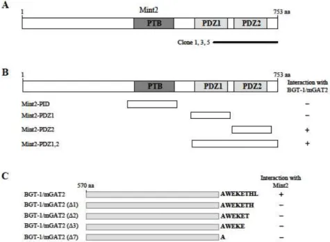
Fig. 1. Identification of the proteins interacting with BGT-1/mGAT2 by yeast two-hybrid screening. (A) The domain structure of Mint2 illustrating that clones 1, 3, and 5 possess a fragment of Mint2 containing the PDZ domain. PTB and PDZ domains are indicated in gray. aa, the amino acid residue number. (B) Minimal BGT-1/mGAT2 binding region in Mint2. Different truncations of Mint2 were constructed by PCR. Several truncated forms of Mint2 were tested in the yeast two-hybrid assay for interaction with BGT-1/mGAT2. +, interaction with BGT-1/mGAT2; -, no interaction with BGT-1/mGAT2. aa, the amino acid residue number. (C) Specific interaction of Mint2 with the C-terminus of BGT-1/mGAT2. Several deletion mutant forms of BGT-1/mGAT2 were tested in the yeast two-hybrid assay for interaction with Mint2. +, interaction with Mint2; -, no interaction with Mint2.
M2 agarose beads (Sigma-Aldrich) for 2 hr at 4
oC with con- stant shaking. The beads were collected by centrifugation at 2,000x g for 30 sec and washed 5 times with ice-cold lysis buffer. The immunoprecipitated proteins were analyzed by Western blotting.
Results
Identification of BGT-1/mGAT2 interacting proteins by yeast two-hybrid screening
To identify BGT-1/mGAT2-interacting proteins, we screened a mouse brain cDNA library through the yeast two-hybrid assays using the C-terminal region of BGT-1/mGAT2 as bait (Fig. 1C). From 6x10
6colonies screened, we obtained 6 positive clones. Three clones (clone 1, 3 and 5) of the 6 clones were identical and possessed a C-terminal fragment of Mint2 containing the PDZ domain (Fig. 1A). Mint2 is composed of several protein-protein inter- action domains, two PDZ domains and one PTB domain (Fig. 1A). To determine the binding domain of Mint2 that is required for the interaction with BGT-1/mGAT2, we con-
structed a series of deletion mutants of Mint2. Yeast two-hy- brid assays with BGT-1/mGAT2 showed that the minimal domain required for binding was critically dependent on the second PDZ domain of Mint2 (Fig. 1B). Next we investigated whether the last seven amino acids of BGT-1/mGAT2 con- tain a functional PDZ-association motif that mediates pro- tein-protein interaction. For this purpose, a series of C-termi- nal deletion mutants of BGT-1/mGAT2 were constructed (Fig. 1C), and co-transfected into yeast cells with pLexA-Mint2. As shown in Fig. 1C, the C-terminal deletion mutants of BGT-1/mGAT2 did not interact with Mint2.
These result indicated that the interaction between BGT-1/mGAT2 and Mint2 is mediated through a PDZ-medi- ated interaction similar to the previously described a class I PDZ interaction [9,27].
When the C-terminal cytoplasmic regions of mGAT1,
BGT-1/mGAT2, mGAT3 and mGAT4 were tested for
Mint2-binding, there was no detectable binding between
Mint2 and the tail domains of mGAT1, mGAT3 and mGAT4
(Fig. 2A). These data indicate that the interaction of Mint2
with GATs is specific to the cytoplasmic region of
Fig. 4. Co-localization of BGT-1/mGAT2 and Mint2 at the plasma membrane. Twenty-four hours after transfection, cells were immu- nostained using anti-BGT-1antibody. (A) BGT-1/mGAT2 and EGFP-Mint2 are seen at the same subcellular region in cells.
(B) Immunofluorescence staining of BGT-1/GAT2 and EGFP-Mint2 shows that both proteins co-localize largely in plasma membrane (arrow).
Fig. 2. Interaction between GATs and Mint proteins. The C-terminal region of each GAT protein and full length Mints were fused to the pLexA DNA binding domain. (A) Mint2 specifically interacted with BGT-1/mGAT2 but not with mGAT1, mGAT3 or mGAT4 (+, interaction with Mint2; -, no interaction with Mint2). (B) BGT-1/mGAT2 interacted with Mint1 and Mint2 (+, interaction with BGT-1/mGAT2;
-, no interaction with BGT-1/mGAT2).
BGT-1/mGAT2 isoform. Interaction was also detected be- tween BGT-1/mGAT2 and other Mint family member, Mint1 (Fig. 2B).
BGT-1/mGAT2 is associated with Mint2 in cells
To assess the interaction of BGT-1/mGAT2 and Mint2 in mammalian cells, HEK-293T cells were co-transfected with constructs expressing myc-EGFP-Mint2 along with FLAG-BGT-1/mGAT2 (Fig. 3). Cell lysates were immunoprecipitated with a monoclonal antibody directed against the FLAG epitope followed by Western blot analysis with anti-BGT-1 antibody. Fig. 3 shows that BGT-1/mGAT2
Fig. 3. BGT-1/mGAT2 and Mint2 were co-immunoprecipitated from mammalian cells. HEK-293Tcells were transiently transfected with Myc-EGFP-Mint2 plasmid and either control vector or FLAG-BGT-1/mGAT2 plasmid as indicated. Cell lysates were incubated with monoclonal anti-FLAG antibody to immunoprecipitate BGT-1/
mGAT2. Western blots were subsequently probed with anti-myc antibody and anti-FLAG antibodies. Mint2 was specifically co-immunoprecipitated with BGT-1/
mGAT2.
co-precipitated Mint2. As expected from our yeast two-hy- brid results, BGT-1/mGAT2 specifically interacts with Mint2.
For a potential interaction between BGT-1/mGAT2 and Mint2 to be physiologically relevant, two proteins must co-localize at the same subcellular region in cells. To de- termine whether BGT-1/mGAT2 and Mint2 co-localize, we generated the N-terminal EGFP-fused Mint2 construct.
BGT-1/mGAT2 was co-transfected with EGFP-Mint2 into
HEK-293T cells. BGT-1/mGAT2 and Mint2 co-localized at
the same subcellular region in cells (Fig. 4A). Co-transfected
BGT-1/mGAT2 and Mint2 revealed a largely plasma mem-
brane expression in a subset cells, forming clusters at the cell surface (Fig. 4B). This result indicates that Mint2 is a specific binding partner of BGT-1/mGAT2 in cell level.
Discussion
In this study, we have shown that the neuronal plasma membrane BGT-1/mGAT2 can associate with Mint2. Using the intracellular carboxyl tail of BGT-1/mGAT2 as bait, we identified Mint2 in a yeast two-hybrid screen of a mouse brain cDNA library. The three C-terminal amino acids con- taining region of BGT-1/mGAT2 can interact with Mint2.
Furthermore, when full-length BGT-1/mGAT2 and Mint2 are expressed in mammalian cells, they co-im- munopresipitate and co-localize in cells. This is the first demonstration of an interaction at the C-terminus of BGT-1/mGAT2 and joins Mint2 as a BGT-1/mGAT2-inter- acting protein.
Interestingly, Mint2 has two PDZ domains that mediate interaction with the other protein [20]. This study demon- strated through domain analysis by yeast two-hybrid assay that the interaction is dependent upon the second PDZ do- main of Mint2 and the C-terminal amino acids of BGT-1/mGAT2. The C-terminal three amino acids (T-H-L) represent a class I PDZ domain ligand [9]. PDZ-domain pro- teins play important roles in establishing and maintaining an asymmetry of membrane proteins in neurons [14].
Proteins containing PDZ domains usually possess multiple protein-protein interaction domains, allowing them to form multimeric complexes [11]. PDZ domains contain a con- served peptide-binding groove that associates with the ex- treme C-terminus of ligands [9]. Although we did not show whether BGT-1/mGAT2 can bind with other PDZ domain containing proteins, our results suggest that the interaction between BGT-1/mGAT2 and Mint2 is mediated through a PDZ interaction.
Mint2 is a multidomain protein composed of PTB and PDZ domains [20]. The N-terminal domain of Mint2 binds to cell-surface receptors [4] and Munc18-1, a protein essential for synaptic vesicle exocytosis [28]. The central phosphotyr- osine binding (PTB) domain binds to the cytoplasmic tail of amyloid-β-protein precursor (APP) [3]. PDZ domains bind to presenilins [16], cell surface receptors, Ca
2+channels [18], and kinesin KIF17 [26]. Thus Mint2 resembles adaptor proteins that connect the N-terminal, PTB domain, and PDZ domain interactions. Protein-protein interactions not only
determine the specific membrane surface expression, but can also affect the membrane surface expression level by altering endocytic rates. In cultured neuron, co-expression of PSD-95 and NMDA receptor NR2B results in decreased endocytosis of NR2B, while deletion of the PDZ-association motif yields an increase in the percentage of receptor that is internalized [22]. Interestingly, the C-terminal PDZ-association motif of BGT-1/mGAT2 is responsible for proper targeting and maintenance at basolateral membrane surface of MDCK cells [1,5].
How might an interaction with Mint2 modulate BGT-1/mGAT2 surface expression? One potential model is the association with Mint2 to prevent internalization from surface membrane. Recent evidence for many different trans- porter systems suggests that direct interacting proteins of the transporter serves as a tag that identifies transporters to be internalized [22]. This might occur because the tag is indicative of a transporter in an appropriate conformational state for internalization. Thus, like PSD-95, it may be that the interaction between BGT-1/mGAT2 and Mint2 induces conformational state that confers a slowing of BGT-1/mGAT2 internalization from membrane surface.
Further functional studies on this and other BGT-1/mGAT2 interacting proteins may help to shed light on the regulating BGT-1/mGAT2 activity.
Acknowledgment
This work was supported by an Inje University Research Grant for 2011.
References
1. Ahn, J., Mundigl, O., Muth, T. R., Rudnick, G. and Caplan, M. J. 1996. Polarized expression of GABA transporters in Madin-Darby canine kidney cells and cultured hippocampal neurons.
J. Biol. Chem
. 271, 6917-6924.2. Amara, S. G. and Kuhar, M. J. 1993. Neurotransmitter trans- porters: recent progress.
Annu. Rev. Neurosci
. 16, 73-93.3. Biederer, T., Cao, X., Südhof, T. C. and Liu, X. 2002.
Regulation of APP-dependent transcription complexes by Mint/X11s: differential functions of Mint isoforms.
J.
Neurosci
. 22, 7340-7351.4. Biederer, T., Sara, Y., Mozhayeva, M., Atasoy, D., Liu, X., Kavalali, E. T. and Südhof, T. C. 2002. SynCAM, a synaptic adhesion molecule that drives synapse assembly.
Science
297, 1525-1531.5. Brown, A., Muth, T. and Caplan, M. 2004. The COOH-termi- nal tail of the GAT-2 GABA transporter contains a novel
motif that plays a role in basolateral targeting.
Am. J. Physiol.
Cell Physiol
. 286, C1071-C1077.6. Burnham, C. E., Buerk, B., Schmidt, C. and Bucuvalas, J.
C. 1996. A liver-specific isoform of the betaine/GABA trans- porter in the rat: cDNA sequence and organ distribution.
Biochim. Biophys. Acta.
1284, 4-8.7. Clausen, R. P., Frølund, B., Larsson, O. M., Schousboe, A., Krogsgaard-Larsen, P. and White, H. S. 2006. A novel se- lective gamma-aminobutyric acid transport inhibitor dem- onstrates a functional role for GABA transporter subtype GAT2/BGT-1 in the CNS.
Neurochem. Int.
48, 637-642.8. Deken, S. L., Beckman, M. L., Boos, L. and Quick, M. W.
2000. Transport rates of GABA transporters: regulation by the N-terminal domain and syntaxin 1A.
Nat. Neurosci
. 3, 998-1003.9. Doyle, D. A., Lee, A., Lewis, J., Kim, E., Sheng, M. and MacKinnon, R. 1996. Crystal structures of a complexed and peptide-free membrane protein-binding domain: molecular basis of peptide recognition by PDZ.
Cell
85, 1067-1076.10. Gether, U., Andersen, P. H., Larsson, O. M. and Schousboe, A. 2006. Neurotransmitter transporters: molecular function of important drug targets.
Trends Pharmacol. Sci
. 27, 375-383.11. Gomperts, S. N. 1996. Clustering membrane proteins: It's all coming together with the PSD-95/SAP90 protein family.
Cell
84, 659-662. 212. Guastella, J., Nelson, N., Nelson, H., Czyzyk, L., Keynan, S., Miedel, M. C., Davidson, N., Lester, H. A. and Kanner, B. I. 1990. Cloning and expression of a rat brain GABA transporter.
Science
249, 1303-1306.13. Iversen, L. 2006. Neurotransmitter transporters and their im- pact on the development of psychopharmacology
. Br. J.
Pharmacol.
147, Suppl. 1, S82-S88.14. Kennedy, M. B. 2000. Signal-processing machines at the postsynaptic density.
Science
290, 750-754.15. Kim, S. J., Lee, C. H., Park, H. Y., Yea, S. S., Jang, W. H., Lee, S. K., Park, Y. H., Cha, O. S., Moon, I. S. and Seog, D. H. 2007. JSAP1 interacts with kinesin light chain 1 through conserved binding segments.
J. Life Sci.
17, 889-895.16. Lau, K. F., McLoughlin, D. M., Standen, C. and Miller, C.
C. 2000. X11 alpha and x11 beta interact with presenilin-1 via their PDZ domains.
Mol. Cell Neurosci.
16, 557-565.17. Liu, Q. R., Mandiyan, S., Nelson, H. and Nelson, N. 1992.
A family of genes encoding neurotransmitter transporters.
Proc. Natl. Acad. Sci. USA
89, 6639-6643.18. Maximov, A., Südhof, T. C. and Bezprozvanny, I. 1999.
Association of neuronal calcium channels with modular adaptor proteins.
J. Biol. Chem.
274, 24453-24456.19. Nelson, N. 1998. The family of Na+/Cl- neurotransmitter transporters.
J. Neurochem
. 71, 1785-1803.20. Okamoto, M. and Südhof, T. C. 1997. Mints, Munc-18 -interacting proteins in synaptic vesicle exocytosis.
J. Biol.
Chem.
272, 31459-31464.21. Radian, R., Ottersen, O. P., Storm-Mathisen, J., Castel, M.
and Kanner, B. I. 1990. Immunocytochemical localization of the GABA transporter in rat brain.
J. Neurosci.
10, 1319-1330.22. Roche, K. W., Standley, S., McCallum, J., Dune Ly, C., Ehlers, M. D. and Wenthold, R. J. 2001. Molecular determi- nants of NMDA receptor internalization.
Nat. Neurosci.
4, 794-802.23. Roettger, V. R. and Amara, S. G. 1999. GABA and glutamate transporters: therapeutic and etiologic implications for epilepsy.
Adv. Neurol.
79, 551-560.24. Sambrook, J., Fritsch, E. F. and Maniatis, T. 1989. Molecular cloning: a laboratory manual.
Cold Spring Habor Laboratory,
Cold Spring Habor, New York.25. Schousboe, A., Larsson, O. M., Sarup, A. and White, H. S.
2004. Role of the betaine/GABA transporter (BGT-1/GAT2) for the control of epilepsy.
Eur. J. Pharmacol
. 500, 281-287.26. Setou, M., Nakagawa, T., Seog, D. H. and Hirokawa, N.
2000. Kinesin superfamily motor protein KIF17 and mLin-10 in NMDA receptor-containing vesicle transport.
Science
288, 1796-1802.27. Sheng, M. and Sala, C. 2001. PDZ domains and the organ- ization of supramolecular complexes.
Annu. Rev. Neurosci.
24, 1-29.
28. Verhage, M., Maia, A. S., Plomp, J. J., Brussaard, A. B., Heeroma, J. H., Vermeer, H., Toonen, R. F., Hammer, R.
E., van den Berg, T. K., Missler, M., Geuze, H. J. and Südhof, T. C. 2000. Synaptic assembly of the brain in the absence of neurotransmitter secretion.
Science
287, 864-869.29. White, H. S., Watson, W. P., Hansen, S. L., Slough, S., Perregaard, J., Sarup, A., Bolvig, T., Petersen, G., Larsson, O. M., Clausen, R. P., Frølund, B., Falch, E., Krogsgaard- Larsen, P. and Schousboe, A. 2005. First demonstration of a functional role for central nervous system betaine/gam- ma-aminobutyric acid transporter (mGAT2) based on syner- gistic anticonvulsant action among inhibitors of mGAT1 and mGAT2.