大韓獸醫學會誌 (2014) 第 54 卷 第 4 號 Korean J Vet Res(2014) 54(4) : 261~263 http://dx.doi.org/10.14405/kjvr.2014.54.4.261
261
<Case Report>
Cricopharyngeal achalasia in an old dog
Ji-Eun Im
1, Hun-Young Yoon
2, Seung-Gon Kim
1, Chang-Min Lee
1, Ki-Dong Eom
3, Hee-Myung Park
1,* Departments of
1Veterinary Internal Medicine,
2Veterinary Surgery, and
3Veterinary Radiology, College of Veterinary Medicine,
Konkuk University, Seoul 143-701, Korea
(Received: August 11, 2014; Accepted: September 12, 2014)
Abstract : A 10-year-old castrated male papillon presented with nasal discharge, coughing and chronic dysphagia. On physical examination, the dog exhibited sneezing, gurgling and movement of the throat with repeated attempts to swallow fluids. A diagnosis of cricopharyngeal achalasia (CPA) was made based on video fluoroscopic demonstration of failure of relaxation of the upper esophageal sphincter. This report describes the diagnosis of CPA in an old dog, which is rarely diagnosed in older animals.
Keywords: cricopharyngeal achalasia, dog, dysphagia, upper esophageal sphincter
Cricopharyngeal achalasia (CPA) is a rare swallowing dis- order of the upper esophageal sphincter (UES) in dogs and other young animals [1, 2, 10]. Swallowing disorders may be classified functionally into oropharyngeal, esophageal and gastroesophageal dysphagia [2, 12]. The oropharyngeal or preesophageal swallowing disorders can be further subdi- vided into oral phase deficits, pharyngeal phase deficits, def- icits of the cricopharyngeal sphincter and lack of coordination between pharyngeal contraction and cricopharyngeal pas- sage [1, 9, 12]. Both of the latter two deficits, achalasia and asynchrony, have been called CPA [6, 11, 12].
Clinical signs of CPA are dysphagia, regurgitation, cough- ing, gurgling, excessive salivation, nasal reflux of fluids, good appetite with poor growth and possible aspiration pneumo- nia [3, 6, 12]. A tentative diagnosis of CPA can be based upon history, clinical signs, and survey radiographic findings but definitive diagnosis is made by using contrast enhanced video fluoroscopy [7, 9, 12]. The conventional and preferred treatment for CPA is surgical intervention, a cricopharyngeal myotomy or myectomy. If there are no underlying diseases that have a negative influence on clinical signs of dysphagia, the prognosis is good for quality of life [1, 3, 10].
The most common signalment of CPA is the development of dysphagia after weaning which is commonly seen in animals under a year of age. In a recent study, the median age of dogs who presented clinical sings of CPA was 15 months [1]. This report describes the diagnosis of CPA in an old papillon dog that has showed clinical signs of dysphagia for almost all his life.
A 10-year-old, castrated male, papillon (2.7 kg) was pre- sented to the referring veterinarian with nasal discharge,
coughing and chronic dysphagia. According to the owner, the patient was reported to have been intermittent vomiting since 4 months of his age. At that time, he was tentatively diag- nosed with megaesophagus. Since then he has eaten fre- quently with small, high-calorie meals in a cranially elevated position. Exclusive of intermittent vomiting, he was well managed before he showed signs of nasal discharge, cough- ing and difficulty respiration. The referring veterinarian was suspicious of aspiration pneumonia, so initial therapy with various antibiotics was performed. But the clinical signs did not improve.
On physical examination the dog appeared bright and alert, but had low body condition score. He exhibited sneezing, gurgling and movement of throat with repeated attempts to swallow saliva. A cough was readily induced on palpating the trachea. The temperature, heart and respiratory rates were within the normal range and normal respiratory sound was found on lung auscultation. Results of complete blood counts (CBC) showed a mild leukocytosis (19.36 × 10
3/ µL; refer- ence range, 6~17 × 10
3/ µL) and results of a biochemical panel displayed mild elevation in alanine aminotransferase (228 U/
L; reference range, 19~70 U/L) and aspartate aminotrans- ferase (72 U/L; reference range, 15~43 U/L). There was also mild hypocholesterolemia (129 mg/dL; reference range, 135~
345 mg/dL).
Plain radiographs did not reveal a dilated esophagus that can be seen on the diagnosis of megasophagus but just showed small gas within the cervical esophagus without its distension. Additionally, moderate and diffuse bronchointer- stitial lung pattern of bilateral caudal lung lobe at perihilar
*Corresponding author
Tel: +82-2-450-4140, Fax: +82-2-444-4396 E-mail: [email protected]
262
Ji-Eun Im, Hun-Young Yoon, Seung-Gon Kim, Chang-Min Lee, Ki-Dong Eom, Hee-Myung Parkregion was found. After survey radiographs were taken a video fluoroscopic swallowing study was perform to find out the causes of dysphasia. Because the dog had a risk of aspi- ration pneumonia, we choose a non-ionic contrast medium, iohexol, for swallowing study. Fluoroscopy showed an accu- mulation of contrast medium within the pharynx despite obvious repeated attempts to swallow. Only very small amounts of contrast medium passed through the pharynx into cervical esophagus and some amounts of it was also regurgitated via the nares and aspirated through the larynx and respiratory tract (Fig. 1). Another contrast radiography was performed so as to observe any anatomical or functional abnormalities of esophagus by injecting contrast medium beyond UES. No anatomical problems were found and esophageal motility was normal. Endoscopic evaluation of larynx, tonsils, epig- lottis and oropharynx revealed no anatomical or obstructive abnormalities. Visualization of the pharynx, oral cavity and laryngeal movement appeared normal. Also, there were no
clinical lesions of esophageal mucosa.
On the basis of the history, clinical signs and fluoroscopic findings, the diagnosis of CPA was made. After 2 weeks of conservative management that consisted of treatment of aspi- ration pneumonia and nutritional support by feeding through esophagostomy tube, cricopharyngeal and thyropharyngeal myectomy were performed. On 4 days after surgery the aspi- ration was aggravated, resulting in death unfortunately.
CPA is a functional abnormality characterized by inade- quate relaxation of UES in relation to other pharyngeal mus- cles during swallowing [1, 7]. Practically speaking, CPA encompasses an abnormality that is a failure of synchroniza- tion between pharyngeal contraction and cricopharyngeal relaxation (asynchrony) as well as nonopening of the cri- copharyngeal muscle (true achalasia) [6, 11, 12].
According to previous studies, dogs ranging in age from 5 weeks to 10 years have reportedly been diagnosed with CPA [2, 4, 6, 8, 12, 13]. The disorder is commonly seen in dogs under a year of age but is rarely diagnosed in older dogs [10, 12]. On account of presentation and diagnosis of CPA at early age, CPA is considered commonly a congenital disor- der. Cricopharyngeal dyssynchrony which is another presen- tation of CPA, however, can be seen in older dogs so a congenital origin is unlikely [7]. This papillon dog was diag- nosed with CPA at 10 years of age, which is considered as rarely reported case. Though a case of a 10-year-old spring spaniel with a diagnosis of CPA was reported in a previous study, duration of dysphagia was just 1 year and video fluo- roscopic finding in this dog was pharyngeal-CPA contraction that could be seen in older dogs [12]. Video fluoroscopic assessment of the dog in the present study revealed inade- quate or minimal opening of UES during deglutition rather than cricopharyngeal asynchrony.
The underlying mechanism of CPA is unknown but appears to be associated with neuromuscular defect such as muscular hypertrophy, myositis, fibrosis and neurogenic defects [4, 5, 8, 13]. The appearance of oral cavity, epiglottis, pharynx and esophagus at endoscopy was normal in this patient. And the appearance of the cricopharyngeal muscle at surgery was also normal. Histopathological examination of the resected muscle revealed no evidence of hypertrophy, myositis or fibrosis. The inappropriate relaxation of the cricopharyngeal muscle, which leads to the clinical signs, is therefore more likely to be attributed to abnormal innervations. The progres- sion of the neurogenic deficits may be worsen the presenting signs that include dysphagia, coughing, gurgling, nasal reflux of fluids and aspiration pneumonia in this dog.
CPA is an uncommon disorder in dogs, but it should be considered as a differential diagnosis in cases of dysphagia and regurgitation. This old papillon dog was diagnosed with CPA, which is rarely reported in old animals. This patient has showed clinical signs such as intermittent regurgitation since 4 months of age. But signs of dysphagia deteriorated with age and a diagnosis of CPA was made by video fluoroscopy at that time.
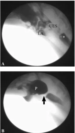
Fig. 1. Digitally captured video fluoroscopic images are shown from the patient. (A) This image is taken when iohexol contrast medium was injected with syringe into the mouth. Most quan- tity of contrast medium is present in the oral cavity with some residual contrast medium in the proximal esophagus from the previous swallow (*). Aspirated contrast medium is also seen in the larynx. (B) The larger portion of the bolus is retained in the relaxing pharynx which contracts vigorously to pass the bolus against the tightly closed cricopharyngeal passage (arrow). UES:
upper esophageal sphincter, LA: larynx, P: pharynx.
Cricopharyngeal achalasia in an old dog
263
References
1. Elliott RC. An anatomical and clinical review of cricopharyngeal achalasia in the dog. J S Afr Vet Ass 2010, 81, 75-79.
2. Ladlow J, Hardie RJ. Cricopharyngeal achalasia in dogs.
Compend Contin Educ Pract Vet 2000, 22, 750-755.
3. Malm C. Souza EM, Ferian PE, Fukushima FB, Macedo SP, Ladeira OHR, Faria ABF, Andrade MGMG. Canine cricopharyngeal achalasia: case report. Arq Bras Med Vet Zootec 2011, 63, 56-60.
4. Niles JD, Williams JM, Sullivan M, Crowsley FE.
Resolution of dysphagia following cricopharyngeal myectomy in six young dogs. J Small Anim Pract 2001, 42, 32-35.
5. Pearson H. The differential diagnosis of persistent vomiting in the young dog. J Small Anim Pract 1970, 11, 403-415.
6. Pfeifer RM. Cricopharyngeal achalasia in a dog. Can Vet J 2003, 44, 993-995.
7. Pollard RE, Marks SL, Davidson A, Hornof WJ.
Quantitative videofluoroscopic evaluation of the pharyngeal function in the dog. Vet Radiol Ultrasound 2000, 41, 409-
412.
8. Sokolovsky V. Cricopharyngeal achalasia in a dog. J Am Vet Med Assoc 1967, 150, 281-285.
9. Suter PF, Watrous BJ. Oropharyngeal dysphagias in the dog: a cinefluorographic analysis of experimentally induced and spontaneously occurring swallowing disorders. I. Oral stage and pharyngeal stage dysphagias. Vet Radiol 1980, 21, 24-39.
10. Warnock JJ, Marks SL, Pollard R, Kyles AE, Davidson A. Surgical management of cricopharyngeal dysphagia in dogs: 14 cases (1989-2001). J Am Vet Med Assoc 2003, 223, 1462-1468.
11. Watrous BJ. Clinical presentation and diagnosis of dysphagia.
Vet Clin North Am Small Anim Pract 1983, 13, 437-459.
12. Watrous BJ, Suter PF. Oropharyngeal dysphagias in the dog: a cinefluorographic analysis of experimentally induced and spontaneously occurring swallowing disorders. II.
Cricopharyngeal stage and mixed oropharyngeal dysphagias.
Vet Radiol 1983, 24, 11-24.
13. Weaver AD. Cricopharyngeal achalasia in Cocker Spaniels.
J Small Anim Pract 1983, 24, 209-214.