식물병연구
Res. Plant Dis. 17(3) : 402−404 (2011)
Research in Plant Disease
© The Korean Society of Plant Pathology
Sclerotium rolfsii에 의한 팔레놉시스 흰비단병
한경숙*·이성찬·한유경·김수·박종한1 국립원예특작과학원 원예특작환경과, 1농촌진흥청 대변인실
Sclerotium blight of Phalaenopsis Orchids Caused by Sclerotium rolfsii in Korea
Kyung-Sook Han*, Seong-Chan Lee, You-Kyoung Han, Su Kim and Jong-Han Park1
Horticultural and Herbal Crop Environment Division, National Institute of Horticultural and Herbal Science, Rural Development Administration, Suwon 441-440, Korea
1Department of Spokesperson, Rural Development Administration, Suwon 441-707, Korea (Received on August 1, 2011; Revised on August 31, 2011; Accepted on September 6, 2011)
The Sclerotium blight was found on Phalaenopsis spp. at Dong-du-cheon city, and Hwa-seong city, Gyenggi- do, Korea in September 2009. The symptom included yellowing on lower leaves and wilt of a whole plant.
Severely infected plants were blighted and died eventually. White mycelial mats appeared on the surface of basal stem and bulbs and the sclerotia were formed on stems, roots, and sphagnum moss. The sclerotia were spherical, 1−3 mm and white to brown. The optimum temperature for the growth and sclerotia formation was 25−30oC on PDA. On the pathogenicity test, the first symptom appeared 5 days after inoculation and developed to severe stem rot and blight. On the basis of mycological characteristics and pathogenicity, the causal fungus was identified as Sclerotium rolfsii. This is the first report on the sclerotium blight on Phalaenopsis spp. caused by Sclerotium rolfsii in Korea.
Keywords : Blight, Phalaenopsis, Sclerotium rolfsii
팔레놉시스(胡蝶蘭; Phalaenopsis spp.)는 재배면적 46 ha, 연간생산액 32,118백만원('09)에 달하는 난과작물로 농업 적 가치가 매우 높은 주요 화훼작물이다(Ministry for Food, Agriculture, Forestry and Fisheries, 2010). 2009년 9월, 경 기도 동두천시와 화성시 소재농가에서 팔레놉시스 재배 중 생육이 부진하고 하엽이 황화되며 시들음증상을 보이 는 식물체의 뿌리에서 흰색 곰팡이와 함께 둥근 형태를 지닌 적갈색 균핵이 형성되는 증상이 발생하였다. 오래 된 병징을 보이는 팔레놉시스는 기부가 심하게 마르고 식물체 전체가 말라죽기도 하였다. 병원균을 순수분리하 기 위하여 병든 식물체의 표면에 부착된 적갈색 균핵을 채취하여, 분리균의 균학적 특징과 병원성을 검정한 결 과 Sclerotium rolfsii에 의한 흰비단병으로 동정되었다. 흰 비단병은 감자를 비롯해서 30여종의 작물에서 발생하며,
난과작물에서는 소엽풍란에 발생한 흰비단병이 보고되었 으며(Han 등, 2010), 해바라기(Kwon, 2010), 옥잠화(Kwon 등, 2010), 애기나리(Kwon과 Jee, 2007) 등 주로 자생화 훼류에서 주로 발생하는 것으로 등록되어 있으나 Sclerotium rolfsii에 의한 팔레놉시스 흰비단병은 아직 보고된 바 없 다(The Korean Society of Plant Pathology, 2009).
본 연구에서는 팔레놉시스에 발생하는 흰비단병의 병 징과 병원균의 균학적 특징 및 병원성 검정 결과를 보고한다.
병징. 팔레놉시스가 처음에는 특이할 만한 병징을 보 이지는 않으나 병징이 진전되면서 식물체가 활기를 잃고 새순이 나오지 않는 증상을 보였다(Fig. 1A). 병징이 진 전됨에 따라 하엽이 시들며 황화되는 증상을 보이다가 차츰 기부가 마르고 뿌리가 썩게 되었다(Fig. 1B). 특히 습한 조건에서는 기부가 옅은 갈색을 띠며 수침상으로 썩고, 표면에 흰색 부챗살 모양의 균사가 형성되며, 심한 경우 식물체 전체를 말라죽게 되는 피해를 나타내기도 하였다. 팔레놉시스 흰비단병 증상은 바크표면과 뿌리에 흰색의 곰팡이가 형성되고 균핵이 밀집할 정도로 심해진
*Corresponding author
Phone) +82-31-290-6233, Fax) +82-31-290-6259 Email) [email protected]
http://dx.doi.org/10.5423/RPD.2011.17.3.402 Note Open Access
Sclerotium rolfsii에 의한 팔레놉시스 흰비단병 403
단계에서야 비로소 뿌리가 썩으면서 기부가 시드는 증상 을 보이기 때문에 균핵이 형성되기 전에는 Fusarium spp.
에 의한 줄기썩음병과 유사한 증상을 나타내기도 하였다.
병든 식물체의 뿌리표면과 기부에도 적갈색 균핵이 쉽게 관찰되었으며 비닐포트 속까지 형성되어 뿌리썩음 증상 까지 유발하였다(Fig. 1C).
균학적 특성. 팔레놉시스에서 병원균을 분리하기 위하 여 병든 조직에 형성된 둥근 적갈색 균핵을 채집하였다.
채집한 균핵은 1% 차아염소산나트륨(NaOCl) 용액에 표 면 살균하고 살균수로 3회 세척한 후 멸균된 여과지에서 물기를 말린 다음 물한천배지(WA) 위에 치상하였다. 병 원균은 25oC 항온기에서 3일간 배양 후 균사 선단부를 떼내어 감자한천배지(PDA, Difco)에 옮겨 시험균주로 사 용하였다.
분리한 병원균 동정을 위해 25oC 항온기에서 PDA에 배양하면 흰색 균사 생장이 왕성하였으며, 배양기간이 길 어지면서 배지표면에 옅은 갈색의 균핵을 많이 형성하였 다(Fig. 1E). 균핵의 모양은 대부분 둥근 형태로 광택이 있고, 처음에는 비교적 옅은 갈색이었다가, 차츰 적갈색 으로 변하였으며, 크기는 1−3 mm였다. 광학현미경으로 관 찰할 때 병원균 특유의 clamp connection 형태가 관찰되 었고(Fig. 1F), 균사폭은 4−7.8 µm였다(Table 1). 병원균의
온도에 따른 배양적 특성을 조사하기 위해 5oC 부터 40oC 까지 5oC 간격으로 균사 생장과 균핵 형성 정도를 조사 하였다. 균사는 5oC에서는 전혀 자라지 않았으며 10oC와 40oC에서는 생장이 극히 저조하였으며, 30oC에서는 25 mm/
24시간으로 가장 양호하였다. 균핵 형성정도는 배양 15일 후에 조사하였는데 10oC 이하와 40oC 이상에서는 균핵이 전혀 형성되지 않았으며 25oC와 30oC에서 갈색 균핵이 가장 많이 형성되었다. 본 연구에서 조사된 병원균의 균 학적 특성은 Mordue 등(1974)이 보고한 Sclerotium rolfsii 와 거의 일치하였다.
Fig. 1. Symptoms of Sclerotium blight on Phalaenopsis spp. caused by Sclerotium rolfsii and mycological characteristics. A: Infected plants showing wilt and yellow lower leaves, B: Typical symptom showing blight on root and bulb surface, C: Screlotia formed on the root in the pot, D: Symptoms occurred by artificial infection, E: Mycelial mat and sclerotia grown on PDA after 9 days incubation at 30oC, F: A clamp connection in the hyphae of the pathogenic fungus (Bar = 7 µm).
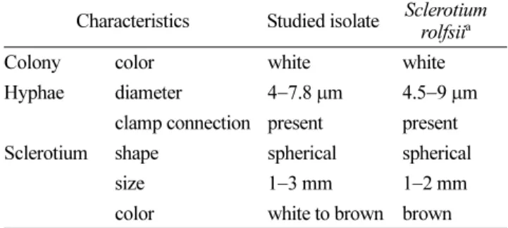
Table 1. Comparison of mycological characteristics between the isolate studied and Sclerotium rolfsii
Characteristics Studied isolate Sclerotium rolfsiia
Colony color white white
Hyphae diameter 4−7.8 µm 4.5−9 µm clamp connection present present Sclerotium shape spherical spherical
size 1−3 mm 1−2 mm color white to brown brown
aDescribed by Mordue
(1974).
404 한경숙·이성찬·한유경·김수·박종한 병원성 검정. 팔레놉시스에 대한 병원성을 확인하기
위하여 직경 10 cm 플라스틱 포트에 심겨진 건전한 팔레 놉시스 식물체를 구입하여 이용하였다. 순수 분리한 병원 균은 PDA에 5일간 배양 후, oat-meal 모래배지(oat-meal:
모래:물=1:5:4)에 균총을 잘게 잘라 접종한 후 27oC에서 10일간 배양하여 접종원을 준비하였다. 이병토양은 그늘 에서 하루 동안 말린 다음 미세하게 부수었으며, 포트 당 5 g씩 지제부에 접종하고 관주하였다. 접종된 식물체는 20−35oC의 유리온실에서 유지하며 발병정도를 관찰하였 다. 접종 5일 후 발병 식물의 수태와 뿌리표면이 흰색의 부챗살 모양의 균사체로 덮혔다. 이후 지제부 기부와 하 엽이 천천히 황화되기 시작하였으며(Fig. 1D), 기부가 수 침상으로 물러지고 썩으면서 접종 20일 만에 말라 죽었 다. 병징을 보이는 팔레놉시스의 기부와 수태속에서 암갈 색의 작고 둥근 균핵이 형성되어 흰비단병 특유의 표징 을 확인할 수 있었다. Sclerotium rolfsi에 의한 흰비단병 은 심비디움, 덴드로비움, 팔레놉시스에 발생하는 것으로 보고되었으며(Kunihei, 1998), 우리나라에서는 소엽풍란에 발생하는 것으로 보고된 바 있다(Han 등, 2010).
요 약
2009년 9월 경기도 동두천시와 화성시 소재 농가에서 팔레놉시스 재배중 생육이 부진하고 하엽이 황화되며 심 한 경우 뿌리와 기부가 마르고 식물체 전체가 말라죽는 증상이 발생하였다. 말라죽은 기부와 수태표면에 흰색 곰 팡이 균사와 함께 둥근 형태의 적갈색 균핵이 관찰되었 다. 균학적 특징으로 균사는 25−30oC에서 가장 활발한 생
장을 보였으며, 균핵은 갈색의 둥근형태로 직경은 1−3 mm 였다. 병원성 검정결과 병징이 접종 후 5일째 관찰되었으 며 그 후 급속하게 진전되어 강한 병원력을 보였다. 팔레 놉시스에서 발생한 병징과 병원균의 균학적 특징 및 병 원성을 검정한 결과 이 증상은 Sclerotium rolfsii에 의한 흰비단병으로 동정되어 본 병을 팔레놉시스 흰비단병으 로 명명하고자 한다.
References
Han, K. S., Lee, S. C., Han, Y. K. and Kim, S. 2010. Sclerotium blight of Neofinetia falcata caused by Sclerotium rolfsii in Korea. Res. Plant Dis. 16: 320–322. (In Korean)
Kwon, J. H. and Jee, H. J. 2007. Occurrence of stem rot of Disporum smilacinum caused by Sclerotium rolfsii in Korea.
Plant Pathology. J. 23: 212–214.
Kwon, J. H. 2010. Occurrence of stem rot of sunflower (Helianthus annuus) caused by Sclerotium rolfsii. Res. Plant Dis. 16: 323–325. (In Korean)
Kwon, J. H., Chi, T. T. P. and Kim, J. W. 2010. First report of stem rot on Hosta plantagina caused by Sclerotium rolfsii in Korea.
Plant Pathology J. 26: 297.
Kunihei, K. 1998. Plant disease in Japan. Association for National Farming Village Education. 1276 pp. (In Japanese) Ministry for Food, Agriculture, Forestry and Fisheries. 2010.
2009 Flower crop production in Korea. 256 pp. (In Korean) Mordue, J. E. M. 1974. CMI descriptions of pathogenic fungi and bacteria. No. 410. Commonwealth Mycological Institute, Kew, Surrey, England.
The Korean Society of Plant Pathology. 2009. List of plant diseases in Korea, 5th ed. 853 pp. (In Korean)