Introduction
Vertebrates with different habitats have diffe- rent proportions of rods and cones in their reti- nas, and some nocturnal species lack cones enti- rely (Nicol, 1989). These facts are among those that lead Schultze (1866) to formulate the dupli- city theory. In accordance with this theory, it is now generally considered that scotopic vision is mediated by rods and photopic vision by cones (Ole, 1984). Indeed, some deep-sea fishes have pure rod retinas (Locket, 1977), but the presence of well-developed double cones in other deep- sea teleosts may serve as further evidence for their ability to function at low light levels. That
is, the quantal response is much faster in cones than in rods, which makes the photopic system better at encoding rapidly changing visual stimu- li (Van der Meer, 1992). In teleosts, most cones are single or double cells, but triple and quadru- ple cones occur occasionally in some species (Engström, 1963; Nicol, 1989). The double cones are divided into equal double cones (twin cones) and unequal double cones by only the cell size.
The equal double cones are subdivided into iden- tical double cones and non-identical double con- es by contained visual pigments (Van der Meer, 1992). Such cones are distributed irregularly in some species. It is usual, however, to find single and double cones arrange in specific patterns, such these cone patterns are only associated wi-
─
─ 183 ──
Comparative Study of Lens and Retinal Tissues in Zacco temmincki and Hemibarbus longirostris
(Cypirinidae, Cyprinifomes)
Jae-Won Lim, Chung-Lyul Lee*, Won-Koo Lee and Sung-Ju Jye-Gal**
Faculty of Biological Science, College of Natural Science, Chonbuk National University, Jeonju 561-756, Korea, *Department of Biology, College of Natural Science,
Kunsan National University, Kunsan, 573-701, Korea,
**Department of Clinical Pathology, College of Wonkwang Health Science, Iksan 570-750, Korea
Retina and lens in Zacco temmincki and Hemibarbus longirostris were comparati- vely investigated using light microscopy and scanning electron microscopy. In radial sections of the two species, the retinal thickness was mostly in the centre of the fundus, and decreased towards the margin. The outer nuclear layer was thicker than the inner nuclear layer. However, the densities of the horizontal cells and ganglion cells in Z. temmincki appeared to be higher than those in H. longirostris. In tangential sections, as one moves away from the centre, the double cone cells and single cone cells increased in size, the densities of these were highest in the centre of the fundus. The cone cells were in a compacted mosaic pattern in Z. temmincki, and the density of the cone cells was higher than that of randomly distributed cone cells in H. longirostris.
It seemed that the width and the interlocking formula of lens fibers differed in each species. In Z. temmincki, the interlocking formula of lens fibers was seen as an
“anchor-and-socket” connection, and the width of the lens fiber was thicker than that of H. longirostris. On the other hand, the interlocking formula of lens fibers was seen as a “ball-and-socket” connection in H. longirostris.
Key words : Lens, Hemibarbus longirostris, SEM, retina, Zacco temmincki
th double cones (Lyall, 1957). Also, the most of the retinas comprises an outer and inner retina.
The former is made up of an external sheet of pi- gment epithelium and a layer of photoreceptors;
the latter is a nervous tissue, comprises 9 or 10 layers; layer of visual cell nuclei, outer nuclear layer, horizontal cell layer, outer plexiform layer, bipolar cell layer, amacrine cell layer, inner nucl- ear layer, inner plexiform layer, ganglion cell la- yer and optic nerve-fiber layer. But bipolar cell layer and amacrine cell layer are located in inner nuclear layer (Nicol, 1989). Dark chub, Z. temmi- ncki and long nose barbel, H. longirostris belong to the abundant fish with pale chub of upper or midstream in Korea, and distributed in Japan and Manju area of China (Kim, 1997). Z. temmin- cki inhabit at swift current, and H. longirostris do at gentle current, all the two species mainly prey on the aquatic insects (Kim, 1997).
The objective of the present work is to identify the differences of the retina and lens by light and scanning electron microscope in two teleosts, Z.
temmincki and H. longirostris that adapted to different habitats. Although the fixed samples were used for electron microscopy, the retina examined was unfortunately not adequately pre- served for a general study of its ultra-structure.
Materials and Methods
In this study, 11 specimens of Z. temmincki (103.7~107.3 mm, TL) and 12 specimens of H.
longirostris (118.9~151.3 mm, TL) were collected at Jeonju stream, Jeonju-si, Jeollabuk-do, Kor- ea in June and July, 2002.
Retina: All specimens were fixed 10% forma-
lin, eye and lens diameter were measured with micrometer in extracted eyeballs. After the corn- ea, iris, and lens removed, retinas were immedia- tely dissected along to the median horizontal and vertical lines. Eight small fragments (2×2 mm) for tangential section and three fragments (3×4 mm) for radial section were cut out mainly from the right eye (Fig. 1). Dissected fragments were subjected to the successive course of dehydration in ethanol for paraffin embedding, clearing in xy- lene, and paraffin infiltration in paraffin wax.
Embedded samples were sectioned at thickness of 3
µm by rotary microtome and retinal tissues were stained with Harris’s haematoxylin. The samples were observed under the light microsco- pe (Olympus BX41), and Image plus pro 4.0 (Me-
dia cyber netics).
Lens: Both sides of lens were cut out to the de-
gree of one-forth and one-third (Nicol, 1989) for scanning electron microscopy (SEM) and prefixed in 2.5% glutaraldehyde. Samples were washed in 0.1 M CB and postfixed in 1% osmium tetroxide.
And samples were washed in 0.1 M CB and carri- ed out the successive course of dehydration in ethanol series. After processing with 3-methyl buthyl (isomyl) acetate and HMDS. They coated with gold and examined with a SEM (Akashi SR -50).
Results
1. The external morphology of the eyeball
In experiments using Z. temmincki, the avera- ge horizontal diameter of the eyeball was 7.5 mm. The average vertical diameter was 7.2 mm (Table 1). And the average ratio of the horizontal and vertical diameter was 0.96 : 1, resulting the eyeball of Z. temmincki close to globular shape.
The pupil was globular in shape and the average diameter was 3.6 mm (Table 1).
In H. longirostris, the average horizontal dia- meter of the eyeball of was 7.6 mm. The average vertical diameter was 6.3 mm (Table 1) and the average ratio of the horizontal and vertical dia- meter was 1.2 : 1. The eyeball of H. longirostris was horizontally ellipse in shape. The average horizontal diameter of pupil,was 4.0 mm. The average vertical diameter of pupil was 3.1 mm (Table 1), leading to the average ratio of the hori- zontal and vertical diameter 1.26 : 1 and the pu- pil of H. longirostris vertically ellipse in shape, opposite of the eyeball.
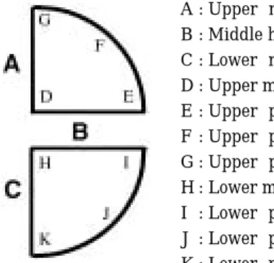
Fig. 1. Idealized position of samples for the radial and ta- ngential sections.
A : Upper-middle verticalness (UMH) B : Middle horizon (MH)
C : Lower-middle verticalness (LMV) D : Upper middle (UM)
E : Upper-peripheral low (UPL) F : Upper-peripheral middle (UPM) G : Upper-peripheral high (UPH) H : Lower middle (LM)
I : Lower-peripheral high (LPH) J : Lower-peripheral middle (LPM) K : Lower-peripheral low (LPL)
2. Morphology of lens
The lens in Z. temmincki was globular in sha- pe, while the average diameter was 3.4 mm and maximum & minimum diameter 4.2~3.0 mm.
In H. longirostris, the lens was globular in sha- pe, the average diameter 3.2 mm, maximum and minimum diameter 3.4~3.0 mm.
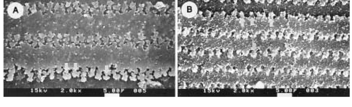
Interlocking interdigitations of the lens fiber were evident at the edges along the length. The interlocking morphology of the lens fibers was shown as an “anchor-and-socket” connection and the width of the lens fibers was about 6.5
µm in Z. temmincki (Fig. 2A). While the interlocking morphology of lens fibers was shown as a “ball- and-socket” connection, and the width of lens fibers was about 4.5
µm in H. longirostris (Fig.
2B). The lens epithelium covered about third- forth of frontal lens surface and the lenses origi- nally occupied a central position within the pupil.
3. Retinal histology
1) Comparative observation of radial sections on the ratinas
The outer nuclear layers of Z. temmincki and H. longirostris were developed more than those of inner nuclear layers. The processes of pigment epithelium were well developed and thus the pig- mented processes penetrated among the visual cells. Totally, the retinal thickness was the thick-
est in the centre of the fundus and decreased towards the margin in both species (Fig. 3A). The total retinal thickness of Z. temmincki was thick- er than that of H. longirostris. The density of the horizontal and ganglion cell of Z. temmincki were 1.7 and 1.3 times higher than those of H. longiro- stris, respectively (Fig. 3B-1, B-2; Table 2).
2) Comparative observation of tangential sections on the ratinas
The cone patterns were shown clearly in tange- ntial sections of the retina in Z. temminck. Of observed eight sections, they were grouped into largely four type arrangements with double cones and single cones (Fig. 3C-1~C-4). The sections of the upper-peripheral high (UPH) and the lower-peripheral low (LPL) were arranged so that largely double cone rows were running ra- dially, a row of single cones being separated by a row of double cones. Each row of double cones contained alternating a little saturated and light stained cells (Fig. 3C-1). The 2nd type of the arrangements was shown in the sections of the upper-peripheral middle (UPM) and the lower- peripheral middle (LPM). In the UPM and the LPM, the arrangements of double and single cells were similar to the sections of the UPH and the LPL, but the size of the double and single cells was smaller than that of cells in the UPH
Fig. 2. Structure and interlocking morphology of (A) Z. temmincki and (B) H. longirostris by the SEM. (×2,000)
Table 2. Measurements of the density of horizontal and ganglion cell layers of Z. temmincki and H. lon-
girostris (Unit: cells/100µm)
Species Z. temmincki H. longirostris
Content Mean Range Mean Range
Den. of Hori. cell* 12.0 13~11 7.22 8~6 Den. of Gang. cell** 16.6 23~11 13.1 20~10
*: Density of horizontal cell
**: Density of ganglion cell Table 1. Measurements of eyeball and pupil in Z. tem-
mincki and H. longirostris (Unit: mm)
Eyeball Pupil
Hori. Dia.* Vert. Dia.** Hori. Dia* Vert. Dia.**
Z. temmincki 7.68~7.35 7.35~7.11 3.02~4.36 H. longirostris 7.73~7.49 6.72~5.77 4.93~3.43 3.43~2.85
*: Horizontal diameter, **: Vertical diameter
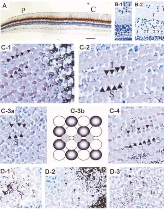
Fig. 3. Structures of retinal tissues of Z. temmincki and H. longirostris.
A, Structure of radial section of retina of H. longirostris. Bar = 60µm. (×40). C, central portion; P, peripheral porti- on
B, Horizontal and ganglion cells (B-1) in Z. temmincki and (B-2) H. longirostris. Bar = 5µm. (×400). G, ganglion cells layer; H, horizontal cells layer
C, Structures of tangential section of retina of Z. temmincki. Bar = 5µm. (×400). (C-1) in the upper-peripheral high and lower-peripheral low. (C-2) in the upper-peripheral middle and lower-peripheral middle. (C-3a) in upper-peripheral low and lower-peripheral high. (C-3b) diagram of cone cells in the upper-peripheral low (al- ternated double cells surround a single cell). (C-4) in the upper middle and lower middle. As, accessory single cone; Cs, central single cone; I, double cone; M, the region that pigment processes surrounding cone cells; S, single cone
D, Structures of tangential section of retina of H. longirostris. Bar = 5µm. (×400). (D-1) in the upper-peripheral high and lower-peripheral low. (D-2) in the upper-peripheral middle and lower-peripheral middle. (D-3) in the upper middle, upper-peripheral low, lower middle and lower-peripheral high. I, identical twin cone; Ls, long single cone; M, the region that pigment processes surrounding cone cells; NI, non-identical twin cone; Ss, short single cone; U, unequal double cone.
and the LPL (Fig. 3C-2). The 3rd type of com- mon patterns was situated in the upper-periph- eral low (UPL) and the lower-peripheral high (LPH). The smallest double and single cones re- gularly alternated in the UPL and the LPH.
Double cones were parallel to each other and seen as row pattern, so it was seen the highest density of cone cell (Fig. 3C-3a, C-3b). The last pattern, the 4th common type was shown at the upper and lower middle (UM & LM) in Z. tem- minck. More clear double cells alternate. Cen- tral single cones (long single cones) and additio- nal single cones (short single cones) existed and they regularly alternated between the double cones (Fig. 3C-4).
In H. longirostris, the visual cells and cone pat- tern were different to those of Z. temminck. The five kinds of cone cells, i.e., short and large sing- le cones, unequal double cones, identical and non -identical twin cones, were found at the eight sections, distinguished into largely three type arrangements in the retinal tissue of H. longiros- tris (Fig. 3D-1~D-4). In the 1st type, all of the five kinds of the cone cells were situated in the UPH and the LPL, but the cone cells were large, mainly non-identical twin cones and long single cones (Fig. 3D-1). The 2nd type of the arrange- ments was shown in the parts of the UPM and the LPM. Three kinds of the cone cells, identical twin cones, short single cones, and some unequal cones were found in the UPM and the LPM. But as the cone cells were smaller than those of the UPH and the LPL, the density of cone cell was appeared higher (Fig. 3D-2). The 3rd type of the arrangement was situated in the UM, the UPL, the LM, and the LPH. And the five kinds of the cone cells were found similarly as the UPH and the LPL. However, since the cone cells were sma- ller than those of the UPM and the LPM, the density of cone cell was appeared higher (Fig. 3D -3).
The cone cells regularly arranged at M region, the long processes of pigment cells surround cone cells, and the rest area in Z. temminck (Fig. 3C- 1). However, the cone cells of H. longirostris were shown irregular arrangements at M region and the rest area (Fig. 3D-2), but only the short sing- le cones were shown comparatively regular arra- ngements (Fig. 3D-2, D-3).
Discussion
The present study was carried out to investiga-
te the kind, shape, arrangement, and distributi- on of cone cells in relation to the positions of reti- na, the shape, and interlocking formula of lens fibers in Z. temmincki and H. longirostris belong- ing to the family Cyprinidae. As a result, several different types were found according to their adaptation of habitat and prey between Z. tem- mincki and H. longirostris.
Nicol (1989) mentioned that horizontal and ganglion cells transmitted the stimuli of photore- ceptor cells to visual cells and inner nuclear lay- er in animal retina tissues. As the cell densities per 100
µm turned out 1.7 and 1.3 times high in Z. temmincki, it was known that Z. temmincki had clearer resolution than that of H. longirostr- is. Also, the good resolution was demanded on the high density of visual cells (Nicol, 1989). The density of visual cell was appeared high with row and square pattern of cone cells in Z. temmincki.
But in the case of H. longirostris, the density of visual cell was emerged lower than that of Z.
temmincki, because cone cells were scattered irr- egularly. In general, middle and long-wave sen- sitivity was subserved by the double cone, while short-wave sensitivity was arched by the single cone (Harosi and Hashimoto, 1983; Bowmaker and Kunz, 1987). Therefore, as the single cones were situated about three times in the retinas of Z. temmincki, double cones were compactly situ- ated in Z. temmincki than the cone cells of H.
longirostris. This showed an adapted phase to more refined freshwater and the eye of Z. tem- mincki had a good resolution for detecting a prey at swift current. But it seemed that visual cells were not well developed, because H. longirostris inhabits at gentle current. And also, interlocking formula of lens fibers was concerned with the transparency of the lens (Kessel and Kardon, 1979). It seemed that the distinctions of the width and the interlocking formula of lens fibers were concerned with a good resolution between Z. temmincki and H. longirostris. Even if the density of visual cells differed in two species, the visual cells became small and it appeared that the densities of visual cells increased from the margin to the centre. It was known that the high densities of the visual cells at the centre and central horizon had been a good resolution to mainly aquatic insects in the front of eye and head (Kim, 1997).
But two specimens, in H. longirostris, collected
near the Wansangyo at Jeonju stream, had only
single cells, short single cells, and long single
cells, in all eight sections. Therefore, it seemed that the phases must be investigated either the changes of the ontogeny or possible deformity due to habitat pollution from sewage in the futu- re.
Acknowledgements
I wish to express my thanks to Prof. Dr. Lee, Moo-Sam, the Medical school and Prof. Dr. Kim, Kyungsik, the Faculty of Biology, Chonbuk Na- tional University for providing the chance to exa- mine the SEM. I am also grateful to the memb- ers of ichthyological laboratory, Faculty of Biolo- gy, Chonbuk National University for help in coll- ecting samples. I particularly thank to the mem- bers of ichthyological laboratory, Department of Biology, Kunsan National University for help in completing the manuscript.
References
Bowmaker, J.K. and Y.W. Kunz. 1987. Ultraviolet receptors, tetrachromatic colour vision and reti- nal mosaics in the brown trout (Salmo trutta):
Age-dependent change. Vision Res., 27(12) : 2101~2108.
Engström, K. 1963. Structure, organization and ul- trastructure of the visual cells in the teleost fa- mily Labridae. Acta Zoologica (Stockbolm), 44 : 1~14.
Harosi, F.I. and Y. Hashimoto. 1983. Ultraviolet visual pigment in a vertebrate: a tetrachromatic cone system in the Dace. Science N. Y., 222 : 1021~1023.
Kim, I.S. 1997. Illustrated encyclopedia of fauna &
flora of Korea. vol. 37. Freshwater fishes. Minis- try of Education, pp. 220~271.
Kessel, R.G. and R.H. Kardon. 1979. Tissues and organs: A text-atlas of scanning electron micro- scopy. W. H. Freeman and company, pp. 84~
105.
Locket, N.A. 1977. Adaptations to the deep sea en- vironment. In: Crescitelli F. (ed.), The visual system in vertebrates. Handbook of sensory phy- siology. Springer Verlag, 7(5) : 67~192.
Lyall, A.H. 1957. Cone arrangements in teleost reti- nae. Quarterly J. Microscopical Sci., 98(2) : 189
~201.
Nicol, J.A.C. 1989. The eye of fishes. Clarendon Press, Oxfors, pp. 1~308.
Ole, M. 1984. Duplex retina in the mesopelagic tele- ost Radiicephalus elongatus osorio, 1917. Viden- sk. Meddr dabnk naturh Foren., 145 : 183~199.
Schultze, M. 1866. Zur anatomie und physiologie der retina. Arch. Mikrosh. Anat., 2 : 175~286.
Van der Meer, H.J. 1992. Constructional morpholo- gy of photoreceptor patterns in Percomorph fish.
Acta Biotheoretica, 40 : 51~85.
Received : June 13, 2002 Accepted : September 8, 2002
갈겨니
(Zacco temmincki )
와 참마자(Hemibarbus longirostris )
의 수정체와 망막조직의 비교 연구임재원∙이충렬*∙이원구∙제갈승주**
전북대학교 자연과학대학 생물과학부
*군산대학교 자연과학대학 생물학과
**원광보건대학 임상병리과
잉어과 어류에 속하는 갈겨니와 참마자의 안구 중 수정체와 망막의 조직을 주사전자현미경과 광학현미경 하에서 조사하였다. 망막조직은 두 종 모두 중심부에서 주변부로 갈수록 얇아지고, outer nuclear layer가 inner nuclear layer보다 발달한 모습을 보였다. 그러나 갈겨니의 경우 horizontal cells과 ganglion cells의 밀도가 참마자보다 높게 나타난다. 그리고 갈겨니 망막 조직 에서 double cone cells과 single cone cells의 밀도는 중심부에서 가장 높게 나타나고, 중심부에서 멀어질수록, 그리고 중심부 수평에서 멀어질수록 세포가 커지면서 세포밀도가 낮아지지만 모자 이크 형태를 띄는 갈겨니의 cone cells은 일정한 규칙성 없이 흩어져 있는 참마자의 cone cells보 다 세포밀도가 높게 나타난다. 수정체 섬유의 폭과 결합방식에서도 두 종이 각기 다른 양상을 보 이는데 갈겨니의 수정체 섬유의 폭은 참마자보다 넓고 수정체 섬유들이 배의 닻과 같은 형태로 맞물려 있으나, 참마자의 수정체 섬유는 공과 같은 형태로 맞물려 있다.