Introduction
Osteoarthritis (OA) is a progressively debilitating condition that causes cartilage erosion of the involved joint with pain and func
tional impairment. Traditional methods to regenerate defects of articular cartilage include microfracture, multiple perforations, abrasions and mosaicplasty, the results of which are not satisfac
tory1,2). Autologous chondrocyte implantation (ACI) could be one method to regenerate an articular cartilage defect. However, ACI is not applicable to articular cartilage defects in OA because chondrocytes from patients suffering from OA have totally differ
ent biological properties3). Additionally, for large articular carti
lage defects in patients with OA, an alternative cell source should be found. As an alternative, mesenchymal stem cells (MSCs) can be used to regenerative articular cartilage defects4). MSCs can be isolated from a variety of sources, such as bone marrow (BM), adipose tissue, umbilical cord, amniotic fluid, dental pulp, sy
novial tissue, peripheral blood and skeletal muscles. It has been reported that the BMderived MSCs contain progenitor cells of
IntraArticular Injection of Bone MarrowDerived Mesenchymal Stem Cells Leading to Better Clinical Outcomes without Difference in MRI Outcomes from Baseline in Patients with Knee Osteoarthritis
YoungSoo Shin, MD, PhD
1,*, JungRo Yoon, MD, PhD
1,*, HeeSun Kim, RN, PhD
2, and SeonHeui Lee, RN, PhD
31Department of Orthopedic Surgery, Veterans Health Service Medical Center, Seoul; 2Chonbuk Research Institute of Nursing Science, College of Nursing, Chonbuk National University, Jeonju; 3Department of Nursing Science, College of Nursing, Gachon University, Incheon, Korea
Purpose: Bone marrow (BM) is frequently used as a source of mesenchymal stem cells (MSCs) because they have a high potential for differentiation.
However, it is unclear whether BMderived MSCs lead to better clinical and magnetic resonance imaging (MRI) outcomes postoperatively.
Materials and Methods: This metaanalysis compared the clinical and MRI outcomes in patients with knee osteoarthritis (OA) treated with BM
derived MSCs. Eight studies comparing the clinical and MRI outcomes assessed with various measurement tools in patients with knee OA treated with BMderived MSCs were included.
Results: The range of motion (95% confidence interval [CI], –13.05 to 4.24; p=0.32) and MRI outcomes (95% CI, –0.16 to 1.40; p=0.12) did not differ significantly between the baseline and final followup. In contrast, pain (95% CI, 0.89 to 1.87; p<0.001) and functional outcomes (95% CI, 0.70 to 2.07;
p<0.001) were significantly improved at the final followup when compared to the baseline.
Conclusions: This metaanalysis found no significant difference in the tested range of motion and MRI outcomes between the baseline and the final followup in patients treated with BMderived MSCs, whereas significant functional improvement and pain relief were noted when compared with the baseline. Thus, BMderived MSCs appear to be a viable alternative for patients with knee OA, although longterm and highquality randomized controlled trials are needed to confirm the clinical benefits.
Keywords: Knee, Osteoarthritis, Bone marrow, Mesenchymal stem cells, Meta-analysis pISSN 2234-0726 · eISSN 2234-2451
Knee Surgery & Related Research
Received December 19, 2017; Revised February 6, 2018;
Accepted April 24, 2018
Correspondence to: SeonHeui Lee, RN, PhD
Department of Nursing Science, College of Nursing, Gachon University, 21 Namdongdaero, 774 beongil, Namdonggu, Incheon 21565, Korea Tel: +82328204230, Fax: +82328204231
Email: [email protected]
*These authors contributed equally to this study.
206
This is an Open Access article distributed under the terms of the Creative Commons Attribution NonCommercial License (http://creativecommons.org/licenses/bync/4.0/) which permits unrestricted noncommercial use, distribution, and reproduction in any medium, provided the original work is properly cited.
Copyright © 2018 KOREAN KNEE SOCIETY www.jksrr.org
some mesenchymal tissues, such as cartilage and other tissues5). Furthermore, BM is frequently used as a source for MSCs be
cause they are relatively easy to isolate and they have a high po
tential for differentiation, even though it needs amount increases over several weeks in culture conditions for effective cellular dos
age6). Although there have been many studies on adiposederived MSCs or adiposederived stromal vascular fraction710), few have assessed BMderived MSCs and the results have been inconclu
sive. Previous studies have conducted a systematic review of the MSCs for the treatment of cartilage lesions11,12); however, evidence is insufficient due to the different location of cartilage defect, dif
ferent cell sources, different etiology, and different MSC dosage.
Few direct comparisons of clinical scores on knee outcome scales and magnetic resonance imaging (MRI) outcomes between the baseline and final followup have been conducted in patients with knee OA treated with BMderived MSCs. In addition, no meta
analyses on this subject have been published.
This metaanalysis was performed to assess clinical and MRI outcomes after surgery in patients with knee OA treated with BMderived MSCs. It was hypothesized that BMderived MSCs would lead to better clinical outcomes and MRI outcomes on fi
nal followup in these patients.
Materials and Methods
1. Data and Literature Sources
This study followed the Cochrane Review Methods. Multiple comprehensive databases, including MEDLINE (January 1, 1976 to September 30, 2017), EMBASE (January 1, 1985 to September 30, 2017), and the Cochrane Library (January 1, 1987 to Septem
ber 30, 2017) were searched for studies of patients with knee OA treated with BMderived MSCs which used the following assess
ments to compare clinical outcome: visual analog scale (VAS) for pain, Western Ontario McMaster Universities Arthritis Index (WOMAC), Lysholm score, Hospital for Special Surgery (HSS) score and range of motion (ROM). Selected studies also com
pared MRI outcomes using the wholeorgan magnetic resonance imaging score (WORMS) and poor cartilage index (PCI). There was no language restriction, and filters of any kind were not ap
plied for the strategy. The following keywords and their compre
hensive combination and pertinent Medical Subject Headings (MeSH) were used to select the relevant articles: ‘mesenchymal stem cells’ OR ‘mononuclear cells’ OR ‘bone marrowderived mesenchymal stem cell’ OR ‘bone marrow stromal cells’ OR
‘muscular skeletal disease’ OR ‘osteoarthritis.’
After the initial electronic search, relevant articles and their bib
liographies were searched manually.
2. Study Selection
Based on the title and abstract, two reviewers independently selected the relevant studies for full review. The full text copy of each article was reviewed if the abstract did not provide enough data to make a decision. Studies were included in the meta
analysis if they (1) assessed clinical outcomes, as determined by VAS for pain, WOMAC, Lysholm score, HSS score and ROM between the baseline and final followup, and MRI outcomes, as determined by WORMS and PCI between the baseline and final followup; (2) reported direct comparisons of surgical outcomes in patients with knee OA treated with intraarticular injections of BMderived MSCs; (3) included data on at least one of the following 7 parameters: VAS for pain, WOMAC, Lysholm score, HSS score, ROM, WORMS and PCI. For the overall functional outcome measure, we combined comparable scores from differ
ent functional outcome tools when these tools scored disability:
the higher the score, the greater the disability. Using the same method, we combined comparable scores of postoperative pain:
the higher the score, the greater the pain. For WORMS, we re
corded total WORMS score as assessed by cartilage thickness, signal intensity, and subchondral bone alteration and volume.
The higher score values indicate more damage13). PCI was evalu
ated using the mean T2 relaxation values sampled in 88 wellde
fined regions of interest: values at 100 present the worst possible PCI, and those at 5 or below are considered healthy14); (4) fully reported the number of patients in each group (baseline and final followup groups) and the means and standard deviations for the 7 parameters; and (5) used adequate statistical methods to com
pare these parameters between groups. Studies were excluded if they (1) were not original articles; (2) were preclinical studies;
(3) had missing or inadequate outcome data, such as standard deviations or ranges of values; and (4) used open surgery as the delivery method.
3. Data Extraction and Methodological Quality Assessment Two reviewers independently recorded data from each study using a predefined data extraction form and resolved any differ
ences by discussion. Variables recorded included those associated with surgical outcomes, such as VAS for pain, WOMAC, Lysholm score, HSS score, ROM, WORMS and PCI. Sample size and the mean and standard deviation of surgical outcomes in each group were also recorded. If these variables were not included in the articles, the standardized mean difference (SMD) was calculated from the pvalue and sample size.
Two reviewers independently assessed the methodological qual
ity of the studies using interrupted time series analyses, as recom
mended by the Cochrane Effective Practical and Organisation of Care Group. Each study was judged based on seven standard criteria to detect whether the intervention has an effect signifi
cantly greater than the underlying secular trend. Any unresolved disagreements between reviewers were resolved by consensus or by consultation with a third investigator.
4. Data Synthesis and Analysis
The main outcomes of the metaanalysis were the SMD for overall clinical outcomes and MRI outcomes at the final follow
up compared to the baseline values due to use of several different measurement tools, including VAS for pain, WOMAC, Lysholm score, HSS score, ROM, WORMS and PCI. For all comparisons, SMD and 95% confidence interval (CI) were calculated for con
tinuous outcomes. Heterogeneity was determined by estimating the proportion of betweenstudy inconsistencies due to actual differences between studies, rather than differences due to ran
dom error or chance, using the I2 statistic, with values of 25%, 50%, and 75% considered low, moderate, and high heterogeneity, respectively. All statistical analyses were performed with RevMan ver. 5.3 (The Cochrane Collaboration, Copenhagen, Denmark).
The risks of bias (low, high, or unclear) were independently as
sessed by two investigators. Subgroup analyses based on differ
ences in the length of followup time were performed for pain scores in an attempt to explore a potential source of heteroge
neity. As a result, two subgroups were created: followup more than 3 years and followup less than 3 years for pain scores. In addition, sensitivity analysis was conducted by excluding one of the eligible studies at a time; two studies with additional surgical procedures, such as cartilage treatment and osteotomy were in
cluded15,16). Pooling of data was feasible for only two outcomes of interest, i.e., pain and function scores.
Results
1. Identification of Studies
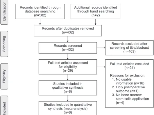
Details on study identification, inclusion, and exclusion are summarized in Fig. 1. An electronic search yielded 270 studies in PubMed (MEDLINE), 309 in EMBASE and 3 in the Cochrane Library. Two additional publications were identified through manual searching. After removing 152 duplicates, 432 studies re
mained; of these, 16 were excluded based on the abstract and full
text article review, and an additional eight studies were excluded because they had unusable information or made inappropriate group comparisons. This eventually resulted in 8 studies that were included in the metaanalysis1522).
IncludedEligibilityScreeningIdentification
Records after duplicates removed (n=432)
Records screened (n=432)
Full-text articles assessed for eligibility
(n=29)
Studies included in qualitative synthesis
(n=8)
Studies included in quantitative synthesis (meta-analysis)
(n=8)
Records excluded after screening of title/abstract
(n=403) Records identified through
database searching (n=582)
Additional records identified through hand searching
(n=2)
Full-text articles excluded (n=21) Reasons for exclusion:
1. No usable (n=16) 2. Only postoperative
outcome (n=1) 3. No bone marrow
stem cells application (n=4)
information
Fig. 1. Preferred Reporting Items for Sys
tematic Reviews and MetaAnalyses (PRIS
MA) flow diagram of literature selection.
2. Study Characteristics and Patient Populations
The 8 studies we examined included 161 patients who under
went surgical treatment for knee OA with BMderived MSCs.
Five studies (5 randomized controlled trials [RCTs]) compared prospectively measured parameters, whereas the other three studies compared parameters measured by retrospective chart review. Seven studies reported pain score, six reported function score, four reported MRI score, and two reported ROM (Table 1).
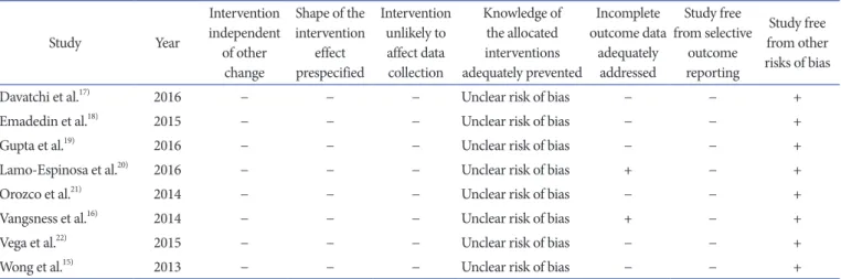
3. Quality and Publication Bias of the Included Studies The quality of the 8 studies included in the metaanalysis is summarized in Table 2. Publication bias could not be assessed in these trials. Tests for funnel plot asymmetry are typically per
formed only when at least 10 studies are included in the meta
analysis. The metaanalysis included only 8 studies, and tests for asymmetry were not performed because these tests would not be able to differentiate asymmetry from chance.
4. Clinical Outcomes
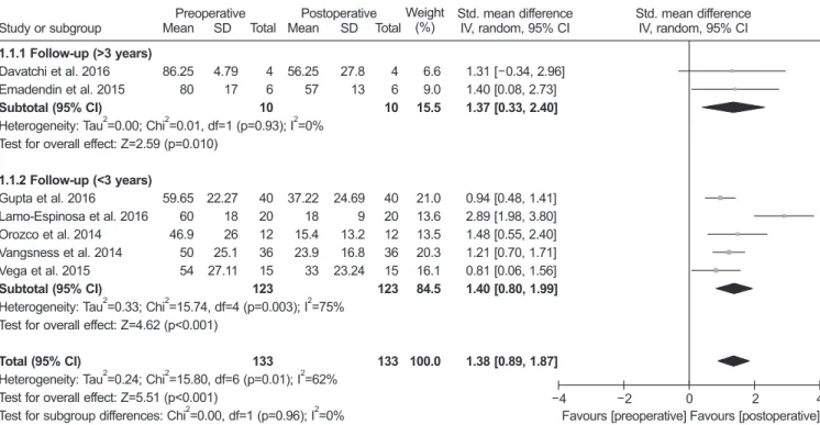
Of the 8 studies, 7 compared pain score and included 133 pa
tients assessed at the baseline and 133 at the final followup. The standardized mean was 1.38 points lower at the final follow
up than at the baseline, and was significantly different between groups (95% CI, 0.89 to 1.87 points; p<0.001; I2=62%) (Fig. 2).
Two studies were assigned to the followup more than 3 years and five studies to the followup less than 3 years. The standard
Table 1. Summary of Study Characteristics Study Year Study
type
No. of patients
(M/F)
Mean age (yr)
BMI (kg/m2)
Mean final F/U
(mo)
Cell
number Amount
(mL) Delivery
method Concomitant
procedure Measured parameter
Davatchi et al.17) 2016 OCS 4 (2/2) 57.8 31.4 60 8–9×10⁶ 30 IA injection No PS, ROM
Emadedin
et al.18) 2015 OCS 6 (0/6) 54.5 NA 60 5×10⁵ 50 IA injection No PS, FS
Gupta et al.19) 2016 RCT 40 (12/28) 56.1 28.1 12 25–150×106 NA IA injection No PS, FS, MRIS LamoEspinosa
et al.20) 2016 RCT 20 (12/8) 61.9 27.8 12 10–100×106 NA IA injection No PS, FS, ROM,
MRIS
Orozco et al.21) 2014 OCS 12 (6/6) 49 NA 12 40×106 86 IA injection No PS, FS, MRIS
Vangsness
et al.16) 2014 RCT 36 (28/11) 46 NA 24 50–150×106 NA IA injection Meniscectomy PS, FS
Vega et al.22) 2015 RCT 15 (6/9) 52.6 NA 12 40×106 103 IA injection No PS, MRIS
Wong et al.15) 2013 RCT 28 (15/13) 53 23.8 24.8 1.46×10⁷ 49 IA injection HTO+
microfracture FS
BMI: bone mineral density, F/U: followup, OCS: observational case series, IA: intraarticular, PS: pain score, ROM: range of motion, NA: not available, FS: function score, RCT: randomized controlled trial, MRIS: magnetic resonance imaging score, HTO: high tibial osteotomy.
Table 2. Risk of Bias Summary: Our Judgment on the Risk for Each Bias Item for Each Included Study
Study Year
Intervention independent of other
change
Shape of the intervention
effect prespecified
Intervention unlikely to affect data collection
Knowledge of the allocated interventions adequately prevented
Incomplete outcome data
adequately addressed
Study free from selective
outcome reporting
Study free from other risks of bias
Davatchi et al.17) 2016 − − − Unclear risk of bias − − +
Emadedin et al.18) 2015 − − − Unclear risk of bias − − +
Gupta et al.19) 2016 − − − Unclear risk of bias − − +
LamoEspinosa et al.20) 2016 − − − Unclear risk of bias + − +
Orozco et al.21) 2014 − − − Unclear risk of bias − − +
Vangsness et al.16) 2014 − − − Unclear risk of bias + − +
Vega et al.22) 2015 − − − Unclear risk of bias − − +
Wong et al.15) 2013 − − − Unclear risk of bias − − +
−: low risk of bias, +: high risk of bias.
ized mean in the more than 3 years of followup subgroup was 1.37 points lower at the final followup than at the baseline, and this difference was significant (95% CI, 0.33 to 2.40 points;
p=0.01; I2=0%) (Fig. 2). Likewise, the value in the less than 3 years of followup subgroup was 1.40 points lower at the final followup than at the baseline, and this difference was significant (95% CI, 0.80 to 1.99 points; p<0.001; I2=62%) (Fig. 2). Six stud
ies, including 142 patients assessed at the baseline and 142 at the final followup, reported function score. The standardized mean was 1.38 points lower at the final followup than at the baseline, and there was significant difference between groups (95% CI, 0.70
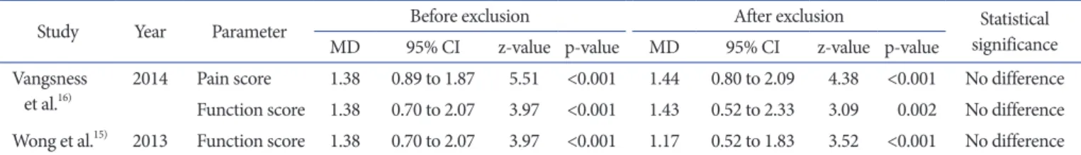
to 2.07 points; p<0.001; I2=83%) (Fig. 3). The results of sensitiv
ity analysis were not materially differentiated from those of the original analysis (Table 3).
5. ROM and MRI Outcomes
Of the 8 studies, two compared ROM and included 24 patients assessed at the baseline and 24 at the final followup. The stan
dardized mean in ROM was –4.41° lower at the baseline than at the final followup (95% CI, –13.05° to 4.24°; p=0.32; I2=98%) (Fig. 4). Of the 8 studies, 4 compared MRI outcome and included 87 patients assessed with MRI at the baseline and 87 at the final
Study or subgroup
Preoperative Postoperative
1.1.1 Follow-up (>3 years)
Subtotal (95% CI)
1.1.2 Follow-up (<3 years)
Subtotal (95% CI)
Total (95% CI) Davatchi et al. 2016 Emadendin et al. 2015
Heterogeneity: Tau =0.00; Chi =0.01, df=1 (p=0.93); I =0%
Test for overall effect: Z=2.59 (p=0.010)
Gupta et al. 2016 Lamo-Espinosa et al. 2016 Orozco et al. 2014 Vangsness et al. 2014 Vega et al. 2015
Heterogeneity: Tau =0.33; Chi =15.74, df=4 (p=0.003); I =75%
Test for overall effect: Z=4.62 (p<0.001)
Heterogeneity: Tau =0.24; Chi =15.80, df=6 (p=0.01); I =62%
Test for overall effect: Z=5.51 (p<0.001)
Test for subgroup differences: Chi =0.00, df=1 (p=0.96); I =0%
2 2 2
2 2 2
2 2 2
2 2
Std. mean difference IV, random, 95% CI
Favours [preoperative] Favours [postoperative]
4
4 2 0 2
Mean
86.25 80
59.65 60 46.9 50 54
SD
4.79 17
22.27 18 26 25.1 27.11
Total
4 6
40 20 12 36 15 10
123
133
Weight (%)
6.6 9.0
21.0 13.6 13.5 20.3 16.1 15.5
84.5
100.0 56.25
57
37.22 18 15.4 23.9 33 Mean
27.8 13
24.69 9 13.2 16.8 23.24 SD
4 6
40 20 12 36 15 10
123
133 Total
1.31 [ 0.34, 2.96]
1.40 [0.08, 2.73]
0.94 [0.48, 1.41]
2.89 [1.98, 3.80]
1.48 [0.55, 2.40]
1.21 [0.70, 1.71]
0.81 [0.06, 1.56]
1.37 [0.33, 2.40]
1.40 [0.80, 1.99]
1.38 [0.89, 1.87]
Std. mean difference IV, random, 95% CI
Fig. 2. Results of aggregate analysis for comparison of pain score between at baseline and at final followup. Std.: standardized, SD: standard deviation, CI: confidence interval.
Study or subgroup
Preoperative Postoperative
Emadendin et al. 2015 Gupta et al. 2016 Lamo-Espinosa et al. 2016 Orozco et al. 2014 Vangsness et al. 2014 Wong et al. 2013
Heterogeneity: Tau =0.57; Chi =29.49, df=5 (p<0.001); I =83%
Test for overall effect: Z=3.97 (p<0.001) Total (95% CI)
2 2 2
Std. mean difference IV, random, 95% CI
Favours [preoperative] Favours [postoperative]
4
4 2 0 2
Mean 70 101.75 22.75 19.1 42.9 58.1
SD 10 67.68 3.7 13.1 22 19.2
Total 6 40 20 12 36 28
142
Weight (%) 10.8 19.6 16.7 16.2 19.1 17.6
100.0 40
66.75 14 9.4 8.4 17 Mean
18 115.83 4.7 11.1 31 16.3 SD
6 40 20 12 36 28
142 Total
1.90 [0.44, 3.37]
0.37 [ 0.08, 0.81]
2.03 [1.25, 2.80]
0.77 [ 0.07, 1.60]
1.27 [0.76, 1.78]
2.28 [1.59, 2.96]
1.38 [0.70, 2.07]
Std. mean difference IV, random, 95% CI
Fig. 3. Results of aggregate analysis for comparison of function score between at baseline and at final followup. Std.: standardized, SD: standard de
viation, CI: confidence interval.
followup. The standardized mean in MRI outcome was 0.62 lower at the final followup than at the baseline (95% CI, –0.16 to 1.40; p=0.12; I2=82%) (Fig. 5).
Discussion
The main findings of the current metaanalysis verified that there were no significant differences in the tested ROM and MRI outcomes when compared to the baseline values in patients treated with BMderived MSCs, whereas significant functional improvement and pain relief from the baseline were observed.
BM stromal cells (BMSCs) can be used as a cell suspension expanded by culture or just as BM concentrate (BMC). There are some differences between these two procedures. Expanded BMSCs require twostep procedures in addition to legal approval
for the clinical application and cost disadvantages. In contrast, BMC contains a mixture of different red blood cells, platelets, and leukocytes23). The adult MSC fraction is present in the leukocytes of the marrow, and their number is very limited compared to cultured MSCs. The marrow MSC and MSC precursors are ex
tremely rare under normal conditions in human marrow before culture24). Some authors suggested that for favorable chondrogen
esis, the optimal count of MSCs per cm3 is an important factor25). However, there is no study of comparison between BMSC and BMC treatment. The treatment using expanded BMSCs might be difficult to manage from a legal point of view because it might be considered as a pharmacological agent administration. In Korea, unlike BMC, expanded BMSCs need additional approval by the Korean Food and Drug Administration for the treatment of OA.
In spite of the complicated procedure, whether the in vitro expan
Table 3. Sensitivity Analysis
Study Year Parameter Before exclusion After exclusion Statistical
significance
MD 95% CI zvalue pvalue MD 95% CI zvalue pvalue
Vangsness
et al.16) 2014 Pain score 1.38 0.89 to 1.87 5.51 <0.001 1.44 0.80 to 2.09 4.38 <0.001 No difference Function score 1.38 0.70 to 2.07 3.97 <0.001 1.43 0.52 to 2.33 3.09 0.002 No difference Wong et al.15) 2013 Function score 1.38 0.70 to 2.07 3.97 <0.001 1.17 0.52 to 1.83 3.52 <0.001 No difference MD: mean difference, CI: confidence interval.
Study or subgroup
Preoperative Postoperative
Davatchi et al. 2016 Lamo-Espinosa et al. 2016
Heterogeneity: Tau =38.09; Chi =46.13, df=1 (p<0.001); I =98%
Test for overall effect: Z=1.00 (p=0.32) Total (95% CI)
2 2 2
Std. mean difference IV, random, 95% CI
Favours [preoperative] Favours [postoperative]
50
50 25 0 25
Mean 106.25 109
SD 33.26
1 Total
4 20
24
Weight (%) 50.4 49.6
100.0 107.5
118.5 Mean
31.75 1.1 SD
4 20
24 Total
0.03 [ 1.42, 1.35]
8.86 [ 10.99, 6.72]
4.41 [ 13.05, 4.24]
Std. mean difference IV, random, 95% CI
Fig. 4. Results of aggregate analysis for comparison of range of motion (ROM) between at baseline and at final followup. Std.: standardized, SD: stan
dard deviation, CI: confidence interval.
Study or subgroup
Preoperative Postoperative
Gupta et al. 2016 Lamo-Espinosa et al. 2016 Orozco et al. 2014 Vega et al. 2015
Heterogeneity: Tau =0.51; Chi =16.49, df=3 (p<0.001); I =82%
Test for overall effect: Z=1.55 (p=0.12) Total (95% CI)
2 2 2
Mean 69.8 67.5 19.5 14
SD 37.1 11.1 4.1 2.3
Total 40 20 12 15
87
Weight (%) 28.5 26.2 22.1 23.3
100.0 67.9
71.5 14.3 9.4 Mean
41 34.1 3.2 3.6 SD
40 20 12 15
87 Total
0.05 [ 0.39, 0.49]
0.15 [ 0.78, 0.47]
1.37 [0.46, 2.27]
1.48 [0.66, 2.30]
0.62 [ 0.16, 1.40]
Std. mean difference IV, random, 95% CI
Std. mean difference IV, random, 95% CI
Favours [preoperative] Favours [postoperative]
4
4 2 0 2
Fig. 5. Results of aggregate analysis for comparison of MRI outcome between at baseline and at final followup. Std.: standardized, SD: standard devia
tion, CI: confidence interval.
sion influenced the effect of clinical application of MSCs was still unclear. For the fate of MSCs in vivo, current studies are not con
clusive on this question: some have suggested MSCs differentiate and survive in vivo up to 6 months, while others suggest MSCs have a chondroinductive role of stimulating cartilage regenera
tion through trophic factors while slowly disappearing from the culture26). BMderived MSCs administered into knee have ad
hered to the surface of a damaged tissue, have differentiated into chondrocyte, and have expressed appropriate extracellular matrix protein, resulting in anatomic restoration on the damaged tissue with a significant relief of pain and disability16,21,22). Our meta
analysis found that MRI outcomes did not show a significant dif
ference from baseline despite the pain and functional advantages of injected BMderived MSCs. The similar results for the MRI outcomes were likely due to the cell dose injected into the knee and culture conditions, suggesting that an optimal cell density and purity could affect the cell expansion. According to the ani
mal study of Agung et al.27), the ideal number of MSCs that are needed for the regeneration of cartilage is known to be 1×10⁷, and a few clinical studies report that 1×10⁷ or more adult stem cells are ideal. Minimal criteria to define expanded multipotent human MSCs, as defined by the International Society for Cellular Therapy, include that they must be plasticadherent when main
tained in standard culture conditions, express CD105, CD73 and CD90, and lack expression of CD45, CD34, CD14 or CD11β, CD79α or CD19 and HLADR surface molecules, and they must be capable of differentiating into osteoblasts, adipocytes and chondroblasts in vitro28). Among our included articles, 6 stud
ies16,1922) met the ideal number of MSCs, 2 studies17,18) used less than 1×10⁷ MSCs. For MSC characterization, 2 studies16,21) did not describe the expression of CD. This could result in poor MRI outcomes than expected. In our metaanalysis, the tested ROM did not demonstrate any significant difference after BMderived MSCs administration. These results may also be partly explained by small patient samples, which can lead to reduced statistical power and less precision. Thus, the ROM outcomes of the cur
rent metaanalysis could not be extended to all knee OA patients and further investigation through a future high volume study is necessary.
This study had several limitations. Of the 8 studies, 3 were ob
servational comparison studies, which are prone to both system
atic and random errors, suggesting some inherent heterogeneity due to uncontrolled bias. In addition, the heterogeneity of the included studies could be explained by slight differences in other factors affecting clinical outcomes, including the use of a wide variety of cell dose and cell processing methods29) as well as vari
ability in functional and pain scores.
Conclusions
This metaanalysis found no significant differences in the tested ROM and MRI outcomes in patients treated with BMderived MSCs. On the other hand, they showed significant functional improvement and pain relief when compared with the baseline.
Thus, BMderived MSCs appear to be a viable alternative for pa
tients with knee OA, although longterm and highquality RCTs are needed to confirm the clinical benefits.
Conflict of Interest
No potential conflict of interest relevant to this article was re
ported.
References
1. Hunziker EB. Articular cartilage repair: basic science and clinical progress: a review of the current status and pros
pects. Osteoarthritis Cartilage. 2002;10:43263.
2. Steadman JR, Rodkey WG, Rodrigo JJ. Microfracture: surgi
cal technique and rehabilitation to treat chondral defects.
Clin Orthop Relat Res. 2001;(391 Suppl):S3629.
3. Davatchi F, Abdollahi BS, Mohyeddin M, Shahram F, Nikbin B. Mesenchymal stem cell therapy for knee osteoarthritis:
preliminary report of four patients. Int J Rheum Dis. 2011;
14:2115.
4. Somoza RA, Welter JF, Correa D, Caplan AI. Chondrogenic differentiation of mesenchymal stem cells: challenges and unfulfilled expectations. Tissue Eng Part B Rev. 2014;20:596
608.
5. Pittenger MF, Mackay AM, Beck SC, Jaiswal RK, Douglas R, Mosca JD, Moorman MA, Simonetti DW, Craig S, Marshak DR. Multilineage potential of adult human mesenchymal stem cells. Science. 1999;284:1437.
6. Mizuno H, Tobita M, Uysal AC. Concise review: adipose
derived stem cells as a novel tool for future regenerative medicine. Stem Cells. 2012;30:80410.
7. Freitag J, Ford J, Bates D, Boyd R, Hahne A, Wang Y, Cicut
tini F, Huguenin L, Norsworthy C, Shah K. Adipose derived mesenchymal stem cell therapy in the treatment of isolated knee chondral lesions: design of a randomised controlled pilot study comparing arthroscopic microfracture versus ar
throscopic microfracture combined with postoperative mes
enchymal stem cell injections. BMJ Open. 2015;5:e009332.
8. Koh YG, Kwon OR, Kim YS, Choi YJ, Tak DH. Adipose
derived mesenchymal stem cells with microfracture versus microfracture alone: 2year followup of a prospective ran
domized trial. Arthroscopy. 2016;32:97109.
9. Bansal H, Comella K, Leon J, Verma P, Agrawal D, Koka P, Ichim T. Intraarticular injection in the knee of adipose derived stromal cells (stromal vascular fraction) and platelet rich plasma for osteoarthritis. J Transl Med. 2017;15:141.
10. Frisbie DD, Kisiday JD, Kawcak CE, Werpy NM, McIlwraith CW. Evaluation of adiposederived stromal vascular fraction or bone marrowderived mesenchymal stem cells for treat
ment of osteoarthritis. J Orthop Res. 2009;27:167580.
11. Afizah H, Hui JH. Mesenchymal stem cell therapy for osteo
arthritis. J Clin Orthop Trauma. 2016;7:17782.
12. Yubo M, Yanyan L, Li L, Tao S, Bo L, Lin C. Clinical efficacy and safety of mesenchymal stem cell transplantation for os
teoarthritis treatment: a metaanalysis. PLoS One. 2017;12:
e0175449.
13. Peterfy CG, Guermazi A, Zaim S, Tirman PF, Miaux Y, White D, Kothari M, Lu Y, Fye K, Zhao S, Genant HK. WholeOrgan Magnetic Resonance Imaging Score (WORMS) of the knee in osteoarthritis. Osteoarthritis Car
tilage. 2004;12:17790.
14. Orozco L, Munar A, Soler R, Alberca M, Soler F, Huguet M, Sentis J, Sanchez A, GarciaSancho J. Treatment of knee os
teoarthritis with autologous mesenchymal stem cells: a pilot study. Transplantation. 2013;95:153541.
15. Wong KL, Lee KB, Tai BC, Law P, Lee EH, Hui JH. Injectable cultured bone marrowderived mesenchymal stem cells in varus knees with cartilage defects undergoing high tibial os
teotomy: a prospective, randomized controlled clinical trial with 2 years’ followup. Arthroscopy. 2013;29:20208.
16. Vangsness CT Jr, Farr J 2nd, Boyd J, Dellaero DT, Mills CR, LeRouxWilliams M. Adult human mesenchymal stem cells delivered via intraarticular injection to the knee following partial medial meniscectomy: a randomized, doubleblind, controlled study. J Bone Joint Surg Am. 2014;96:908.
17. Davatchi F, Sadeghi Abdollahi B, Mohyeddin M, Nikbin B.
Mesenchymal stem cell therapy for knee osteoarthritis: 5 years followup of three patients. Int J Rheum Dis. 2016;19:
21925.
18. Emadedin M, Ghorbani Liastani M, Fazeli R, Mohseni F, Moghadasali R, Mardpour S, Hosseini SE, Niknejadi M, Moeininia F, Aghahossein Fanni A, Baghban Eslaminejhad R, Vosough Dizaji A, Labibzadeh N, Mirazimi Bafghi A,
Baharvand H, Aghdami N. Longterm followup of intra
articular injection of autologous mesenchymal stem cells in patients with knee, ankle, or hip osteoarthritis. Arch Iran Med. 2015;18:33644.
19. Gupta PK, Chullikana A, Rengasamy M, Shetty N, Pandey V, Agarwal V, Wagh SY, Vellotare PK, Damodaran D, Viswana
than P, Thej C, Balasubramanian S, Majumdar AS. Efficacy and safety of adult human bone marrowderived, cultured, pooled, allogeneic mesenchymal stromal cells (Stempeucel®):
preclinical and clinical trial in osteoarthritis of the knee joint. Arthritis Res Ther. 2016;18:301.
20. LamoEspinosa JM, Mora G, Blanco JF, GraneroMolto F, NunezCordoba JM, SanchezEchenique C, Bondía JM, Aquerreta JD, Andreu EJ, Ornilla E, Villaron EM, Valenti
Azcarate A, SanchezGuijo F, Del Canizo MC, ValentiNin JR, Prosper F. Intraarticular injection of two different doses of autologous bone marrow mesenchymal stem cells versus hyaluronic acid in the treatment of knee osteoarthritis: mul
ticenter randomized controlled clinical trial (phase I/II). J Transl Med. 2016;14:246.
21. Orozco L, Munar A, Soler R, Alberca M, Soler F, Huguet M, Sentis J, Sanchez A, GarciaSancho J. Treatment of knee osteoarthritis with autologous mesenchymal stem cells: two
year followup results. Transplantation. 2014;97:e668.
22. Vega A, MartinFerrero MA, Del Canto F, Alberca M, Gar
cía V, Munar A, Orozco L, Soler R, Fuertes JJ, Huguet M, Sanchez A, GarciaSancho J. Treatment of knee osteoarthri
tis with allogeneic bone marrow mesenchymal stem cells: a randomized controlled trial. Transplantation. 2015;99:1681
90.
23. Martin I, Baldomero H, BocelliTyndall C, Passweg J, Saris D, Tyndall A. The survey on cellular and engineered tissue ther
apies in Europe in 2010. Tissue Eng Part A. 2012;18:226879.
24. Simmons PJ, TorokStorb B. Identification of stromal cell precursors in human bone marrow by a novel monoclonal antibody, STRO1. Blood. 1991;78:5562.
25. Skowronski J, Rutka M. Osteochondral lesions of the knee reconstructed with mesenchymal stem cells: results. Ortop Traumatol Rehabil. 2013;15:195204.
26. de Windt TS, Hendriks JA, Zhao X, Vonk LA, Creemers LB, Dhert WJ, Randolph MA, Saris DB. Concise review: unrav
eling stem cell cocultures in regenerative medicine: which cell interactions steer cartilage regeneration and how? Stem Cells Transl Med. 2014;3:72333.
27. Agung M, Ochi M, Yanada S, Adachi N, Izuta Y, Yamasaki T, Toda K. Mobilization of bone marrowderived mesen
chymal stem cells into the injured tissues after intraarticular injection and their contribution to tissue regeneration. Knee Surg Sports Traumatol Arthrosc. 2006;14:130714.
28. Dominici M, Le Blanc K, Mueller I, SlaperCortenbach I, Marini F, Krause D, Deans R, Keating A, Prockop Dj, Hor
witz E. Minimal criteria for defining multipotent mesen
chymal stromal cells: the International Society for Cellular Therapy position statement. Cytotherapy. 2006;8:3157.
29. Xia P, Wang X, Lin Q, Li X. Efficacy of mesenchymal stem cells injection for the management of knee osteoarthritis: a systematic review and metaanalysis. Int Orthop. 2015;39:
236372.