• 검색 결과가 없습니다.

Non-Operative Management with Angioembolization of Grade IV and V Renal Injuries in a Hybrid Emergency Room System

N/A
N/A
Protected

Academic year: 2021

Share "Non-Operative Management with Angioembolization of Grade IV and V Renal Injuries in a Hybrid Emergency Room System"

Copied!
7
0
0

로드 중.... (전체 텍스트 보기)

전체 글

(1)

Received: May 3, 2021 Revised: June 22, 2021 Accepted: June 28, 2021 Trauma and InJury

Correspondence to

Chan Yong Park, M.D., Ph.D.

Division of Trauma Surgery, Department of Surgery, Seoul National University Hospital, 101 Daehak-ro, Jongno-gu, Seoul 03080, Korea

Tel: +82-2-2072-2817 Fax: +82-2-766-3975

E-mail: [email protected] ORCID: https://orcid.org/0000-0002- 5111-3270

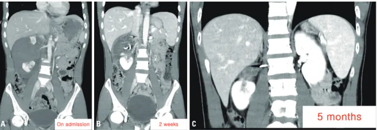
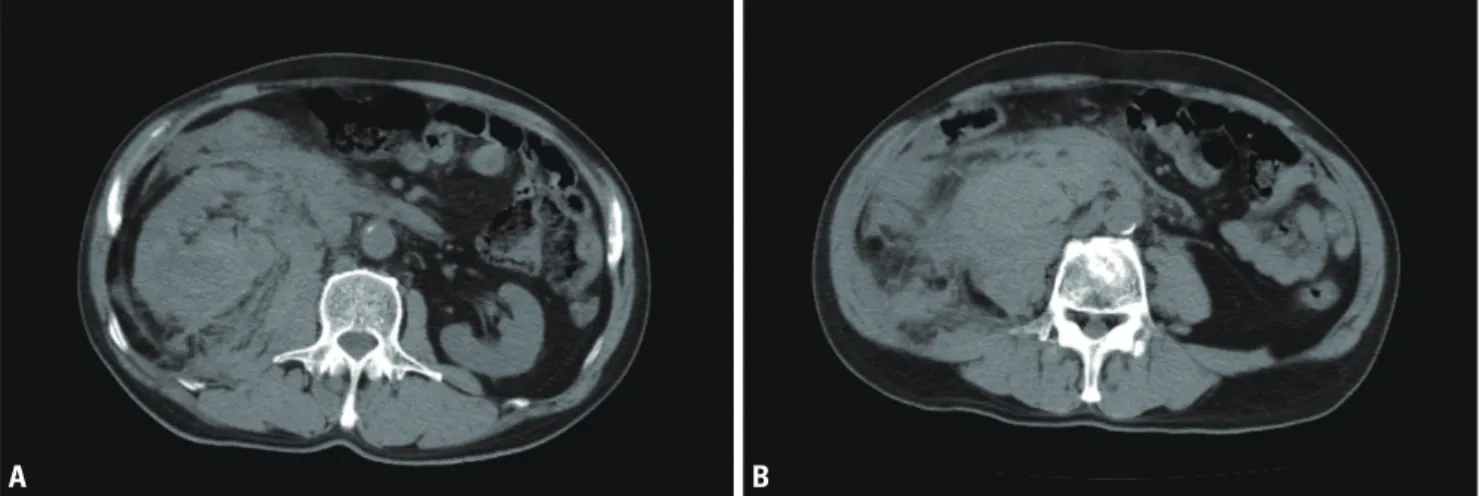
non-operative management with angioembolization of Grade IV and V renal Injuries in a Hybrid Emergency room System

So Ra Ahn, M.D.

1

, Sang Hyun Seo, M.D.

2

, Joo Hyun Lee, M.D.

1

, Chan Yong Park, M.D., Ph.D.

3

1

Department of Trauma Surgery, Wonkwang University Hospital Regional Trauma Center, Iksan, Korea

2

Department of Radiology, Wonkwang University Hospital Regional Trauma Center, Iksan, Korea

3

Division of Trauma Surgery, Department of Surgery, Seoul National University Hospital, Seoul, Korea

Renal injuries occur in more than 10% of patients who sustain blunt abdominal inju- ries. Non-operative management (NOM) is the established treatment strategy for low- grade (I–III) renal injuries. However, despite some evidence that NOM can be success- fully applied to high-grade (IV, V) renal injuries, it remains unclear whether NOM is appropriate in such cases. The authors report two cases of high-grade renal injuries that underwent NOM after embolization in a hybrid emergency room (ER) system with a 24/7 in-house interventional radiology (IR) team. A 29-year-old male visited Wonk- wang University Hospital Regional Trauma Center complaining of right abdominal pain after being hit by a rope. Computed tomography (CT) was performed 16 minutes after arrival, and the CT scan indicated a grade V right renal injury. Arterial emboli- zation was initiated within 31 minutes of presentation. A 56-year-old male was trans- ferred to Wonkwang University Hospital Regional Trauma Center with a complaint of right flank pain. He had initially presented to a nearby hospital after falling from a 3-m height. Thanks to the key CT images sent from the previous hospital prior to the patient’s arrival, angiography was performed within 8 minutes of the patient’s arrival and arterial embolization was completed within 25 minutes. Both patients were treated successfully through NOM with angioembolization and preserved kidneys. Hematoma in the first patient and urinoma in the second patient resolved with percutaneous cath- eter drainage. The authors believe that the hybrid ER system with an in-house IR team could contribute to NOM and kidney preservation even in high-grade renal injuries.

Keywords: Renal injury; Non-operative management; High-grade; Hybrid emergency

room system; Embolization

(2)

INTRODUCTION

Renal injuries occur in more than 10% of patients who sustain blunt abdominal injuries. For decades, non-op- erative management (NOM) has been the established treatment strategy for low-grade (I–III) renal injuries;

however, studies investigating the efficacy of NOM for high-grade (IV, V) renal injuries are limited. Despite some evidence that NOM can be successfully applied to high-grade renal injuries, it remains unclear whether this approach is appropriate [1] The decision to perform in- terventional or conservative treatment depends on each patient’s overall treatment strategy and the circumstances of each hospital.

The regional trauma center of Wonkwang University Hospital operates a dual-room hybrid emergency room (ER) system (Fig. 1) with a continuously staffed, in-house interventional radiology (IR) team. The following cases

describe the treatment of high-grade renal injuries within this system.

CASE REPORT

Case 1. Grade V renal injury

A 29-year-old male presented to Wonkwang Universi- ty Hospital Regional Trauma Center with complaints of gross hematuria and right abdominal pain. The pain started after he sustained a blow to his right abdomen from a rope while working on a ship, approximately 90 minutes prior to being seen at the hospital. The patient was immediately transferred to the dual-room hybrid ER where a focused assessment with sonography in trau- ma was performed; the assessment conveyed positive findings. The patient’s Glasgow Coma Scale (GCS) score was 15 and his initial vital signs were stable: blood pres- sure, 120/70 mmHg; pulse, 61 beats/min. The patient demonstrated right lower abdominal tenderness, and his computed tomography (CT) results were available with- in 16 minutes of his arrival at the center. The chest CT results indicated fractures of ribs 10 and 11; however, the abdominopelvic CT scan indicated a grade V right renal injury and a massive retroperitoneal hematoma (Fig. 2).

Contrast extravasation from the upper pole of the right kidney was observed, and the patient underwent emer- gency angiography. An ultrasound-guided, right common femoral artery puncture was performed 31 minutes after the patient’s arrival. Extravasation from and multiple pseudoaneurysms in the superior segmental artery were identified using right renal arteriography. Following coil embolization, further bleeding was not observed on fol- low-up renal arteriography (Fig. 3). Since the patient’s Fig. 1. Dual-room hybrid emergency room.

A b C D

Fig. 2. (A-D) Abdominal computed tomography (axial and coronal images) of the patient with a grade V injury, showing the separated vascularized

kidneys and a hypodense hematoma. Contrast extravasation is seen in the upper pole of the right kidney (arrows of A, B).

(3)

initial hemoglobin (Hb) level was 12.0 g/dL and his vital signs were stable, an emergency transfusion was not per- formed. His initial laboratory tests also showed blood concentrations of ALT 681 U/L, AST 122 U/L, creatine kinase 648 IU/L, blood urea nitrogen (BUN) 25.0 mg/dL, and creatinine 0.86 mg/dL; urine microscopy revealed 1,000,000 red blood cells (RBCs)/high-power field (HPF).

After embolization, the patient’s follow-up Hb level was 11.9 g/dL, which was not significantly different from the initial level. Three days after embolization, active bleeding was not observed on the follow-up abdominopelvic CT

scan. Although the right upper renal parenchyma was completely devascularized, the right inferior residual renal parenchyma showed contrast enhancement. Significant changes were not observed on a 1-week postadmission ab- dominopelvic CT scan. However, the patient complained of abdominal pain and exhibited abdominal distension due to the hematoma. Percutaneous catheter drainage (PCD) was performed to remove the perirenal fluid collection. Af- ter 3 weeks of hospitalization, the patient was discharged without any further complications. Six weeks after its in- sertion, the PCD catheter was removed in the outpatient

Fig. 3. (A) Right renal angiography of the patient with a grade V injury shows separation of the upper and lower poles. (B) Contrast extravasation is seen during selective angiography (arrowhead). (C) The upper polar artery is shown after complete embolization using a coil.

A b C

Fig. 4. (A-C) These computed tomography images show the postprocedural course of the patient with a grade V injury. The large hematoma, shown in the upper pole of the right kidney, is observed to have gradually decreased in size. (C) Five months after the accident, the hematoma is no longer evident.

A b C

(4)

clinic (Fig. 4). In this case, the patient required transfusion of only one unit of packed RBCs (pRBCs) and one unit of fresh frozen plasma (FFP) during hospitalization.

Case 2. Grade IV renal injury

A 56-year-old male was transferred to Wonkwang Uni- versity Hospital Regional Trauma Center with complaints of right flank pain. He had initially present to a nearby

hospital after falling from a 3-m height. There, he had undergone non-contrast-enhanced abdominopelvic CT that suggested severe right renal injury. During the patient’s transfer to Wonkwang University Hospital Re- gional Trauma Center, the previous hospital was asked to provide a key CT image. This allowed the trauma team to review the image (Fig. 5) and prepare a radiologic intervention before the patient’s arrival. As soon as the

Fig. 5. (A) Abdominal non-contrast computed tomography (axial image) in the patient with a grade IV injury shows large right perirenal and pararenal hematomas. (B) A right psoas muscle hematoma and a retroperitoneal hematoma are also seen.

A b

Fig. 6. (A) Right renal angiography of the patient with a grade IV injury shows a small pseudoaneurysm in an arcuate artery in the lower pole (arrow). (B) This image was obtained after injecting Histoacryl® (B. Braun, Melsungen, Germany) and Lipiodol® (Jubilant HollisterStier, Spokane, WA, USA). Emboli- zed blood vessels are seen in a lower polar artery (arrowheads).

A b

(5)

patient arrived, he was taken to the hybrid ER where his GCS score was 15 and his initial vital signs were unstable (blood pressure 76/48 mmHg; pulse 75 beats/min); his abdomen was soft and mildly distended but not tender.

Ultrasound-guided right common femoral artery punc- ture was performed 8 minutes after the patient’s arrival.

Arteriography revealed extravasation from the lower po- lar artery of the right kidney and multiple extravasations from the right lumbar artery. Therefore, embolization was performed using Histoacryl

®

(B. Braun, Melsungen, Ger- many) and Lipiodol

®

(Jubilant HollisterStier, Spokane, WA, USA) in the right lower polar artery and Gelfoam

®

(Pfizer, New York, NY, USA) particles in the right lum- bar artery (Fig. 6). Postembolization, contrast-enhanced CT was performed, but focal bleeding was not observed.

A grade IV right renal injury, a large hemoperitoneum, hemoretroperitoneum (Fig. 7A), and a left tibia fracture were identified. The patient’s initial Hb level was 11.0 g/dL, but he was hemodynamically unstable; an emergency transfusion of two units of O

+

pRBCs was started within 2 minutes of his arrival. Ten minutes after presentation, his systolic blood pressure was 103 mmHg, and an additional two units of unmatched ABO type-specific pRBCs and four units of FFP were transfused; after the transfusion, his Hb level was 11.6 g/dL. The patient received a total of seven units of pRBCs and four units of FFP while he was hospitalized. Laboratory tests showed a mildly elevated BUN concentration (21.0 mg/dL) and a creatinine level

(0.97 mg/dL) that was within the normal range. Gross hematuria was observed during Foley catheterization, and urine microscopy showed 22,499 RBCs/HPF. Two days after embolization, the patient’s hemoretroperitoneum had decreased according to the follow-up CT. However, the hematoma in the renal pelvis had increased in size, leading to suspicion of a collecting duct injury. However, the continued mild pain in the patient’s right flank and the increasing size of the hematoma led to a suspicion of a collecting duct injury. Fortunately, the hemoretro- peritoneum had sharply decreased in size on a follow-up CT scan performed 2 weeks after admission. Right peri- ureteral fluid collection that was thought to be urinoma was observed around the psoas muscle, and a PCD was performed. After 1 week, the amount of fluid draining through the PCD had decreased, and the urine turbidity reduced and it became clear. Follow-up CT indicated that the fluid collection along the right psoas muscle had markedly decreased, and the PCD catheter was re- moved. The urinoma around the kidney was not present on follow-up CT performed 40 days after the accident (Fig. 7B-D), and the patient was discharged without fur- ther complications.

DISCUSSION

Renal injuries account for approximately 1–5% of all

Fig. 7. (A) Immediately after the procedure in the patient with a grade IV injury, large hematomas are seen in the right perirenal space, pararenal space, and right psoas muscle. (B, C) The hematomas are seen to gradually decrease in size, and percutaneous catheter drainage was performed during fol- low-up. (D) A maximum intensity projection image shows that the shape of the kidney is well maintained.

A b C D

(6)

trauma cases, and the kidney is the most commonly in- jured genitourinary organ [2]. Approximately 90–95%

of renal injuries are caused by blunt injuries [3], such as those caused by high-energy deceleration collisions (e.g., motor vehicle accidents, falls, and assaults) [2]. The most commonly used measure of renal injury is the American Association for the Surgery of Trauma (AAST) renal inju- ry scale. The renal injury scale is divided into five grades, with grades IV and V usually being referred to as high- grade injuries [4]. According to the 2018 AAST revised grading, grade IV renal injuries include segmental vascu- lar injuries, segmental or complete parenchymal infarc- tions, and active bleeding beyond the Gerota fascia; grade V injuries include main renal artery or vein lacerations, hilum avulsion, kidney devascularization with active bleeding, and shattered kidney with loss of identifiable parenchymal renal anatomy [5,6].

Previously, intravenous urography and renal arteriog- raphy were used to evaluate hemodynamically stable pa- tients [7]. Currently, however, abdominopelvic multi-de- tector CT (MDCT) with contrast enhancement is the imaging modality of choice for hemodynamically stable patients with genitourinary injuries. MDCT is fast, widely available, and provides anatomical and functional data [8].

During the past 3 decades, the treatment strategy for patients with renal injuries has shifted from operative management to NOM [9]. Depending on the guideline, conservative treatment is recommended for most patients who are hemodynamically stable. However, conservative management of patients with grade IV or V injuries who are hemodynamically unstable remains controversial [2]. In most high-grade renal injury cases, nephrectomy remains a possibility even though successful NOM is possible [10]. However, recent developments in imaging technology and in the available treatment options have decreased the need for surgical interventions, allowing kidney preservation in most cases [11]. In a recent review, 90% of 3,528 surveyed patients with renal injuries recov- ered without open surgical interventions [12]. Altman et al. [13] conservatively treated hemodynamically stable patients with grade V renal injuries and reported that the intensive care unit stays were shorter, the required blood transfusion amounts were smaller, and the numbers of complications were lower than in patients undergoing

surgical interventions.

Renal artery embolization is an effective, minimally invasive treatment for renal injury that can prevent gross hematuria and preserve renal function [11]. According to the 2018 AAST revised grading, active bleeding and vas- cular injuries (e.g., pseudoaneurysms and arteriovenous fistulas) are included, and interventions may be required in cases of hemodynamic instability [8]. Breyer et al. [14]

reported that renal artery embolization was effective in patients with grade IV renal injuries when conservative treatment failed. According to them, the indications for renal artery embolization include renal injuries with per- sistent gross hematuria, increasing perirenal hematoma, renal arteriovenous fistulas, and pseudoaneurysm forma- tion.

Kidney-related complications (excluding renal fail- ure) occur in approximately 5.2% of patients with renal injuries [15]. The most common complication is per- sistent urinary extravasation, resulting in the formation of a urinoma [16]. In both cases above, fluid collection occurred and was treated through PCD insertion. Fluid collection around the kidneys, such as urinoma and he- matoma, can be a source of infection. PCD insertion to drain a fluid collection may result in a prolonged hospital stay compared to surgical treatment. Furthermore, simple contrast enhancement on CT does not necessarily mean that kidney function has recovered, so additional imaging tests may be needed to accurately confirm kidney func- tion recovery [17].

The regional trauma center of Wonkwang University

Hospital is equipped with a hybrid ER system that in-

cludes a continuously staffed, in-house IR team. There-

fore, we can very quickly perform arterial embolization,

which is the treatment of choice for solid organ (e.g.,

kidney, spleen, and liver) injuries in the abdominal cav-

ity, even in hemodynamically unstable patients. In our

patient with a grade V renal injury, a CT scan was per-

formed 16 minutes after arrival, and arterial embolization

was initiated within 31 minutes of presentation. In our

patient with a grade IV renal injury, common femoral

artery puncture was performed within 8 minutes of the

patient’s arrival, renal artery embolization was completed

within 25 minutes, and lumbar artery embolization was

completed within 35 minutes of arrival; an additional CT

(7)

evaluation was performed within 42 minutes of arrival.

Thanks to the availability of this hybrid ER system and the IR team, bleeding can be controlled within a very short time. As a result, surgery can be avoided and good results can be obtained, including kidney preservation. However, gathering additional cases and comparing the features of these cases with those of previous patients remains neces- sary. The features that should be compared among cases include blood transfusion volume, intensive care unit stay duration, total hospital stay duration, and complications.

The authors believe that the hybrid ER system with in- house IR team could contribute to NOM and kidney preservation even in hemodynamically unstable patients with high-grade renal injuries. In the future, a compara- tive study on the treatment results of the conventional ER and hybrid ER systems is considered to be necessary.

REFERENCES

1. van der Wilden GM, Velmahos GC, Joseph DK, Jacobs L, Debusk MG, Adams CA, et al. Successful nonoperative man- agement of the most severe blunt renal injuries: a multicenter study of the research consortium of New England Centers for Trauma. JAMA Surg 2013;148:924-31.

2. Chouhan JD, Winer AG, Johnson C, Weiss JP, Hyacinthe LM.

Contemporary evaluation and management of renal trauma.

Can J Urol 2016;23:8191-7.

3. Santucci RA, McAninch JW. Diagnosis and management of re- nal trauma: past, present, and future. J Am Coll Surg 2000;191:

443-51.

4. Moore EE, Shackford SR, Pachter HL, McAninch JW, Browner BD, Champion HR, et al. Organ injury scaling: spleen, liver, and kidney. J Trauma 1989;29:1664-6.

5. Chien LC, Vakil M, Nguyen J, Chahine A, Archer-Arroyo K, Hanna TN, et al. The American Association for the Surgery of Trauma Organ Injury Scale 2018 update for computed tomog- raphy-based grading of renal trauma: a primer for the emergen- cy radiologist. Emerg Radiol 2020;27:63-73.

6. Kozar RA, Crandall M, Shanmuganathan K, Zarzaur BL, Coburn M, Cribari C, et al. Organ injury scaling 2018 up-

date: spleen, liver, and kidney. J Trauma Acute Care Surg 2018;85:1119-22.

7. Santucci RA, Wessells H, Bartsch G, Descotes J, Heyns CF, McAninch JW, et al. Evaluation and management of renal in- juries: consensus statement of the renal trauma subcommittee.

BJU Int 2004;93:937-54.

8. Hosein M, Paskar D, Kodama R, Ditkofsky N. Coming together:

a review of the American Association for the surgery of trau- ma’s updated kidney injury scale to facilitate multidisciplinary management. AJR Am J Roentgenol 2019;213:1091-1099.

9. Kuan JK, Wright JL, Nathens AB, Rivara FP, Wessells H; Amer- ican Association for the Surgery of Trauma. American Associ- ation for the surgery of trauma organ injury scale for kidney injuries predicts nephrectomy, dialysis, and death in patients with blunt injury and nephrectomy for penetrating injuries. J Trauma 2006;60:351-6.

10. Salem MS, Urry RJ, Kong VY, Clarke DL, Bruce J, Laing GL.

Traumatic renal injury: five-year experience at a major trauma centre in South Africa. Injury 2020;51:39-44.

11. Vozianov S, Sabadash M, Shulyak A. Experience of renal artery embolization in patients with blunt kidney trauma. Cent Euro- pean J Urol 2015;68:471-7.

12. McClung CD, Hotaling JM, Wang J, Wessells H, Voelzke BB.

Contemporary trends in the immediate surgical management of renal trauma using a national database. J Trauma Acute Care Surg 2013;75:602-6.

13. Altman AL, Haas C, Dinchman KH, Spirnak JP. Selective non- operative management of blunt grade 5 renal injury. J Urol 2000;164:27-30; discussion 30-1.

14. Breyer BN, McAninch JW, Elliott SP, Master VA. Minimally in- vasive endovascular techniques to treat acute renal hemorrhage.

J Urol 2008;179:2248-52; discussion 2253.

15. Starnes M, Demetriades D, Hadjizacharia P, Inaba K, Best C, Chan L. Complications following renal trauma. Arch Surg 2010;145:377-81; discussion 381-2.

16. Lee YJ, Oh SN, Rha SE, Byun JY. Renal trauma. Radiol Clin North Am 2007;45:581-92.

17. Yao YL, Gao Y. Present situation and research progress of kidney

function recoverability evaluation of acute kidney injury pa-

tient. Int J Gen Med 2021;14:1919-25.

수치

Fig. 4. (A-C) These computed tomography images show the postprocedural course of the patient with a grade V injury
Fig. 5. (A) Abdominal non-contrast computed tomography (axial image) in the patient with a grade IV injury shows large right perirenal and pararenal  hematomas
Fig. 7. (A) Immediately after the procedure in the patient with a grade IV injury, large hematomas are seen in the right perirenal space, pararenal space,  and right psoas muscle

참조

관련 문서

Therefore, this study aims to investigate the relationship between explicit grammar instruction and English speaking of lower-grade students in third-grade

Parents of the children completed a questionnaire, The questionnaire was consisted of the prevalence and types of alternative medicine used in the children,

Haemorragic fever with renal syndrome (HFRS) caused by Hantanvirus has been one of the principal acute febrile disease in Korea. Hantaviruses are carried by numerous

The locations of aneurysms were middle cerebral artery in 15 patients, cerebral artery in 15 patients, cerebral artery in 15 patients, cerebral artery in

In this paper, a methodology for estimating the parameters of non-linear system including stabilizing system(Night Vision Pedestal System) was presented.. To

At the end of the study, a reevaluation of each study case was performed with the same questionnaire. The result shows there has been a meaningful result in the group

Patients with breast cancer; ovarian cancer; renal cell carcinoma; pancreatic neuroendocrine cancer; colorectal cancer; head and neck cancer; non-small cell lung

Intermittent hemodialysis(IDH) and Continuous renal replacement therapy (CRRT) has been used widely for treating critically ill patients with acute renal failure, We