Received August 18, 2021, Revised September 14, 2021, Accepted September 14, 2021 Corresponding author: Ji-Yeun Park
College of Korean Medicine, Daejeon University, 62 Daehak-ro, Dong-gu, Daejeon 34520, Korea Tel: +82-42-280-2615, Fax: +82-42-280-2609, E-mail: jypark@dju.kr
*These authors have contributed equally to this work.
This research was supported by the Basic Science Research Program through the National Research Foundation of Korea (NRF) funded by the Ministry of Science, ICT & Future Planning (NRF-2018R1C1B3004424 and NRF-2018R1A 6A1A03025221).
CCThis is an open access article distributed under the terms of the Creative Commons Attribution Non-Commercial License (http://creativecommons.org/licenses/ by-nc/4.0) which permits unrestricted non-commercial use, distribution, and reproduction in any medium, provided the original work is properly cited.
간정격과 사관혈 침 치료의 우울 행동 개선 효과 및 뇌신경 반응성 분석 연구
엄근향* ㆍ류재상* ㆍ박지연
대전대학교 한의과대학
Antidepressant Effect of Liver Tonification and Four Gate Acupuncture Treatments and Its Brain Neural Activity
Geun-Hyang Eom*, Jae-Sang Ryu*, Ji-Yeun Park
College of Korean Medicine, Daejeon University
Objectives : We aimed to identify the antidepressant effect of liver tonification acupuncture treatment (ACU (LT); KI10, LR8, LU8, LR4) and four gate acupuncture treatment (ACU (FG); LI4, LR3) and its brain neural activity in the normal and chronic restraint stress (CRS)-induced mouse model. Methods : Firstly, normal mice were given ACU (LT) or ACU (FG) and the c-Fos expressions in each brain region were analyzed to examine brain neural activity. Secondly, CRS was administered to mice for 4 weeks, then ACU (LT) or ACU (FG) was performed for 2 weeks. The depression-like behavior was evaluated using open field test (OFT) before and after acupuncture treatment. Then, the c-Fos expressions in each brain region were analyzed to examine brain neural activity.
Results : In normal mice, ACU (FG) regulated brain neural activities in the hypothalamus, hippocampus, and periaqueductal gray.
ACU (LT) changed more brain regions in the prefrontal cortex, insular cortex, striatum, and hippocampus, including those altered by ACU (FG). In CRS-induced model, ACU (LT) alleviated depression-like behavior more than ACU (FG). Also, brain neural activities in the motor cortex area 2 (M2), agranular ventral part and piriform of insular cortex (AIV and Pir), and cornu ammonis (CA) 1 and CA3 of hippocampus were changed by ACU (LT), and those of AIV and CA3 were also changed by ACU (FG). As in normal mice, ACU (LT) resulted in changes in more brain regions, including those altered by ACU (FG) in CRS model. M2, Pir, and CA1 were only changed by ACU (LT) in depression model, suggesting that these brain regions reflect the specific effect of ACU (LT). Conclusions : ACU (LT) relieved depression-like behavior more than ACU (FG), and this acupuncture effect was associated with modulation of brain neural activities in the motor cortex, insular cortex, and hippocampus.
Key words : depression, sa-am acupuncture, liver tonification acupuncture, four gate acupuncture, brain neural activity
서 론
우울증은 기쁨이나 즐거움과 같은 감정의 손실, 급격한 체중 변
화, 수면 장애, 초조 및 격정 등의 감정 변화, 에너지 감소, 무기력 함, 자살 및 죽음에 대한 생각 등의 증상을 동반하는 심리적 장애이 다1,2). 우울증은 만성적으로 진행될수록 다양한 심리적 문제 뿐 아
니라 신체적 장애 및 고통 등 심각한 병적인 상태로 이어져 사망률 증가 등 여러 사회적 문제를 야기하게 된다3). 우울증의 발생에는 유전적, 사회적, 심리적, 신체적 요인 등 다양한 원인들이 관련되어 있으며, 최근에는 뇌가 우울증의 핵심 기전 중 하나로 연구되고 있다4,5). 많은 연구들이 cortex, striatum, amygdala, hypothalamus, hippocampus, thalamus, midbrain 등 여러 뇌 영역이 우울증과 밀접한 연관이 있음을 보고하고 있으며, 특히 insular cortex, amygdala, hippocampus 등의 뇌 영역에서 발생된 병리적 변화 가 우울증과 관련되어 있음이 보고된 바 있다6-8). 우울증 치료를 위해서는 주로 삼환계 항우울제(tricyclic antidepressant), 선택적 세로토닌 재흡수 억제제(selective serotonin reuptake inhibitors), 모노아민 산화효소 억제제(monoamine oxidase inhibitors) 등의 항우울제가 사용되고 있으나9), 우울증 환자의 20∼30%는 약물 치 료에 대한 저항성을 나타내는 것으로 보고되고 있으며, 치료 효과 또한 부족한 것이 현실이다10). 따라서 최근에는 운동, 독서치료, 인지치료, 대인관계치료 등과 같은 인지 행동 치료를 비롯하여 침 치료 등 비약물적 치료법에 대한 관심이 증대되고 있다3,11-14).
침 치료는 질환의 특성에 따라 특정 경혈을 선택하여 물리적으 로 자극하는 방법으로, 단일 경혈 또는 여러 경혈 조합이 치료에 활용된다. 침 치료는 다양한 유형의 통증15-18) 및 퇴행성 뇌질환19,20) 을 비롯하여 우울증 등 심리적 질환에도 효과적인 치료법으로 활 용되고 있다21-24). 최근 연구들에서 백회(GV20) 경혈에 시행된 전 침 치료가 우울증 환자의 뇌 활성 변화를 야기함이 보고된 바 있으
며25,26), 곡천(LR8), 기문(LR14), 거궐(CV14) 경혈에 레이저 침 치
료를 시행한 후 우울증 환자의 frontal cortex, limbic cortex, cerebellum 등 뇌 영역에서의 활성도 변화가 관찰되었다27). 동물을 활용한 연구에서는 주로 내관(PC6), 삼음교(SP6), 족삼리(ST36), 신문(HT7) 등 단일 경혈을 활용한 연구가 많이 이루어지고 있었는
데24,28-30), 내관(PC6) 침 치료는 대뇌 hypothalamus의 paraven-
tricular hypothalamic nucleus (PVN) 영역에서 c-Fos 발현을 조 절하였으며30), 삼음교(SP6) 침 치료는 대뇌 hippocampus 영역에 서 brain-derived neurotrophic factor (BDNF)와 neuropeptide Y (NPY)의 발현을 조절하였다29). 이 외에도 최근에는 다양한 경혈 조합 또한 시도되고 있는데, 백회(GV20)와 인당(EX-HN3) 경혈 조 합의 전침 치료는 5-hydroxytryptamine (5-HT), glutamate, gamma aminobutyric acid (GABA) 발현을 증가시키고, BDNF, cyclic adenosine monophosphate (cAMP) response element- binding protein (CREB), extracellular signal-regulated kinase (ERK) 신호전달 경로를 조절함으로써 우울 행동 개선에 기여함이 보고되었다31-33). 그 외에도 사관혈에 시행된 침 치료가 cAMP-
protein kinase A (PKA) 신호 전달 경로 조절을 통해 우울증 개선 에 관여함이 보고되었으며34), 간정격에 시행된 침 치료가 동물의 우울 행동을 개선하고 뇌의 신경반응성 및 5-HT 관련 기전을 조절 함이 보고된 바 있다22).
이렇듯 침 치료의 우울증 개선 효과 및 관련 기전 규명을 위한 많은 연구들이 수행되고 있지만, 임상에서 주로 활용되는 경혈 조 합을 이용한 연구는 많이 부족한 상황이며, 특히 경혈 조합 간의 효과를 비교하는 연구는 거의 이루어져 있지 않다. 따라서 우리는 우울증 치료에 활용 가능한 경혈 조합인 간정격과 사관혈을 활용 하여 정상 동물에서의 뇌신경 반응성을 확인하고자 하였다. 또한 만성 우울증 동물모델에서 간정격과 사관혈 침 치료의 우울 행동 개선 효과 및 뇌신경 반응성을 비교 분석함으로써 우울증 치료에 효과적인 경혈 조합 도출 및 관련 기전을 규명하고자 하였다.
재료 및 방법
1. 실험동물
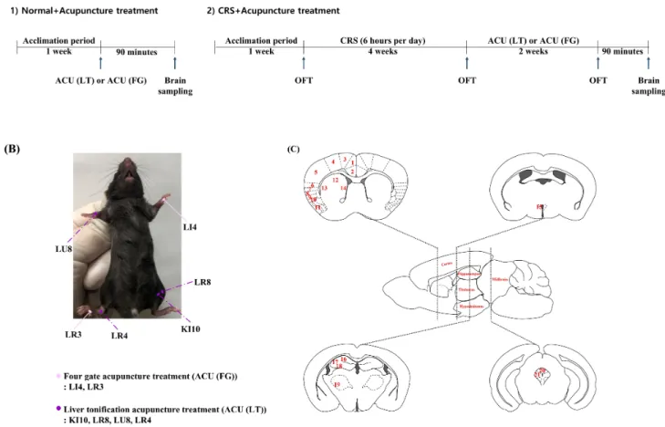
실험에 사용된 동물은 체중 20∼25 g의 7주령 수컷 C57BL/6N 마우스(DaeHan Biolink, Inc.)로, 항온 및 항습(실내온도 22∼2 6℃, 습도 60%)이 유지되는 사육장에서 일주일 동안 적응시킨 후 실험에 사용되었다(Fig. 1A). 실험기간 동안 스트레스를 최소화 할 수 있는 환경을 유지하였으며, 식수 및 사료는 자유롭게 섭취할 수 있도록 하였다. 실험동물은 케이지당 4마리씩 배정하여 유지하 였으며, 각 그룹당 4마리씩 배정되었다. 모든 동물 실험은 실험 동 물 관리 및 사용의 가이드라인(Care and Use of Laboratory Animals of the National Institutes of Health)에 따라 진행되었 으며, 대전대학교 실험동물윤리위원회(Daejeon University Insti- tutional Animal Care and Use Committee)의 승인을 거쳐 수행 되었다(DJU-ARB-2016-E017).
2. 침 치료
간정격 침 치료(Liver tonification acupuncture treatment;
ACU (LT)) 및 사관혈 침 치료(Four gate acupuncture treatment;
ACU (FG))는 한의사에 의해 시행되었으며, ACU (LT)는 음곡(KI10), 곡천(LR8), 경거(LU8), 중봉(LR4)에, ACU (FG)는 합곡(LI4), 태충 (LR3)에 시행되었다(Fig. 1B). 침 치료를 위하여 마우스의 목과 꼬 리를 잡아 움직임을 최소화한 후, 침(0.18 mm×8 mm, Dongbang medical, Korea)을 피부 표면에 수직이 되도록 2∼3 mm 깊이로 자입하였으며, 각각 1분씩 좌우 염전하여 처치하였다. 정상 쥐에는
Fig. 1. Experimental schedule and methods of this study.
(A) Experimental schedule in normal and CRS mice. (B) Locations of liver tonification acupuncture treatment (ACU (LT); KI10, LR8, LU8, LR4) and four gate acupuncture treatment (ACU (FG); LI4, LR3). (C) Brain regions used to analyze brain neural activity with c-Fos expression.
The detailed names of each brain region are shown in Table 1. CRS : chronic restraint stress, OFT : open field test.
1회 침 치료를 시행하였으며, 우울증 동물 모델에서는 2주간 총 14회 침 치료를 시행하였다.
3. 우울증 동물 모델 확립 및 우울 행동 평가
우울증 동물 모델 확립을 위하여, 마우스에 4주간 만성 구금 스 트레스(chronic restraint stress; CRS)를 시행하였다. CRS는 일정 간격으로 숨 구멍이 있는 50 ml falcon-tube에 마우스를 매일 6시 간씩 4주간 유지하였으며, 움직임을 최소화하기 위하여 tube 입구 에 거즈를 넣어 압박하였다.
동물의 우울 행동은 open field test (OFT)를 통해 평가하였으 며, CRS 시행 전(BASE), 4주간 CRS 시행 후(CRS), 이후 2주간 침 치료 시행 후(ACU)에 진행되었다. 모든 동물은 OFT를 실시하는 방에서 30분 이상 안정화한 후, 실험 박스(30×30×30 cm)에 넣고 행동테스트 기기(SMART 3.0; Panlab S.1., Barcelona, Spain)를 사용하여 10분간 총 이동거리(total distance)를 측정하였다. 모든
테스트는 동일한 시간에 실시되었다(Fig. 1A).
4. 면역조직화학염색법(Immunohistochemistry) 뇌 조직에서의 신경 반응성을 확인하기 위하여, 실험 종료 후 동물을 Zoletil50로 마취한 후, phosphate-buffered saline (PBS) 을 심장을 통해 관류시켜 혈액을 모두 제거한 다음 뇌를 적출하였 다. 적출된 뇌는 조직 고정을 위하여 하루 동안 4% paraformaldehyde 에 보관한 후 10%, 20%, 30% sucrose 용액에 순차적으로 보관되 었다. 뇌는 냉동절편기(LEICA, Germany)를 이용하여 −20℃에 서 40 μm의 두께로 절편되었으며, storing solution에 넣어 4℃
에 보관되었다. 면역조직화학염색을 위하여 각 뇌 영역(frontal cortex, insular cortex, striatum, thalamus, hypothalamus, hippocampus, midbrain)이 포함된 뇌 조직 절편에 1% H2O2를 처리하여 endogenous peroxidase를 제거하였다. 이후 뇌 조직을 blocking buffer (1% bovine serum albumin in PBS)에 90분간
Table 1. Observed brain regions in this study
No. Abbreviation Brain region
Prefrontal cortex
1 Cg1 Cingulate cortex, area 1 2 Cg2 Cingulate cortex, area 2
3 M2 Motor cortex, area 2
4 M1 Motor cortex, area 1
5 S1 Somatosensory cortex, area 1 6 S2 Somatosensory cortex, area 2 Insular cortex
7 GI Granular insular cortex 8 DI Dysgranular insular cortex
9 AID Agranular insular cortex dorsal part 10 AIV Agranular insular cortex ventral part
11 Pir Piriform cortex
Striatum
12 ST-DM Dorsal medial striatum 13 ST-DL Dorsal lateral striatum 14 ST-VL Ventral lateral striatum Hypothalamus
15 PVN Paraventricular hypothalamic nucleus Hippocampus
16 CA1 Cornu ammonis 1 of hippocampus 17 CA2 Cornu ammonis 2 of hippocampus 18 CA3 Cornu ammonis 3 of hippocampus Thalamus
19 VP Ventral posterior thalamic nucleus Midbrain
20 DMAPG Dorsomedial periaqueductal gray 21 LPAG Lateral periaqueductal gray
처리하였으며, 이어서 1차 항체(anti-mouse c-Fos, Santa Cruz, diluted 1:100 in blocking solution) 처리 후 4℃에서 하루 동안 유지하였다. 다음날 2차 항체(Biotinylated anti-mouse IgG H+L, Vector Laboratories, diluted 1:1000 in blocking solution)를 상 온에서 1시간 동안 처리한 후 ABC reagent (Avidinbiotin horse- radish peroxidase complex, Vector Laboratories, USA)을 90분 동안 처리하였으며, DAB Peroxidase substrate kit (Vector Laboratories, USA)에 10초간 노출하였다. 각 실험 단계 사이에는 PBS를 사용하여 조직을 세척한 후 진행하였다. 이후 슬라이드 글 라스에 뇌 조직 절편을 붙이고 15분간 상온 건조 후, 70-80-90- 95-100% ethyl alcohol (EtOH)에 5분씩 처리하여 수분을 제거하 였다. 이후 xylene을 3분간 처리하여 EtOH를 제거하였으며, 슬라 이드 글라스에 Permount (Fisher Scientific, USA)을 소량 분주한 후 커버글라스(22×60 mm, Marienfeld-Superior, Germany)를 덮어주었다. 면역조직화학염색을 통해 염색된 뇌 부위는 광학 현 미경(Nikon, DS-Ri1, Japan)을 통해 관찰하였으며, NIS-Elements F 3.2 Software를 이용하여 사진을 저장하였다. 각 뇌 부위의
c-Fos 반응성은 IPP software (Image-Pro plus 6.0, ipwin32, USA)를 사용하여 32 μm×32 μm 크기의 영역에서 측정되었다.
각 뇌 부위가 지정된 영역보다 넓을 경우 총 3개의 영역을 지정한 후 평균값을 산출하였으며, 좌우로 나뉜 부위의 경우 좌우 평균값 을 산출하여 분석에 사용하였다(Fig. 1C).
5. 분석
실험 결과는 GraphPad Prism 7.0 software (GraphPad Software Inc., USA)을 이용하여 One-way ANOVA (Newman Keuls post-hoc test) 또는 unpaired t-test을 통해 유의성을 검증하였 다. 모든 결과값은 mean±standard error of mean (SEM)으로 기 록하였으며, p값 0.05 이하를 기준으로 유의성을 평가하였다.
결 과
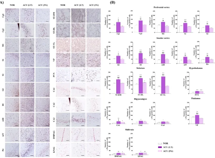
1. 정상 쥐에 간정격 및 사관혈 침 처치 후 대뇌의 c-Fos 발현 분석
정상 쥐에 간정격 또는 사관혈 침 치료를 1회 시행한 후, 각 뇌 영역에서의 신경세포 활성도를 c-Fos 발현을 통해 관찰하였다.
ACU (LT)는 prefrontal cortex의 cingulate cortex area 1 and 2 (Cg1, p<0.05; Cg2, p<0.01), motor cortex area 1 (M1, p< 0.01), somatosensory cortex area 1 and 2 (S1, p<0.001; S2, p<0.01), insular cortex의 granular insular cortex (GI, p< 0.05), dysgranular insular cortex (DI, p<0.001), agranular insular cortex dorsal part (AID, p<0.001), agranular insular cortex ventral part (AIV, p<0.01), piriform cortex (Pir, p< 0.01), striatum (ST)의 dorsal medial part (ST-DM, p<0.001), dorsal lateral part (ST-DL, p<0.001), ventral lateral part (ST- VL, p<0.001), hypothalamus의 PVN (p<0.001), hippocampus의 cornu ammonis (CA) 1, 2, 3 (CA1, p<0.01; CA2, p<0.01; CA3, p<0.05), thalamus의 ventral posterior thalamic nucleus (VP, p<0.01), periaqueductal gray (PAG)의 dorsomedial part와 lateral part (DMPAG, p<0.01; LPAG, p<0.05) 영역에서 침 치 료를 하지 않은 그룹(NOR)에 비하여 c-Fos 발현을 유의미하게 증 가시켰으며, 이는 PVN, CA2, CA3, DMPAG, LPAG를 제외한 모든 뇌 부위에서 ACU (FG)에 비하여 높은 증가를 보였다(Fig. 2).
ACU (FG)는 ST-DL (p<0.05), PVN (p<0.001), CA1 (p< 0.05), DMPAG (p<0.05), LPAG (p<0.01) 영역에서 NOR에 비 하여 c-Fos 발현이 유의미하게 증가하였다(Fig. 2).
Fig. 2. The changes of c-Fos expression levels in each brain region after ACU (LT) and ACU (FG).
(A, B) Mouse brains were extracted 90 min after acupuncture treatment, and the c-Fos positive cells were observed using immunohistochemistry. Statistical analysis was performed using one-way ANOVA followed by Newman-Keuls tests (*p<0.05, **p<0.01,
***p<0.001 vs. NOR; $p<0.05, $$p<0.01, $$$p<0.001 vs. ACU (FG), each n=4). Data are shown as means±SEM. Scar bar : 200 μm.
Abbreviations of each brain region are shown in Table 1. ACU (FG) : four gate acupuncture treatment, ACU (LT) : liver tonification acupuncture treatment, NOR: normal.
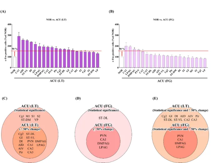
보다 주요한 뇌 부위를 도출하기 위하여, 침 치료 후 NOR 대비 c-Fos 변화량을 증가순으로 나열하였으며(Fig. 3A, B), 50% 이상 변화량을 보인 뇌 부위와 NOR 대비 통계적으로 유의한 차이를 보인 뇌 부위 모두에 해당하는 뇌 부위를 도출하였다(Fig. 3C, D).
ACU (LT)에 의하여 50% 이상 변화량 및 통계적 유의성을 보인 뇌 부위는 Cg2, GI, DI, AID, AIV, Pir, ST-DL, ST-VL, PVN, CA1, CA2, CA3, DMPAG, LPAG였다(Fig. 3C). ACU (FG)에 의하여 50% 이상 변화량 및 통계적 유의성을 보인 뇌 부위는 PVN, CA1, DMPAG, LPAG였는데, 이들은 모두 ACU (LT)에 의하여 50% 이상 변화량 및 통계적 유의성을 보인 뇌 부위에 포함되어 있었다(Fig.
3D). 분석을 통해 우리는 ACU (LT)가 ACU (FG)에 비하여 보다 많은 뇌 부위에서 신경 반응성의 변화를 야기하고 있음을 알 수
있었다(Fig. 3E).
2. 우울증 동물모델에서 간정격 및 사관혈 침 치료의 우 울 행동 개선 효과
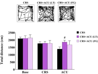
간정격과 사관혈 침 치료가 동물의 우울 행동을 개선하는지 확 인하기 위하여, 4주간의 CRS로 우울 행동을 유발한 후, 2주간 침 치료를 시행하여 동물의 행동 변화를 관찰하였다. CRS 유발 후 모든 동물의 총 이동거리는 감소하였으며, 침 치료 후 ACU (LT)는 동물의 이동거리를 유의하게 증가시켰다(p<0.05 vs. CRS). ACU (FG)도 동물의 이동거리를 소폭 증가시키는 경향을 보였으나, 통계 적 유의성은 보이지 않았다(Fig. 4).
Fig. 3. Altered brain regions of c-Fos positive cells expression after ACU (LT) and ACU (FG).
(A, B) The changed brain regions compared to the NOR were represented in descending order after ACU (LT) and ACU (FG). (C, D) After ACU (LT) and ACU (FG), the brain regions corresponding to both showing a statistically significant difference compared to the NOR and greater than 50% change compared to the NOR were analyzed. (E) The brain regions corresponding to showing a statistically significant difference and greater than 50% change compared to the NOR after ACU (LT) and ACU (FG). Abbreviations of each brain region are shown in Table 1. ACU (FG) : four gate acupuncture treatment, ACU (LT) : liver tonification acupuncture treatment, NOR : normal, ↑50% : more than 50% change.
3. 우울증 동물 모델에 간정격, 사관혈 침 치료 후 뇌신 경 변화 양상 관찰
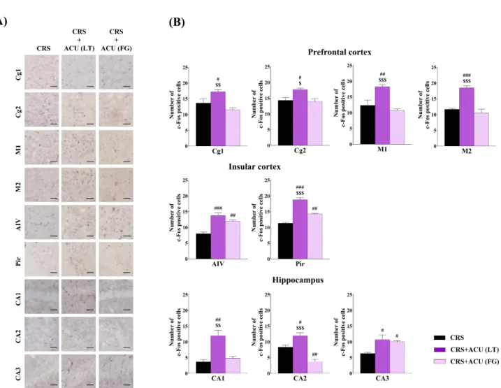
우울증 동물 모델에서 간정격과 사관혈 침 치료의 뇌신경 반응 성 변화를 관찰한 결과, CRS+ACU (LT)는 prefrontal cortex의 Cg1 (p<0.05), Cg2 (p<0.05), M1 (p<0.01), motor cortex area 2 (M2, p<0.001), insular cortex의 AIV (p<0.001), Pir (p<0.001), hippocampus의 CA1 (p<0.01), CA2 (p<0.05), CA3 (p<0.05) 영역에서 CRS군에 비하여 c-Fos 발현이 유의하게 증가하였으며, 이는 AIV, CA3를 제외한 모든 뇌 부위에서 CRS+
ACU (FG)에 비하여 높은 증가를 보였다(Fig. 5).
CRS+ACU (FG)는 AIV (p<0.01), Pir (p<0.01), CA3 (p< 0.05) 영역에서 CRS 군에 비하여 c-Fos 발현이 증가하였으며, CA2 (p<0.01 vs. CRS)에서는 CRS군에 비하여 발현이 감소하였
다(Fig. 5).
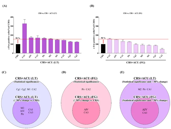
우울 행동 개선과 관련된 보다 핵심적인 뇌 부위를 도출하기 위하여, 침 치료 후 CRS 대비 c-Fos 변화량을 증가순으로 나열하 였으며(Fig. 6A, B), 50% 이상 변화량을 보인 뇌 부위와 CRS 대비 통계적으로 유의한 차이를 보인 뇌 부위 모두에 해당하는 뇌 부위 를 도출하였다(Fig. 6C, D). CRS+ACU (LT)에 의하여 50% 이상 변화량 및 통계적 유의성을 보인 뇌 부위는 M2, AIV, Pir, CA1, CA3 였다(Fig. 6C). CRS+ACU (FG)에 의하여 50% 이상 변화량 및 통계적 유의성을 보인 뇌 부위는 AIV와 CA3였는데, 이들은 모 두 CRS+ACU (LT)에 의하여 50% 이상 변화량 및 통계적 유의성을 보인 뇌 부위에 포함되어 있었다(Fig. 6D). 본 분석을 통해 우리는 CRS+ACU (LT)가 CRS+ACU (FG)에 비하여 보다 많은 뇌 부위에 서 신경 반응성의 변화를 야기하고 있음을 알 수 있었으며,
Fig. 4. Change of depression-like behavior after ACU (LT) and ACU (FG) in a 4-weeks CRS model.
Depression-like behavior of mice was analyzed using open field test. After CRS, total traveled distance of all mice was decreased.
After 2 weeks of acupuncture treatment, CRS+ACU (LT) signifi- cantly increased total traveled distance, but CRS+ACU (FG) did not.
Statistical analysis was performed using unpaired t-test (#p<0.05 vs. CRS, each n=4). Data are shown as means±SEM. CRS : chronic restraint stress, CRS+ACU (FG) : CRS followed by four gate acu- puncture treatment, CRS+ACU (LT) : CRS followed by liver tonifi- cation acupuncture treatment.
CRS+ACU (LT)에 의해 추가로 변화된 뇌 부위는 M2, Pir, CA1으 로 확인되었다(Fig. 6E).
4. 정상 쥐와 우울증 동물모델에서 간정격 침 치료에 의 해 변화된 핵심 뇌 부위 도출
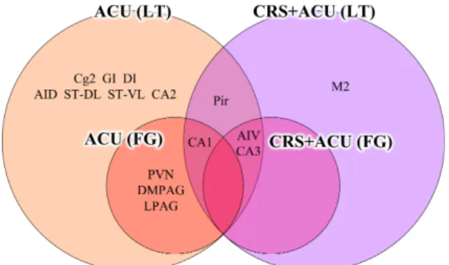
ACU (LT)는 정상 쥐와 우울증 쥐 모두에서 ACU (FG)에 비하여 많은 뇌 부위에서 신경 반응성의 변화를 야기하였으며, ACU (LT) 에 의해 변화된 뇌 부위에는 ACU (FG)에 의해 변화된 뇌 부위가 모두 포함되어 있었다. 이들 뇌 부위 중 M2는 오직 CRS+ACU (LT) 에 의해서만 변화되었으며, ACU (LT)와 CRS+ACU (LT)에서만 공 통적으로 변화된 뇌 부위는 Pir이었다(Fig. 7).
고 찰
본 연구에서는 우울증 치료에 활용 가능한 경혈 조합인 간정격 과 사관혈을 이용하여 정상 동물에서의 뇌신경 반응성을 확인하였 다. 또한 만성 우울증 동물모델에서 ACU (LT)와 ACU (FG)의 우울 행동 개선 효과 및 뇌신경 반응성을 비교 분석함으로써 우울증 치 료에 보다 효과적인 경혈 조합을 도출하고, 치료 효과와 관련된 주요 뇌 부위를 도출하였다. 정상 쥐와 우울증이 유발된 쥐 모두에
서 ACU (LT)는 ACU (FG)에 의해 변화된 뇌 부위를 포함하여 더 많은 뇌 부위에서 신경 반응성 변화를 야기하였다. 또한 ACU (LT) 는 만성 스트레스로 유도된 동물의 우울 행동을 유의하게 개선하 였으며, ACU (LT)의 우울 행동 개선 효과와 밀접하게 관련된 뇌 부위는 prefrontal cortex의 M2, hippocampus의 CA1, insular cortex의 Pir로 도출되었다.
우울증은 뇌의 여러 영역에서의 구조적 또는 기능적 결함이 주 요 발병 원인으로 작용하는 것으로 알려져 있다35,36). 우울증 환자는 prefrontal cortex, anterior cingulate cortex, striatum, hippo- campus 등의 영역에서 뇌의 용적 감소 및 병리적 단백질 발현을 보이는 것으로 보고되어 있으며6,7), 우울증 환자에게서 흔히 보이 는 hypothalamic-pituitary-adrenal axis 기능 장애는 hypo- thalamus의 PVN과 hippocampus의 병리적 변화와 밀접한 관련 이 있다고 알려져 있다. 심리적 스트레스로 인하여 PVN이 활성화 되면 부신피질자극호르몬 방출호르몬(corticotrophin releasing hormone)에 의해 glucocorticoid가 생성되며, 이는 hippocampus 의 신경세포 생존과 발생에 영향을 미쳐 용적 감소로 이어지며 우 울증을 유발하게 된다6,37,38). 최근에는 대뇌 각 영역의 신경 활성도 를 관찰함으로써 각 영역의 기능적 변화와 질환과의 연관성을 규 명하기 위한 연구가 활발하게 진행되고 있는데, 임상 연구에서는 주로 양전자 방출 단층촬영(positron emission tomography;
PET), 기능성 자기공명영상(functional magnetic resonance imaging; fMRI), 뇌전도(electroencephalogram; EEG) 등의 방법 을 사용하여 뇌 영역의 활성 정도를 관찰한다6). 이러한 방법들은 비침습적이라는 장점으로 임상에서는 활용도가 높으나, 뇌신경 활 성도를 직접적으로 측정하지는 못한다는 한계가 있다. 이러한 한 계를 보완하기 위한 방법으로 동물 연구에서는 뇌신경 활성도를 보다 직접적으로 관찰할 수 있는 신경 활성 마커인 c-Fos, early growth response 1 (Egr1) 등을 사용하여 보다 심화된 기전 연구 를 진행하고 있다39-42). 그 중 c-Fos는 초기반응 유전자(imme- diate early genes) 중 하나로 다양한 외부 자극에 반응하여 활성화 된다는 특성이 있어 신경세포의 활동을 측정하는 마커로 널리 활 용되고 있다39,43). 따라서 본 연구에서도 침 치료에 의한 뇌신경 활 성도를 관찰하기 위한 방법으로, 각 대뇌 영역에서의 c-Fos 발현을 관찰하였다.
침 치료는 대상 질환 및 환자의 증상에 따라 변증 과정을 거쳐 단일 경혈을 선정하거나 몇 개의 경혈로 이루어진 경혈 조합을 구 성하여 치료하게 된다. 임상에서는 주로 단일 경혈 보다는 여러 개의 경혈이 함께 활용되는데, 사암침법을 비롯하여 사관혈, 중풍 칠처혈, 육총혈 등이 널리 활용되고 있다. 그 중 사암침법은 조선시
Fig. 5. The number of c-Fos positive cells in each brain region after ACU (LT) and ACU (FG) in a 4-weeks CRS model.
(A, B) The c-Fos positive cells in each brain region of CRS, CRS+ACU (LT), and CRS+ACU (FG) groups were analyzed using immunohistochemistry. Statistical analysis was performed using one-way ANOVA followed by Newman-Keuls tests (#p<0.05, ##p<0.01,
###p<0.001 vs. CRS; $p<0.05, $$p<0.01, $$$p<0.001 vs. CRS+ACU (FG)). Data are shown as means±SEM. Scar bar : 200 μm.
Abbreviations of each brain region are shown in Table 1. CRS : chronic restraint stress (n=3), CRS+ACU (FG) : CRS followed by four gate acupuncture treatment (n=4), CRS+ACU (LT) : CRS followed by liver tonification acupuncture treatment (n=4).
대 사암도인에 의해 창시되었다고 알려져있으며, 오수혈의 음양오 행 속성을 활용하여 12경맥의 정격, 승격, 한격, 열격 등의 다양한 조합으로 활용되고 있다. 그 중 가장 널리 활용되는 정격의 경우, 해당 경맥 또는 장부의 기(氣)를 보하거나, 균형을 바로잡기 위해 사용되며, 승격의 경우 해당 경맥 또는 장부의 편승된 기운을 제어 하기 위하여 사용된다44,45). 본 연구에서는 우울증 치료를 위하여 다양한 사암침법 조합 중 간정격을 선정하였는데, 한의학에서 스 트레스로 인한 우울증은 주로 간(肝)의 기운이 울결되어 발생하는 것으로 보기 때문에 간(肝)의 기운을 북돋아 울결된 기운을 해소해 줄 수 있는 간정격이 적합하다고 생각하였다. 간정격은 사암침법 중에서 가장 많이 활용되는 조합 중 하나로, 임상적으로는 간기능 및 비만도 개선, 근시, 바르톨린선염, 이명, 딸국질과 불면증, 전립
선 암, 진전 등 다양한 질환에 활용되고 있다45). 그러나 이러한 임 상적 활용성에도 불구하고 간정격을 활용한 동물 기전 연구는 매 우 부족한 상황이며, 다른 경혈 조합과의 비교 연구 또한 거의 이루 어져 있지 않다. 따라서 본 연구에서는 간정격과 유사하게 신체의 막힌 기운을 소통해주는 의미로 주로 사용되는 사관혈을 선정하여 치료 효과 및 관련 기전을 비교하기 위한 연구를 수행하였다. 사관 혈은 수양명대장경의 원혈인 합곡(LI4)과 족궐음간경의 원혈인 태 충(LR3)으로 구성되어 있으며, 기혈의 흐름을 원활하게 하고, 장기 의 막힌 기운을 해소하는 역할을 한다46). 따라서 임상적으로 소화 기 계통 장애나 호흡 부전, 만성 피로 증후군, 월경통 등의 질환에 사용되며47), 우울증 치료에도 사용되고 있다34).
본 연구 결과, 정상 쥐와 우울증 유발 쥐 모두에서 ACU (LT)는
Fig. 6. Altered brain regions of c-Fos positive cells expression after ACU (LT) and ACU (FG) in 4-weeks CRS model.
(A, B) The changed brain regions compared to the CRS were represented in descending order after ACU (LT) and ACU (FG). (C, D) After ACU (LT) and ACU (FG), the brain regions corresponding to both showing a statistically significant difference compared to the CRS and greater than 50% change compared to the CRS were analyzed. (E) The brain regions corresponding to showing a statistically significant difference and greater than 50% change compared to the CRS after ACU (LT) and ACU (FG). Abbreviations of each brain region are shown in Table 1. ACU (FG) : four gate acupuncture treatment, ACU (LT) : liver tonification acupuncture treatment, CRS : chronic restraint stress, CRS+ACU (FG) : CRS followed by four gate acupuncture treatment, CRS+ACU (LT) : CRS followed by liver tonifi- cation acupuncture treatment, ↑50% : more than 50% change.
ACU (FG)에 의해 변화된 뇌 부위를 포함하여 더 많은 뇌 부위에서 신경 반응성 변화를 야기하였다. 정상 쥐에서는 PVN, CA1, DMPAG, LPAG 영역이 ACU (LT)와 ACU (FG)에 의해 공통적으로 변화하였 는데, 이들은 ACU (LT)와 ACU (FG)의 공통 효과를 반영하는 뇌 부위로 볼 수 있다. Hypothalamus의 PVN은 스트레스, 대사, 면역 등을 조절하며 심혈관과 신장 및 위장관과 같은 자율 기능을 조절 하는데 관여한다48). Hippocampus의 CA1은 기억 및 인지 기능과 관련이 있으며, 주로 스트레스에 의한 우울증이나 불면증 등의 질 환과 밀접한 관련이 있다49,50). 또한, hippocampus는 PVN 뿐 아 니라 brain stem과 nucleus tractus solitarius (NTS) 등과 밀접하 게 관련되어 자율 신경계 조절에 관여한다51). Midbrain의 PAG 영 역(DMAPG와 LPAG)은 모두 통증 조절과 연관된다52). 따라서 ACU (LT)와 ACU (FG)는 모두 스트레스로 인한 우울증, 인지 기능,
자율신경계, 통증 관련 질환에 적용될 수 있다. 또한 ACU (LT)에 의해 추가로 변화된 뇌 영역인 Cg2, GI, DI, AID, AIV, Pir, ST-DL, ST-VL, CA2, CA3 영역은 ACU (LT)만의 특이적인 효과를 반영하 는 뇌 부위라고 생각된다. Prefrontal cortex의 anterior cingulate cortex (Cg1, Cg2)는 정서적 기능, 기억력, 학습능력, 기억력, 통증 과 관련되어 있으며, insular cortex (GI, DI, AID, AIV, Pir)는 pre- frotal cortex 및 hippocampus 등과 연결되어 감정, 운동 정보, 통증 등에 관여한다53,54). Striatum (ST-DL, ST-VL)은 주로 운동 기능과 관련된 신경 반응 조절을 통해 운동 기능을 억제하거나 향 상하는데 관여하며, 목표 지향적인 행동과 습관 학습에도 관여한 다55). Hippocampus의 CA2/3는 CA1과 마찬가지로 인지기능, 스 트레스, 우울증 등의 질환과 밀접한 관련성이 있다49,50). 따라서 ACU (LT)는 정서적 기능, 운동 기능, 감각 정보 처리 기능, 학습
Fig. 7. Schematic venn diagram of brain regions altered by ACU (LT) and ACU (FG) in normal and CRS mice.
The brain regions corresponding to showing a statistically sig- nificant difference and greater than 50% change compared to the NOR or CRS after ACU (LT) and ACU (FG). Abbreviations of each brain region are shown in Table 1. ACU (FG) : four gate acupuncture treatment, ACU (LT) : liver tonification acupuncture treatment, CRS : chronic restraint stress, CRS+ACU (FG) : CRS followed by four gate acupuncture treatment, CRS+ACU (LT) : CRS followed by liver tonification acupuncture treatment, NOR : normal.
및 기억, 통증 조절, 자율신경 및 내분비 조절을 통한 항상성 유지 에 활용될 수 있을 것이다.
간정격과 사관혈의 역할을 보다 직접적으로 규명하기 위하여 우울증 동물모델에서 동물의 행동개선 및 뇌신경 반응성을 비교 분석한 결과, ACU (LT)는 동물의 우울 행동을 유의하게 개선한 반면, ACU (FG)는 소폭 개선효과를 보였으나 유의성은 없었다. 뇌 신경 반응 분석에서도 ACU (LT)와 ACU (FG)는 공통적으로 AIV와 CA3 영역의 변화를 야기하였는데, 이들 부위는 모두 정서적 변화 및 운동 기능과 밀접한 관련이 있는 부위이므로 사관혈이 소폭 우 울 행동을 개선한 것에 기여한 것으로 생각된다. ACU (LT)는 이 외에도 M2, Pir, CA1 영역의 변화를 추가로 야기하였는데, motor cortex는 운동 신호를 생성하고 근육에 전달하여 운동 행동을 제 어하는데 관여하며53), insular의 Pir과 hippocampus의 CA1은 상 기 기술한 것처럼 모두 정서적 변화 및 운동 기능과 관련되어 있다.
따라서 ACU (FG)보다 ACU (LT)가 우울 행동 개선 효과가 더 명확 하게 나타난 것은 이러한 보다 많은 뇌 영역의 활성화와 관련성이 높다고 생각된다.
본 연구는 간정격과 사관혈의 우울 행동 개선 효과 및 뇌신경 반응성을 비교 분석함으로써 우울증 치료에 보다 효과적인 경혈 조합과 관련 기전을 규명하였다는 데 의의가 있다. 그러나 이러한 차이는 간정격과 사관혈의 혈자리 개수 차이에 따른 자극량의 차 이 및 치료 횟수의 차이에 의해서 발생했을 가능성이 존재하며, 보다 많은 뇌 부위를 관찰하지 못했다는 한계가 여전히 존재한다.
따라서 향후 동일한 치료 기간 및 자극량을 가진 다른 경혈 조합과
의 비교 연구 또한 진행될 필요가 있으며, 전체 뇌 부위를 대상으로 한 종합적인 기전 도출 및 주요 뇌 부위에서의 우울증 관련 심화 기전 규명이 진행되어야 할 것이다.
결 론
간정격과 사관혈 침 치료의 우울 행동 개선 효과 및 뇌신경 반응 성을 분석한 결과, 정상 쥐에서 ACU (LT)와 ACU (FG)는 공통적으 로 hypothalamus, hippocampus, PAG 영역의 변화를 야기하였 으며, ACU (LT)는 이와 더불어 prefrontal cortex, insular cortex, striatum, hippocampus 영역에서도 변화를 야기하였다.
ACU (LT)는 스트레스로 유도된 동물의 우울 행동을 유의하게 개선 하였으며, ACU (LT)에 의하여 대뇌의 M2, AIV, Pir, CA1, CA3 영역에서 신경 세포 활성이 관찰되었다. 이 중, AIV와 CA3는 ACU (FG)에서도 공통적으로 변화되었으며, M2, Pir, CA1 영역은 ACU (LT)에 의해서만 변화되었다. 따라서 ACU (LT)에 의해서만 변화된 이들 뇌 부위가 ACU (LT)의 우울 행동 개선 효과와 관련된 핵심 뇌 부위로 작용함을 알 수 있었다. 향후 보다 많은 뇌 영역의 신경 활성도 관찰 및 다양한 경혈 조합과의 비교가 이루어질 필요가 있 으며, 핵심 부위로 도출된 뇌 부위에서 우울증과 관련된 다른 주요 기전들의 연구 또한 이루어져야 할 것이다.
Acknowledgement
None.
Funding
This research was supported by the Basic Science Re- search Program through the National Research Foundation of Korea (NRF) funded by the Ministry of Science, ICT &
Future Planning (NRF-2018R1C1B3004424 and NRF-2018R1A 6A1A03025221).
Data availability
The authors can provide upon reasonable request.
Conflicts of interest
저자들은 아무런 이해 상충이 없음을 밝힌다.
References
1. Reddy M. Depression: the disorder and the burden. Indian J Psychol Med. 2010 ; 32(1) : 1-2. https://doi.org/10.4103/0253- 7176.70510
2. Semenkovich K, Brown ME, Svrakic DM, Lustman PJ. Depression in type 2 diabetes mellitus: prevalence, impact, and treatment.
Drugs. 2015 ; 75(6) : 577-87. https://doi.org/10.1007/s40265- 015-0347-4
3. Blumenthal JA, Babyak MA, Doraiswamy PM, Watkins L, Hoffman BM, Barbour KA, et al. Exercise and pharmacotherapy in the treatment of major depressive disorder. Psychosom Med. 2007 ; 69(7) : 587-96. https://doi.org/10.1097/PSY.0b013e318148c19a 4. Bachmann S. Epidemiology of suicide and the psychiatric
perspective. Int J Environ Res Public Health. 2018 ; 15(7) : 1425.
https://doi.org/10.3390/ijerph15071425
5. Newman DWA. Dorland’s pocket medical dictionary. 25th ed.
Philadelphia : Saunders; 1995.
6. Yang L, Zhao Y, Wang Y, Liu L, Zhang X, Li B, et al. The effects of psychological stress on depression. Curr Neuropharmacol. 2015 ; 13(4) : 494-504. https://doi.org/10.2174/1570159x1304150831150507 7. Bennett M. The prefrontal–limbic network in depression: Modu- lation by hypothalamus, basal ganglia and midbrain. Prog Neurobiol.
2011 ; 93(4) : 468-87. https://doi.org/10.1016/j.pneurobio.2011.
01.006
8. Pandya M, Altinay M, Malone DA, Anand A. Where in the brain is depression? Curr Psychiatry Rep. 2012 ; 14(6) : 634-42.
https://doi.org/10.1007/s11920-012-0322-7
9. Winokur A, Gary KA, Rodner S, Rae‐Red C, Fernando AT, Szuba MP. Depression, sleep physiology, and antidepressant drugs. Depress
Anxiety. 2001 ; 14(1) : 19-28. https://doi.org/10.1002/da.1043 10. Liu Q, Li B, Zhu HY, Wang YQ, Yu J, Wu GC. Glia atrophy in the hippocampus of chronic unpredictable stress-induced depres- sion model rats is reversed by electroacupuncture treatment. J Affect Disord. 2011 ; 128(3) : 309-13. https://doi.org/10.1016/
j.jad.2010.07.007
11. Wu J, Yeung AS, Schnyer R, Wang Y, Mischoulon D. Acupuncture for depression: a review of clinical applications. Can J Psychiatry. 2012 ; 57(7) : 397-405. https://doi.org/10.1177/070674371205700702 12. Gualano M, Bert F, Martorana M, Voglino G, Andriolo V, Thomas
R, et al. The long-term effects of bibliotherapy in depression treatment: Systematic review of randomized clinical trials. Clin Psychol Rev. 2017 ; 58 : 49-58. https://doi.org/10.1016/j.cpr.
2017.09.006
13. David HB. Clinical handbook of psychological disorders: A step- by-step treatment manual. 4th ed. The Guilford Press. 2008 : 275-331.
14. de Mello MF, de Jesus Mari J, Bacaltchuk J, Verdeli H, Neugebauer R. A systematic review of research findings on the efficacy of interpersonal therapy for depressive disorders. Eur Arch Psychiatry Clin Neurosc. 2005 ; 255(2) : 75-82. https://doi.
org/10.1007/s00406-004-0542-x
15. Lee IS, Cheon S, Park JY. Central and Peripheral Mechanism of Acupuncture Analgesia on Visceral Pain: A Systematic Review.
Evid Based Complement Alternat Med. 2019 ; 2019 : 1304152.
https://doi.org/10.1155/2019/1304152
16. Park JY, Park JJ, Jeon S, Doo AR, Kim SN, Lee H, et al. From pe- ripheral to central: the role of ERK signaling pathway in acu- puncture analgesia. J Pain. 2014 ; 15(5) : 535-49. https://doi.
org/10.1016/j.jpain.2014.01.498
17. Zhang R, Lao L, Ren K, Berman BM. Mechanisms of acupuncture- electroacupuncture on persistent pain. Anesthesiology. 2014 ; 120(2) : 482-503. https://doi.org/10.1097/aln.0000000000000101 18. Goldman N, Chen M, Fujita T, Xu Q, Peng W, Liu W, et al.
Adenosine A1 receptors mediate local anti-nociceptive effects of acupuncture. Nat Neurosci. 2010 ; 13(7) : 883-8. http://doi.
org/10.1038/nn.2562
19. Park JY, Choi H, Baek S, Jang J, Lee A, Jeon S, et al. p53 signalling mediates acupuncture-induced neuroprotection in Parkinson’s disease. Biochem Biophys Res Commun. 2015 ; 460(3) : 772-9.
http://doi.org/S0006-291X(15)00571-9
20. Park JY, Kim SN, Yoo J, Jang J, Lee A, Oh JY, et al. Novel Neuroprotective Effects of Melanin-Concentrating Hormone in Parkinson’s Disease. Mol Neurobiol. 2017 ; 54(10) : 7706-21.
http://doi.org/10.1007/s12035-016-0258-8
21. Jung J, Lee SM, Lee MJ, Ryu JS, Song JH, Lee JE, et al. Lipidomics reveals that acupuncture modulates the lipid metabolism and in- flammatory interaction in a mouse model of depression. Brain Behav Immun. 2021 ; 94 : 424-36. http://doi.org/10.1016/j.bbi.
2021.02.003
22. Lee MJ, Ryu JS, Won SK, Namgung U, Jung J, Lee SM, et al. Effects of Acupuncture on Chronic Stress-Induced Depression-Like Behavior and Its Central Neural Mechanism. Front Psychol. 2019 ; 10 : 1353. http://doi.org/10.3389/fpsyg.2019.01353
23. Dong B, Chen Z, Yin X, Li D, Ma J, Yin P, et al. The Efficacy of Acupuncture for Treating Depression-Related Insomnia Com- pared with a Control Group: A Systematic Review and Meta- Analysis. BioMed Research International. 2017 ; 2017 : 9614810.
http://doi.org/10.1155/2017/9614810
24. Park H, Yoo D, Kwon S, Yoo TW, Park HJ, Hahm DH, et al. Acu- puncture stimulation at HT7 alleviates depression-induced be- havioral changes via regulation of the serotonin system in the pre- frontal cortex of maternally-separated rat pups. J Physiol Sci.
2012 ; 62(4) : 351-7. http://doi.org/10.1007/s12576-012-0211-1 25. Duan G, He Q, Pang Y, Chen W, Liao H, Liu H, et al. Altered
amygdala resting-state functional connectivity following acu- puncture stimulation at BaiHui (GV20) in first-episode drug- Naïve major depressive disorder. Brain Imaging Behav. 2020 ; 14(6) : 2269-80. https://doi.org/10.1007/s11682-019-00178-5 26. Deng D, Liao H, Duan G, Liu Y, He Q, Liu H, et al. Modulation of the default mode network in first-episode, drug-naive major de- pressive disorder via acupuncture at Baihui (GV20) acupoint.
Front Hum Neurosci. 2016 ; 10 : 230. https://doi.org/10.3389/
fnhum.2016.00230
27. Quah-Smith I, Wen W, Chen X, Williams MA, Sachdev PS. The brain effects of laser acupuncture in depressed individuals: an fMRI investigation. Medical Acupuncture. 2012 ; 24(3) : 161-71.
https://doi.org/10.1089/acu.2011.0870
28. Lottering B, Lin YW. TRPV1 Responses in the Cerebellum Lobules VI, VII, VIII Using Electroacupuncture Treatment for Chronic
Pain and Depression Comorbidity in a Murine Model. Int J Mol Sci. 2021 ; 22(9) : 5028. http://doi.org/10.3390/ijms22095028 29. Seo SY, Moon JY, Kang SY, Kwon OS, Kwon S, Bang SK, et al. An
estradiol-independent BDNF-NPY cascade is involved in the an- tidepressant effect of mechanical acupuncture instruments in ovariectomized rats. Sci Rep. 2018 ; 8(1) : 5849. http://doi.
org/10.1038/s41598-018-23824-2
30. Kim H, Park HJ, Han SM, Hahm DH, Lee HJ, Kim KS, et al. The effects of acupuncture stimulation at PC6 (Neiguan) on chronic mild stress-induced biochemical and behavioral responses.
Neurosci Lett. 2009 ; 460(1) : 56-60. https://doi.org/10.1016/
j.neulet.2009.05.008
31. Duan Dm, Tu Y, Liu P, Jiao S. Antidepressant effect of electro- acupuncture regulates signal targeting in the brain and increases brain-derived neurotrophic factor levels. Neural Regen Res. 2016 ; 11(10) : 1595-602. https://doi.org/10.4103/1673-5374.193238 32. Zhang X, Song Y, Bao T, Yu M, Xu M, Guo Y, et al. Antidepres- sant-like effects of acupuncture involved the ERK signaling pathway in rats. BMC Complement Altern Med. 2016 ; 16(1) : 380.
https://doi.org/10.1186/s12906-016-1356-x
33. Kou RZ, Chen H, Yu Ml, Xu TC, Fu SP, Lu SF. Acupuncture for be- havioral changes of experimental depressive disorder: a system- atic review and meta-analysis. Sci Rep. 2017 ; 7(1) : 9669.
https://doi.org/10.1038/s41598-017-09712-1
34. Liu JH, Wu ZF, Sun J, Jiang L, Jiang S, Fu WB. Role of AC-cAMP- PKA cascade in antidepressant action of electroacupuncture treatment in rats. Evid Based Complement Alternat Med. 2012 ; 2012 : 932414. https://doi.org/10.1155/2012/932414 35. Paelecke-Habermann Y PJ, Leplow B. Attention and executive
functions in remitted major depression patients. J Affect Disord.
2005 ; 89(1-3) : 125-35. http://doi.org/https://doi.org/10.1016/
j.jad.2005.09.006
36. Vythilingam M, Vermetten E, Anderson GM, Luckenbaugh D, Anderson ER, Snow J, et al. Hippocampal volume, memory, and cortisol status in major depressive disorder: effects of treatment.
Biol Psychiatry. 2004 ; 56(2) : 101-12. https://doi.org/10.1016/
j.biopsych.2004.04.002
37. Belmaker R, Agam G. Major depressive disorder. N Engl J Med.
2008 ; 358(1) : 55-68. https://doi.org/10.1056/NEJMra073096 38. Massart R, Mongeau R, Lanfumey L. Beyond the monoaminergic
hypothesis: neuroplasticity and epigenetic changes in a trans- genic mouse model of depression. Philos Trans R Soc Lond B Biol Sci.
2012 ; 367(1601) : 2485-94. https://doi.org/10.1098/rstb.2012.0212 39. Wheeler AL, Teixeira CM, Wang AH, Xiong X, Kovacevic N, Lerch
JP, et al. Identification of a functional connectome for long-term fear memory in mice. PLoS Comput Biol. 2013 ; 9(1) : e1002853.
http://doi.org/10.1371/journal.pcbi.1002853
40. Aggleton JP, Brown MW, Albasser MM. Contrasting brain activity patterns for item recognition memory and associative recog- nition memory: insights from immediate-early gene functional imaging. Neuropsychologia. 2012 ; 50(13) : 3141-55. http://doi.
org/10.1016/j.neuropsychologia.2012.05.018
41. Park JY, Cho SJ, Lee SH, Ryu Y, Jang JH, Kim SN, et al. Peripheral ERK modulates acupuncture-induced brain neural activity and its functional connectivity. Sci Rep. 2021 ; 11(1) : 5128.
https://doi.org/10.1038/s41598-021-84273-y
42. Gallo FT, Katche C, Morici JF, Medina JH, Weisstaub NV. Imme- diate early genes, memory and psychiatric disorders: focus on c-Fos, Egr1 and Arc. Front Behav Neurosci. 2018 ; 12 : 79.
https://doi.org/10.3389/fnbeh.2018.00079
43. Dragunow M, Faull R. The use of c-fos as a metabolic marker in neuronal pathway tracing. J Neurosci Methods. 1989 ; 29(3) : 261-5. https://doi.org/10.1016/0165-0270(89)90150-7 44. Park JY, Lee SH, Kim SY, Park HJ. Literature Review and Network
Analysis on the Pain Disease Approach of Saam Acupuncture Method. Korean J Acupunct. 2017 ; 34(2) : 88-99. https://doi.
org/10.14406/acu.2017.011
45. Yoon MJ, Kim SY, Park JY. Recent Study Trends of the Liver-toni- fication and Liver-sedation of Saam Acupuncture. Korean J Acu- punct. 2018 ; 35(1) : 1-17. https://doi.org/10.14406/acu.2018.004 46. Fan L, Fu WB, Xu NG, Liu JH, Ou A. Impacts of acupuncture and moxibustion on outcome indeices of depression patients’
subjective reports. Zhongguo Zhen Jiu. 2012 ; 32(5) : 385-9.
https://doi.org/10.1016/s1003-5257(13)60039-2
47. Shin KM, Park JE, Lee S, Choi SM, Ahn YC, Lee JW, et al. Effect of siguan acupuncture on gastrointestinal motility: a randomized,
sham-controlled, crossover trial. Evid Based Complement Alternat Med. 2013 ; 2013 : 918392. https://doi.org/10.1155/2013/918392 48. Ferguson AV, Latchford KJ, Samson WK. The paraventricular nu- cleus of the hypothalamus–a potential target for integrative treat- ment of autonomic dysfunction. Expert Opin Ther Targets 2008 ; 12(6) : 717-27. https://doi.org/10.1517/14728222.12.6.717 49. Fanselow MS, Dong HW. Are the dorsal and ventral hippocampus
functionally distinct structures? Neuron. 2010 ; 65(1) : 7-19.
https://doi.org/10.1016/j.neuron.2009.11.031
50. MacQueen GM, Campbell S, McEwen BS, Macdonald K, Amano S, Joffe RT, et al. Course of illness, hippocampal function, and hippocampal volume in major depression. Focus. 2005 ; 3(1) : 146-155. https://doi.org/10.1176/foc.3.1.146
51. Cui S, Zhou Y, Wu S, Cao J, Zhu G, Zhou M. Electroacupuncture improved the function of myocardial ischemia involved in the hippocampus-paraventricular nucleus-sympathetic nerve path- way. Evid Based Complement Alternat Med. 2018 ; 2018 : 2870676. https://doi.org/10.1155/2018/2870676
52. de Freitas RL, de Oliveira RC, de Oliveira R, Paschoalin‐Maurin T, de Aguiar Corrêa FM, Coimbra NC. The role of dorsomedial and ventrolateral columns of the periaqueductal gray matter and in situ 5‐HT2A and 5‐HT2C serotonergic receptors in post‐ictal antinociception. Synapse. 2014 ; 68(1) : 16-30. https://doi.
org/10.1002/syn.21697
53. Teka WW, Hamade KC, Barnett WH, Kim T, Markin SN, Rybak IA, et al. From the motor cortex to the movement and back again.
PLoS One. 2017 ; 12(6) : e0179288. https://doi.org/10.1371/
journal.pone.0179288
54. Nagai M, Kishi K, Kato S. Insular cortex and neuropsychiatric disorders: a review of recent literature. Eur Psychiatry. 2007 ; 22(6) : 387-94. https://doi.org/10.1016/j.eurpsy.2007.02.006 55. Vink M, Kahn RS, Raemaekers M, Van Den Heuvel M, Boersma M,
Ramsey NF. Function of striatum beyond inhibition and ex- ecution of motor responses. Hum Brain Mapp. 2005 ; 25(3) : 336-44. https://doi.org/10.1002/hbm.20111