www.kjpp.net 217 Korean J Physiol Pharmacol 2021;25(3):217-225
Author contributions: Conceptualization, methodology, software, and writing-original draft preparation: H.B. Data curation: S.C. Visualization and investigation: T.Y. Supervision: D.X. Writing-reviewing and editing: S.C.2 and X.L.
This is an Open Access article distributed under the terms of the Creative Commons Attribution Non-Commercial License, which permits unrestricted non-commercial use, distribution, and reproduction in any medium, provided the original work is properly cited. Copyright © Korean J Physiol Pharmacol, pISSN 1226-4512, eISSN 2093-3827
INTRODUCTION
Neuropathic pain (NP) is a chronic pain caused by central or peripheral nerve damage, long-term diabetes, or chemotherapy drugs [1]. NP is often accompanied by decreased cognitive func-tion, anxiety, depression, and insomnia; these symptoms may lead to a decline in the quality of life [2]. The comorbidity be-tween pain and depression is considered the bottleneck of NP clinical treatment [3]. Accumulating evidence indicates that neu-roinflammation is related to the pathophysiology of depression [4]. A study reported that proinflammatory cytokines were elevated
in the hippocampus of patients with major depression [5]. Some studies showed that a variety of proinflammatory cytokines ac-tivated by hippocampal microglia are involved in the occurrence and maintenance of both anxiety and depression-like behaviors [6,7].
The toll-like receptor 4 (TLR4)/nuclear factor-kappa B (NF-B) is a Ca2+-independent signaling pathway that activates microglia and enhances pain signal transduction. When peripheral nerves are injured, nociceptive transmitters combine with TLR4 to ac-tivate it, thereby activating microglia, which in turn acac-tivate NF-B and downstream factors, promoting the synthesis and release
Original Article
Paeoniflorin ameliorates neuropathic pain-induced
depression-like behaviors in mice by inhibiting hippocampal
neuroinflammation activated
via TLR4/NF-
B pathway
Hualei Bai1, Shize Chen1, Tiezheng Yuan1, Dongyuan Xu1, Songbiao Cui2,*, and Xiangdan Li1,*
1
Center of Morphological Experiment, Medical College of Yanbian University, 2
Department of Neurology, Affiliated Hospital of Yanbian University, Yanji, Jilin 133000, China ARTICLE INFO Received October 29, 2020 Revised February 7, 2021 Accepted February 15, 2021 *Correspondence Xiangdan Li E-mail: [email protected] Songbiao Cui E-mail: [email protected] Key Words Depression Hippocampus Neuralgia Paeoniflorin TLR4/NF-B pathway
ABSTRACT Neuropathic pain (NP) that contributes to the comorbidity between pain and depression is a clinical dilemma. Neuroinflammatory responses are known to have potentially important roles in the initiation of NP and depressive mood. In this study, we aimed to investigate the effects of paeoniflorin (PF) on NP-induced depression-like behaviors by targeting the hippocampal neuroinflammation through the toll-like receptor 4 (TLR4)/nuclear factor-kappa B (NF-B) signaling pathway. We used a murine model of NP caused by unilateral sciatic nerve cuffing (Cuff ). PF was injected intraperitoneally once a day for a total of 14 days. Pain and depression-like behavior changes were evaluated via behavioral tests. Pathological changes in the hippocampus of mice were observed by H&E staining. The levels of proinflammatory cytokines in the hippocampus were detected using ELISA. Activated microglia were measured by immunohistochemical staining. The TLR4/NF-B signaling pathway-associated protein expression in the hippocampus was detected by western blot-ting. We found that the PF could significantly alleviate Cuff-induced hyperalgesia and depressive behaviors, lessen the pathological damage to the hippocampal cell, reduce proinflammatory cytokines levels, and inhibit microglial over-activation. Fur-thermore, PF downregulated the expression levels of TLR4/NF-B signaling pathway-related proteins in the hippocampus. These results indicate that PF is an effective drug for improving the comorbidity between NP and depression.
of inflammatory mediators, such as tumor necrosis factor- (TNF-) and interleukin-6 (IL-6), and thus, inducing the pro-duction and enhancement of pain [8]. Cheng et al. [9] found that TLR4 and NF-B were highly expressed in the brain tissue of mouse depression model. They found intense brain tissue inflam-mation and the severity of the inflaminflam-mation was related to the degree of depression-like symptoms in mice. This suggests that the inflammatory cascade caused by the activation of the TLR4/ NF-B pathway may be associated with the induction of depres-sion.
Paeoniflorin (PF) is the main bioactive component of Paeonia lactiflora pall [10]. PF can ameliorate central nervous system dis-eases, such as cognitive impairment, Parkinson’s disease, and de-pression [11-13]. It also possesses sedative, anti-inflammatory, and analgesic effects [14,15]. Studies have found that PF can inhibit the activation of spinal cord microglia and exert anti-inflammatory effects [16]. However, in the NP model, it has not been reported whether PF improves pain-related depression by regulating the TLR4/NF-B signaling pathway in the hippocampus. Therefore, this study aimed to investigate the effect of PF on depression-like behaviors induced by NP, and its relationship with the TLR4/ NF-B signaling pathway in the hippocampus. This will further provide a new experimental basis for PF to treat the comorbidity between NP and depression.
METHODS
Drugs and reagents
PF (purity ≥ 98%) was acquired from Sigma-Aldrich (Product Number: P0038 CAS Number: 23180-57-6, Saint Louis, MO, USA), the chemical structure of PF is shown in Fig. 1, it is a white powder, has a formula: C23H28O11 and formula weight: 480.46 g/ mol, dissolved in methanol for use, the concentration is 1 mg/ml. TNF-, IL-6, and IL-1 enzyme-linked immunosorbent assay (ELISA) kits were produced by Nanjing SenBeiJia Biological Tech-nology Co. Ltd. (Nanjing, China). All antibodies were provided by Cell Signaling Technology (Danvers, MA, USA).
Animals
Specific pathogen free grade male Balb/c mice (n = 40), weighing 18–22 g, were supplied by the Animal Science Ex-perimental Center of Yanbian University (certificate number: SCXK(J)2011-0007). Animals were maintained in room tempera-ture (22°C ± 2°C) and humidity, with food and water available ad libitum. This study was approved by the Laboratory Animal Eth-ics Committee of Yanbian University (study approval number: 20190712, Yanji, China).
Preparation of model and treatment
Generation of sciatic nerve cuffing (Cuff) mouse models: The mice were anesthetized by intraperitoneal injection of 5% chloral hydrate (0.1 ml/10 g). The method of exposing the sciatic nerve trunk was carried out according to Benbouzid et al. report [17]. The nerve trunk was carefully separated by a glass minute needle, and a 2 mm polyethylene tube (inside diameter = 0.38 mm, out-side diameter = 1.09 mm; PE-20) with one opening inserted into the sciatic nerve trunk. The wound was sutured after treatment with penicillin. The mice in the Sham group received the same surgical procedure as above, but only the main sciatic nerve was isolated, and the cuff was not implanted.
The mice were randomly classified into four groups (n = 10/ group): Sham surgery group (Sham), Cuff group (Cuff), Cuff + 50 mg/kg PF group (PF 50 mg/kg), and Cuff + 100 mg/kg PF group (PF 100 mg/kg). The dosages of PF were selected based on our preliminary data. Mice in the PF group were injected with 50 and 100 mg/kg PF intraperitoneally once a day for 14 consecutive days starting on the first day after surgery (the time of administration was 10–12 am). The Sham surgery and Cuff groups were injected with the same volume of normal saline.
Mechanical allodynia test and thermal hyperalgesia
test
The method of measuring the paw withdrawal threshold (PWT) of the hind limbs was done according to the method published by Chaplan et al. [18]. After administration, the mice were placed in a plexiglass box in a quiet room to adapt for 20 min and to main-tain immobility. We used von Frey filaments with different stim-ulation intensities to stimulate the soles of the mice. The stimula-tion intensity generally starts from 0.4 g. If a mouse did not lift its foot when the von Frey filaments were bent by more than 90°, it was considered unresponsive. At this time, the adjacent von Frey filaments with a higher stimulation intensity should be used; if there is a response, the adjacent von Frey filaments with a lower stimulation intensity should be used. The maximum stimulation intensity in this experiment was 4.0 g. We recorded the stimulus intensity value of the tested mice that caused the foot-lifting re-sponse 50% of the time, which was recorded as the PWT (g).
According to Wang et al.’s method [19], a pathway-cheps ther-mal pain stimulator (Beijing Hudoba Biotechnology Co., Ltd., Beijing, China) was used to assess thermal hyperalgesia. The mice were individually placed in a plexiglass box and were allowed to adapt for 30 min. Radiant heat was then applied to the surface of the sole of the hind paw on the surgical side. The paw withdrawal latency (PWL), defined as the time taken for the mice to lick or withdraw their paws was taken and recorded. In order to avoid tissue damage, we set an automatic delay cutoff time of 30 s, with an interval of 10 min between each measurement.
Depression-related behavior test
Sucrose preference test (SPT): The mice were trained to adapt drinking 1% sucrose solution 2 days before the test. The sucrose solution was then replaced with distilled water and left it for 24 h. After fasting for 23 h, the mice were each given one bottle of the pre-weighed 1% sucrose solution and distilled water. After 1 h, the consumption of sugar and pure water was measured. The sugar water preference (SP) was calculated according to the following formula: SP = sugar water intake (g)/[sugar water intake (g) + pure water intake (g)] × 100%.
Forced swimming test (FST): The mice were placed in a trans-parent bucket with a height of 25 cm and a diameter of 15 cm, with a water depth of 15 cm, so that no part of the mice limbs could touch the bottom of the tank. The water temperature was maintained at 24°C for 6 min, and the immobility time of the mice was recorded in the last 4 min of the test.
Tail suspension test (TST): The mice were suspended 15 cm from the ground with a tape fixed about 1 cm from the tail tip for 6 min. The time of immobility (s) was recorded in the last 4 min of the test.
Hematoxylin & eosin (H&E) staining
The brains of the animals in each group were fixed in 4% para-formaldehyde, dehydrated with 70%, 85%, 90%, 95%, and 100% ethanol. The brains were then embedded in paraffin and sliced at a thickness of 5 m. H&E staining kit was used and H&E were used according to the instructions; the stained sections were de-hydrated and sealed for preservation.
Immunological staining
Sections were dried, deparaffinized, and hydrated. The antigen unmasking solution was heated at high temperature and the sec-tions were incubated for 20 min. The solution was then allowed to stand to return to room temperature. Endogenous peroxidase blocker was added and incubated at room temperature for 10 min. The sections were incubated with Iba-1 (0.5% Blocking buf-fer, 1:500 dilution) primary antibody at 4°C overnight, reheated for 30 min, and then incubated with goat anti-rabbit secondary
antibody (0.5% BSAT, 1:400 dilution) at room temperature for 30 min. They were washed with 0.5% PBST (Phosphate Buffered Saline with 0.1% Tween-20). Freshly prepared DAB (3,3’-diami-nobenzidine) color developing solution was used for 2–10 min to patch and cover film. The photomicrograph system (Leica Cam-era AG, Solms, Germany) system was used to collect images, and the image-Proplus software (Media Cybernetics Image Technol-ogy Company, Orlando, FL, USA) was used to draw the cell body area and analyze the statistics.
ELISA
The hippocampal tissue was weighed to make a 10% tissue ho-mogenate. The supernatant was collected and stored in a freezer at –80°C for later use. The assay was performed according to the instructions of the ELISA kit, and the optical density value of the final reaction product at 450 nm in a microplate reader was plot-ted using GraphPad Prism 8.4.2 (GraphPad Software, San Diego, CA, USA). The concentrations of TNF-, IL-6, and IL-1 were calculated based on the absorbance readings of TNF-, IL-6, and IL-1 standard curve.
Western blot analysis
The total protein extraction kit (Millipore, Billerica, MA, USA) was used to extract hippocampal tissue proteins and to determine protein concentration. The proteins were separated on SDS-PAGE gel (50 g/lane) and were transferred to a polyvinylidene difluoride membrane. After blocking with 5% skim milk for 1 h, the TLR4, MyD88, IB, p-NF-Bp65, and NF-Bp65 antibod-ies (dilution 1:1,000) were incubated overnight at 4°C. The mem-branes were washed and probed with corresponding horseradish peroxidase-conjugated secondary antibody, and finally, were imaged with the Electro-Chemi-Luminescence kit (Kaiji Bio Co., Nanjing, China). ImageJ software (http://rsbweb.nih.gov) was used for semi-quantitative analysis of western blot bands.
Statistical analysis
ImageJ software was used to process the pictures collected above as required. GraphPad Prism 8 software was used to per-form the statistical analysis on the data. All data were expressed as mean ± standard deviation. The comparison between groups was performed by one-way analysis of variance. A p-value of < 0.05 means that the difference is statistically significant.
RESULTS
Effect of PF on Cuff-induced hyperalgesia
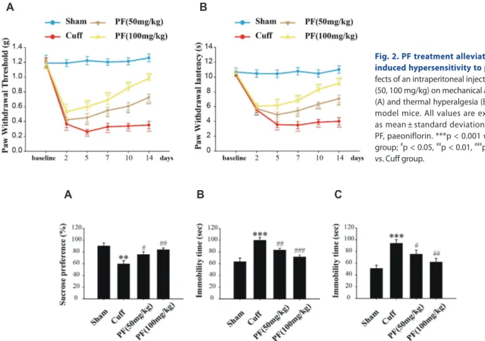
la-tency of all the groups were detected 1 day before model creation and at days 2, 5, 7, 10, and 14 after model creation. The results are shown in Fig. 2. The PWT and PWL in the Cuff group were significantly lower than those in the Sham group (PWT, p < 0.001; PWL, p < 0.001). After PF treatment, the PWT and PWL of mice were significantly increased (PWT, p < 0.001; PWL, p < 0.001). In addition, it was positively correlated with increasing concentra-tion of PF. The results suggest that the Cuff model successfully induced NP in mice. The PF reduced the mechanical hyperalgesia and thermal hyperalgesia in a dose-dependent manner and has a strong analgesic effect on NP.
Effect of PF on depressive-like behavior of NP mice
The SPT is a test to check the response of the mice to reward stimuli, and a lack of interest in reward stimuli is an important manifestation of depression. As shown in Fig. 3A, there were sig-nificant differences seen in the rate of sucrose consumption in the four groups of mice in SPT. The mice in the Cuff group showed a statistically significant decrease (p < 0.01) in sugar preference 14 days after surgery, compared with the Sham group. The sugar preference of the PF 50 mg/kg and PF 100 mg/kg groups wassig-nificantly increased (p < 0.05, p < 0.01) compared with the Cuff group. Results suggested that PF could inhibit the anhedonia successfully induced by the Cuff model in mice, indicating its an-tidepressant effects.
The increased immobility time of mice in the FST and TST is considered to be a manifestation of hopelessness. As shown in Fig. 3B, C, there were significant differences in the immobility time in FST and TST between the four groups of mice. The immobil-ity time of mice in the Cuff group was significantly increased compared with that in the Sham group (FST, p < 0.001; TST, p < 0.001). The treatment with PF shortened the time of Cuff mice (FST, p < 0.01, p < 0.001; TST, p < 0.05, p < 0.01), and the response was found to be dose dependent. The results suggest that the Cuff model successfully enhanced emotions of despair in mice, and PF can significantly improve despair-like emotions.
Effect of PF on morphological changes in
hippocampal CA3 structure in NP mice
H&E staining results are displayed in Fig. 4A. The pyramidal cells in the CA3 region of the Sham group were arranged neatly and tightly, without vacuoles and obvious nucleoli. In the Cuff
Fig. 2. PF treatment alleviates Cuff-induced hypersensitivity to pain. Ef-fects of an intraperitoneal injection of PF (50, 100 mg/kg) on mechanical allodynia (A) and thermal hyperalgesia (B) in Cuff model mice. All values are expressed as mean ± standard deviation. n = 10. PF, paeoniflorin. ***p < 0.001 vs. Sham group; #p < 0.05, ##p < 0.01, ###p < 0.001 vs. Cuff group.
B A
Fig. 3. Effects of PF on the depressive behavior of NP model mice. Effects of PF (50, 100 mg/kg) on sucrose preference test (A), forced swimming test (B), and tail suspension test (C) in Cuff model mice. Results are presented as mean ± standard deviation. n = 10. PF, paeoniflorin; NP, neuropathic pain. **p < 0.01, ***p < 0.001 vs. Sham group; #p < 0.05, ##p < 0.01, ###p < 0.001 vs. Cuff group.
group, the pyramidal cells in the CA3 region were arranged loosely, with vacuolar changes and inflammatory cell infiltration. The number of necrotic and inflammatory cells were signifi-cantly reduced and the cell arrangement was neat in the PF (50 mg/kg) group. After 14 days of treatment with PF (100 mg/kg), the morphology of the pyramidal cells in the CA3 region normal-ized, and the arrangement was also basically normal, with no sig-nificant difference compared with the Sham group. These results suggest that PF can reduce pyramidal cell damage and inhibit the inflammatory response in the CA3 region of the hippocampus.
Effect of PF on the expression of pro-inflammatory
cytokines in the hippocampus of NP mice
We used ELISA to detect the levels of pro-inflammatory cy-tokines TNF-, IL-6, and IL-1 in the hippocampus of mice. As shown in Fig. 4B–D, compared with the Sham group, the Cuff group caused a significant increase in the levels of TNF-, IL-6, and IL-1 in the hippocampus (p < 0.001). After PF treatment,
the levels of TNF-, IL-1, and IL-6 were significantly reduced (TNF-, p < 0.05, p < 0.01; IL-6, p < 0.01, p < 0.001; IL-1, p < 0.05, p < 0.01). These results suggest that the abnormal increase of pro-inflammatory cytokines in the hippocampus may be involved in the influence of NP-related depression, and PF can inhibit the expression of pro-inflammatory cytokines in the hippocampus.
Effect of PF on the activation of microglia induced by
NP mice
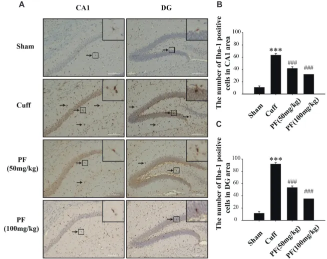
We used Iba-1 as a microglia activation marker to observe whether PF played a role in the activation of hippocampal mi-croglia in NP mice induced by the Cuff model. The Iba-1 positive cells were scattered in the hippocampal CA1 and DG regions of the Sham group (Fig. 5A). The cell bodies were elongated with dendritic protrusions, indicating a resting state. Compared with the Sham group, the Iba-1 positive cell bodies of the mice in the Cuff group became larger, the protrusions were short and thick, and the branch retraction was reduced, showing an activated
Fig. 4. PF improves the inflammatory infiltration of hippocampal pyramidal cells and reduces the expression of pro-inflammatory cytokines.
Representative photomicrographs of H&E staining in the CA3 (Bar = 100 m) region of hippocampus in different groups (A). ELISA showed that PF treatment decreased the expression of TNF- (B), IL-6 (C), and IL-8 (D) caused by Cuff. The data represent the mean ± standard deviation. n = 4. PF, pae-oniflorin. ***p < 0.001 vs. Sham group; #p < 0.05, ##p < 0.01, ###p < 0.001 vs. Cuff group.
B C D
state. After treatment with PF, the activated morphology of Iba-1 positive cells in mice was significantly improved compared with the Cuff group, leaning to a resting morphology.
The results of immunohistochemistry were analyzed using Image-ProPlus software. The expression level of Iba-1 in the CA1 and DG regions of the Cuff group significantly increased (p < 0.001) compared with the Sham group (Fig. 5B, C). The expres-sion of Iba-1 was significantly reduced after PF treatment in a dose-dependent manner (p < 0.001, p < 0.001). These results sug-gest that the activation of hippocampal microglia may be involved in NP-related depression, and PF can inhibit the activation of hip-pocampal microglia.
Effect of PF on the TLR4/NF-
B pathway in NP mice
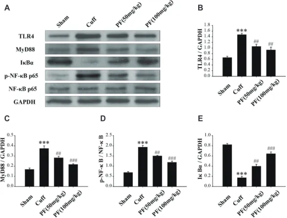
Western blot was used to analyze the expression levels of TLR4, MyD88, IB, p-NF-Bp65, and NF-Bp65 in the hippocam-pus. As shown in Fig. 6A–C, the expression levels of TLR4 and MyD88 in Cuff group mice were significantly increased (TLR4, p < 0.001; MyD88, p < 0.001). PF treatment downregulated theexpression of (TLR4, p < 0.01, p < 0.01; MyD88, p < 0.01, p < 0.001). NF-Bp65 is a nuclear protein related to the NF-B signaling pathway. As shown in Fig. 6D, PF inhibited the increase of p-NF-Bp65 expression caused by the Cuff model (p < 0.01, p < 0.001). IB is known to inhibit the activation and nuclear translocation of NF-B molecules, and the production of NF-Bp65 molecules is usually accompanied by the degradation of IB molecules [20]. As shown in Fig. 6E, the expression of IB was reduced in the Cuff group of mice (p < 0.001). The expression of IB was significantly upregulated after PF intervention (p < 0.01, p < 0.001). These results suggest that PF has an inhibitory effect on the TLR4/NF-B signaling pathway, and the TLR4/NF-B signaling pathway may be related to the antihyperalgesic and antidepres-sant‐like effects of PF.
DISCUSSION
According to epidemiological reports, the prevalence of depres-sion among patients with NP is approximately 34% [21]. Although
Fig. 5. PF treatment suppressed overactivation of microglia in the CA1 and DG induced by Cuff. Representative images of CA1 and DG (Bar = 100 m) region sections stained with Iba-1 antibodies (A; black arrows indicate positive microglia). Effect of PF on the number of positive cells for Iba-1 in CA1 (B) and DG (C) regions. Mean ± standard deviation of three experiments. n = 4. PF, paeoniflorin. ***p < 0.001 vs. Sham group; ###p < 0.001 vs. Cuff group.
B
C A
many drugs have been developed to treat pain and depression, most drugs have multiple adverse effects [22]. Previous studies have shown that PF, as a natural medicine, has the advantage of multiple targets and multiple pathways, and has significant effects in pain relief and anti-depression [23-26]. In this study, intraperi-toneal injection of PF for 14 days could attenuate hyperalgesia in mice, suggesting that PF has analgesic effects. After modeling, the chronic pain of mice may interfere with the results of the FST and TST; however, it has little effect on the increase in immobility time in the experiment, whether it was in the SPT, FST, or TST, it was concluded that the depression of mice increased. It should be noted that PF treatment can improve depression-like behavior.
The hippocampus is an important anatomical structure where NP is found to be associated with depression. Pathological studies have shown that hippocampal atrophy is detected in both NP and depressed patients [27-29]. H&E staining of the mouse hippocam-pus showed a large amount of inflammatory infiltration in the pyramidal cells of the CA3 region after Cuff modeling, and some cells were atrophied or lost. We speculate that this is precisely due to the neuroinflammation that caused changes in the hippocam-pus after Cuff modeling, which in turn increased depression in mice. PF can improve the damage in the hippocampal structure of the Cuff mice, suggesting that PF has neuroprotective and
antidepressant-like effects.
The mechanism of neuroinflammation has been reported in studies of NP and depression [4,30], and the occurrence and de-velopment of neuroinflammation are vital in the involvement of pro-inflammatory cytokines. Several clinical studies have found that patients with pain and depression have significantly higher concentrations of pro-inflammatory factors, such as TNF- and IL-6 in the peripheral blood than those not in pain and non-depressed patients [31,32]. In addition, it has also been found that animal models can manifest depression-like behaviors after re-ceiving intraventricular injection of pro-inflammatory cytokines [33]. The injection of an antagonist to interfere with the inflam-matory pathway can simultaneously alleviate hyperalgesia and depressive symptoms in the pain model [34]. Thus, the reduction of pro-inflammatory cytokines may have a therapeutic effect on NP and depression, which allowed us to evaluate the expression of several inflammation-related factors in the hippocampus. We found that PF significantly inhibited the overexpression of TNF-, IL-6, and IL-1 in the hippocampus of the Cuff group in a dose-dependent manner. We concluded that the analgesic and antidepressant effects of PF may be mediated, at least in part, by inhibiting hippocampal inflammation.
Microglia are widely distributed in the central nervous system
Fig. 6. PF inhibits the increase in expression of TLR4/NF-B pathway induced by Cuff. The expression of TLR4, MyD88, IB, p-Bp65, and NF-Bp65 were determined by western blot analysis in the hippocampus (A). Statistical analysis of relative levels of TLR4 (B), MyD88 (C), p-NF-NF-Bp65 (D), and IB (E). Quantitative results are expressed as mean ± standard deviation. n = 3. PF, paeoniflorin. ***p < 0.001 vs. Sham group; ##p < 0.01, ###p < 0.001
vs. Cuff group.
B
C D E
and can mediate a series of changes in the process of pain signal transmission. Their role has been widely recognized in mediat-ing pain. Under normal circumstances, microglia are considered to be dormant cells of the central nervous system, which activate rapidly after the nervous system is subjected to noxious stimula-tion. Activated microglia can release a large amount of neuroac-tive substances and pro-inflammatory cytokines [35]. These sub-stances and cytokines can act on neurons to enhance the release of nociceptive neurotransmitters from the primary afferent nerve endings and can further activate the glial cells to release more cytokines, forming a positive feedback effect, which causes per-sistent NP. Previous studies have found that in the NP model, mi-croglia activation participates and promotes pain hypersensitivity [36]. The activation of microglia has also been reported in human studies on depression [37]. It can be seen that abnormal activation of microglia is involved in the pathogenesis of NP and depression. Our results show that hippocampal microglia in Cuff mice are in an activated state, and PF can significantly inhibit the activation of hippocampal microglia.
The TLR4/NF-B signaling pathway is an important target for inhibiting microglial activation [38]. There are reports of high ex-pression of TLR4 and NF-B in both pain and deex-pression models [8,9]. First, we detected the expression of TLR4 and its down-stream MyD88 in the hippocampus by western blotting; PF in-hibited the expression of TLR4 and MyD88 in a dose-dependent manner. IB inhibits the transcription factor activity of NF-B by binding to NF-B [39]. Once stimulated, IB kinase partici-pates in the phosphorylation and ubiquitin-mediated degrada-tion of NF-B through the proteasome pathway, so that NF-B can be released and enter the nucleus to perform transcriptional activation, thereby activating the release of various inflammatory factors [40,41]. We continue to explore whether PF inhibits NF-B phosphorylation and INF-B degradation. Our research shows that PF blocks IB degradation in a dose-dependent manner. It effectively inhibits NF-B phosphorylation, thereby inhibiting the release of inflammatory factors and improving NP and its re-lated depression-like behaviors. In summary, our research proved that PF can reduce hippocampal inflammation by inhibiting the TLR4/NF-B signaling pathway in the hippocampus and allevi-ate Cuff-induced NP and depression-like symptoms. This study suggests that PF is a potentially effective treatment option for patients with NP and depression and is worthy of further clinical research.
We believe that neuralgia causes increased expression of TLR4/ NF-B pathway-related proteins, and activation of hippocampal microglia to release pro-inflammatory cytokines which causes hippocampal inflammation leading to the maintenance of NP and production of depression-like behaviors. This study shows that PF can inhibit the hippocampal inflammatory response through the TLR4/NF-B pathway and display an analgesic and antidepressant effect.
ACKNOWLEDGEMENTS
This work was supported by Morphology Experimental Center of Yanbian University. We are grateful for the financial support from the National Natural Science Foundation of China (No. 81860461).
CONFLICTS OF INTEREST
The authors declare no conflicts of interest.REFERENCES
1. Jensen TS, Baron R, Haanpää M, Kalso E, Loeser JD, Rice ASC, Treede RD. A new definition of neuropathic pain. Pain. 2011;152: 2204-2205.
2. Conrad R, Wegener I, Geiser F, Kleiman A. Temperament, charac-ter, and personality disorders in chronic pain. Curr Pain Headache Rep. 2013;17:318.
3. Radat F, Margot-Duclot A, Attal N. Psychiatric co-morbidities in patients with chronic peripheral neuropathic pain: a multicentre cohort study. Eur J Pain. 2013;17:1547-1557.
4. Mechawar N, Savitz J. Neuropathology of mood disorders: do we see the stigmata of inflammation? Transl Psychiatry. 2016;6:e946. 5. Mahajan GJ, Vallender EJ, Garrett MR, Challagundla L, Overholser
JC, Jurjus G, Dieter L, Syed M, Romero DG, Benghuzzi H, Stock-meier CA. Altered neuro-inflammatory gene expression in hippo-campus in major depressive disorder. Prog Neuropsychopharmacol Biol Psychiatry. 2018;82:177-186.
6. Wang YL, Han QQ, Gong WQ, Pan DH, Wang LZ, Hu W, Yang M, Li B, Yu J, Liu Q. Microglial activation mediates chronic mild stress-induced depressive- and anxiety-like behavior in adult rats. J Neu-roinflammation. 2018;15:21.
7. Lei Y, Chen CJ, Yan XX, Li Z, Deng XH. Early-life lipopolysaccha-ride exposure potentiates forebrain expression of NLRP3 inflam-masome proteins and anxiety-like behavior in adolescent rats. Brain Res. 2017;1671:43-54.
8. Liu S, Liu YP, Song WB, Song XJ. EphrinB-EphB receptor signaling contributes to bone cancer pain via Toll-like receptor and proin-flammatory cytokines in rat spinal cord. Pain. 2013;154:2823-2835. 9. Cheng Y, Pardo M, Armini RS, Martinez A, Mouhsine H, Zagury
JF, Jope RS, Beurel E. Stress-induced neuroinflammation is mediat-ed by GSK3-dependent TLR4 signaling that promotes susceptibility to depression-like behavior. Brain Behav Immun. 2016;53:207-222. 10. Zhou YX, Gong XH, Zhang H, Peng C. A review on the
pharmaco-kinetics of paeoniflorin and its anti-inflammatory and immuno-modulatory effects. Biomed Pharmacother. 2020;130:110505. 11. Wang D, Liu L, Li S, Wang C. Effects of paeoniflorin on
neurobe-havior, oxidative stress, brain insulin signaling, and synaptic altera-tions in intracerebroventricular streptozotocin-induced cognitive impairment in mice. Physiol Behav. 2018;191:12-20.
12. Zheng M, Liu C, Fan Y, Yan P, Shi D, Zhang Y. Neuroprotection by Paeoniflorin in the MPTP mouse model of Parkinson's disease. Neuropharmacology. 2017;116:412-420.
13. Gu X, Cai Z, Cai M, Liu K, Liu D, Zhang Q, Tan J, Ma Q. Protective effect of paeoniflorin on inflammation and apoptosis in the cerebral cortex of a transgenic mouse model of Alzheimer's disease. Mol Med Rep. 2016;13:2247-2252.
14. Zhou J, Wang L, Wang J, Wang C, Yang Z, Wang C, Zhu Y, Zhang J. Paeoniflorin and albiflorin attenuate neuropathic pain via MAPK pathway in chronic constriction injury rats. Evid Based Comple-ment Alternat Med. 2016;2016:8082753.
15. Yin D, Liu YY, Wang TX, Hu ZZ, Qu WM, Chen JF, Cheng NN, Huang ZL. Paeoniflorin exerts analgesic and hypnotic effects via adenosine A1 receptors in a mouse neuropathic pain model. Psy-chopharmacology (Berl). 2016;233:281-293.
16. Hu B, Xu G, Zhang X, Xu L, Zhou H, Ma Z, Shen X, Zhu J, Shen R. Paeoniflorin attenuates inflammatory pain by inhibiting microglial activation and Akt-NF-B signaling in the central nervous system. Cell Physiol Biochem. 2018;47:842-850.
17. Benbouzid M, Pallage V, Rajalu M, Waltisperger E, Doridot S, Pois-beau P, Freund-Mercier MJ, Barrot M. Sciatic nerve cuffing in mice: a model of sustained neuropathic pain. Eur J Pain. 2008;12:591-599. 18. Chaplan SR, Bach FW, Pogrel JW, Chung JM, Yaksh TL.
Quantita-tive assessment of tactile allodynia in the rat paw. J Neurosci Meth-ods. 1994;53:55-63.
19. Wang Z, Huang H, Yang S, Huang S, Guo J, Tang Q, Qi F. Long-term effect of ropivacaine nanoparticles for sciatic nerve block on postoperative pain in rats. Int J Nanomedicine. 2016;11:2081-2090. 20. Schneiderhan J, Orizondo C. Chronic pain: how to approach these 3
common conditions. J Fam Pract. 2017;66:145-157.
21. Gustorff B, Dorner T, Likar R, Grisold W, Lawrence K, Schwarz F, Rieder A. Prevalence of self-reported neuropathic pain and impact on quality of life: a prospective representative survey. Acta Anaes-thesiol Scand. 2008;52:132-136.
22. Kuthati Y, Lin SH, Chen IJ, Wong CS. Melatonin and their analogs as a potential use in the management of Neuropathic pain. J Formos Med Assoc. 2019;118:1177-1186.
23. Qiu F, Zhong X, Mao Q, Huang Z. The antidepressant-like effects of paeoniflorin in mouse models. Exp Ther Med. 2013;5:1113-1116. 24. Qiu FM, Zhong XM, Mao QQ, Huang Z. Antidepressant-like effects
of paeoniflorin on the behavioural, biochemical, and neurochemical patterns of rats exposed to chronic unpredictable stress. Neurosci Lett. 2013;541:209-213.
25. Nam KN, Yae CG, Hong JW, Cho DH, Lee JH, Lee EH. Paeoniflo-rin, a monoterpene glycoside, attenuates lipopolysaccharide-in-duced neuronal injury and brain microglial inflammatory response. Biotechnol Lett. 2013;35:1183-1189.
26. Wu YM, Jin R, Yang L, Zhang J, Yang Q, Guo YY, Li XB, Liu SB, Luo XX, Zhao MG. Phosphatidylinositol 3 kinase/protein kinase B is responsible for the protection of paeoniflorin upon H2O2-induced neural progenitor cell injury. Neuroscience. 2013;240:54-62. 27. Nguyen L, Kakeda S, Katsuki A, Sugimoto K, Otsuka Y, Ueda I,
Igata R, Watanabe K, Kishi T, Iwata N, Korogi Y, Yoshimura R. Re-lationship between VEGF-related gene polymorphisms and brain morphology in treatment-naïve patients with first-episode major de-pressive disorder. Eur Arch Psychiatry Clin Neurosci. 2019;269:785-794.
28. Taylor WD, Deng Y, Boyd BD, Donahue MJ, Albert K, McHugo M, Gandelman JA, Landman BA. Medial temporal lobe volumes in late-life depression: effects of age and vascular risk factors. Brain Imaging Behav. 2020;14:19-29.
29. Mutso AA, Radzicki D, Baliki MN, Huang L, Banisadr G, Centeno MV, Radulovic J, Martina M, Miller RJ, Apkarian AV. Abnormali-ties in hippocampal functioning with persistent pain. J Neurosci. 2012;32:5747-5756.
30. Jha MK, Jeon S, Suk K. Glia as a Link between neuroinflammation and neuropathic pain. Immune Netw. 2012;12:41-47.
31. Burston JJ, Valdes AM, Woodhams SG, Mapp PI, Stocks J, Watson DJG, Gowler PRW, Xu L, Sagar DR, Fernandes G, Frowd N, Mar-shall L, Zhang W, Doherty M, Walsh DA, Chapman V. The impact of anxiety on chronic musculoskeletal pain and the role of astrocyte activation. Pain. 2019;160:658-669.
32. Ünal Ö, Akyol Y, Tander B, Ulus Y, Terzi Y, Kuru Ö. The relation-ship of illness perceptions with demographic features, pain sever-ity, functional capacsever-ity, disabilsever-ity, depression, and quality of life in patients with chronic low back pain. Turk J Phys Med Rehabil. 2019;65:301-308.
33. Kaster MP, Gadotti VM, Calixto JB, Santos AR, Rodrigues AL. De-pressive-like behavior induced by tumor necrosis factor- in mice. Neuropharmacology. 2012;62:419-426.
34. Dellarole A, Morton P, Brambilla R, Walters W, Summers S, Ber-nardes D, Grilli M, Bethea JR. Neuropathic pain-induced depres-sive-like behavior and hippocampal neurogenesis and plasticity are dependent on TNFR1 signaling. Brain Behav Immun. 2014;41:65-81.
35. Bachtell RK, Jones JD, Heinzerling KG, Beardsley PM, Comer SD. Glial and neuroinflammatory targets for treating substance use dis-orders. Drug Alcohol Depend. 2017;180:156-170.
36. Mika J, Zychowska M, Popiolek-Barczyk K, Rojewska E, Przewlocka B. Importance of glial activation in neuropathic pain. Eur J Pharma-col. 2013;716:106-119.
37. Krabbe KS, Reichenberg A, Yirmiya R, Smed A, Pedersen BK, Bru-unsgaard H. Low-dose endotoxemia and human neuropsychologi-cal functions. Brain Behav Immun. 2005;19:453-460.
38. Chen J, Wang Z, Zheng Z, Chen Y, Khor S, Shi K, He Z, Wang Q, Zhao Y, Zhang H, Li X, Li J, Yin J, Wang X, Xiao J. Neuron and mi-croglia/macrophage-derived FGF10 activate neuronal FGFR2/PI3K/ Akt signaling and inhibit microglia/macrophages TLR4/NF-B-dependent neuroinflammation to improve functional recovery after spinal cord injury. Cell Death Dis. 2017;8:e3090.
39. Zhou Z, Lin J, Huo R, Huang W, Zhang J, Wang L, Sun Y, Shen B, Li N. Total glucosides of paeony attenuated functional maturation of dendritic cells via blocking TLR4/5 signaling in vivo. Int Immuno-pharmacol. 2012;14:275-282.
40. Wium-Andersen MK, Ørsted DD, Nielsen SF, Nordestgaard BG. El-evated C-reactive protein levels, psychological distress, and depres-sion in 73, 131 individuals. JAMA Psychiatry. 2013;70:176-184. 41. Dahl J, Ormstad H, Aass HC, Malt UF, Bendz LT, Sandvik L,
Brun-din L, Andreassen OA. The plasma levels of various cytokines are increased during ongoing depression and are reduced to normal levels after recovery. Psychoneuroendocrinology. 2014;45:77-86.